Growing up
In some insects - think flies, beetles, moths and butterflies - the creature that emerges from the egg bears little resemblance to the adult. In others - such as cockroaches, grasshoppers and mantids - it is quite recognisable as the mature insect, but has a lot of growing up to do.
Katydids (family Tettigoniidae) belong to the latter group of insects. You may recall my account of the hatching of a clutch of katydid eggs we discovered in early Spring last year. We decided to follow these young nymphs right through to the adult stage.


Why? First, we were keen to find out exactly what species of katydid these nymphs were. We were pretty sure they belonged to the genus Torbia. But species identity generally can’t be determined with confidence at nymphal or larval stages. We had to find out what they looked like as adults.
Second, in few cases have the changes that particular katydid species undergo during nymphal development been described in detail (David Rentz 2010 “A Guide to the Katydids of Australia”. CSIRO Publishing).
What was clear at the outset was that our baby katydids would undergo a series of moults on their journey to adulthood. This involves shedding the entire body covering, the exoskeleton, to allow continued growth. You can see this process happening here in a stick insect.
We sought answers to a number of specific questions:
How long does a freshly hatched nymph take to grow into an adult?
How many moults does a katydid nymph make and how long is the period between moults (an instar)?
How does a nymph change in appearance and behaviour as it develops?
When do the wings and other external organs become visible?
When can one first distinguish the sexes?
And so it begins - 1st instar
Our clutch of katydid eggs hatched out over a 4 day period between 20-23 October. This asynchronony in development became more pronounced as the nymphs progressed further.
25 of the 29 eggs in the clutch hatched successfully. Four of the nymphs escaped soon after hatching and 2 died in the first couple of days from an apparent fungal infection. So we were left with 19 animals to foster.
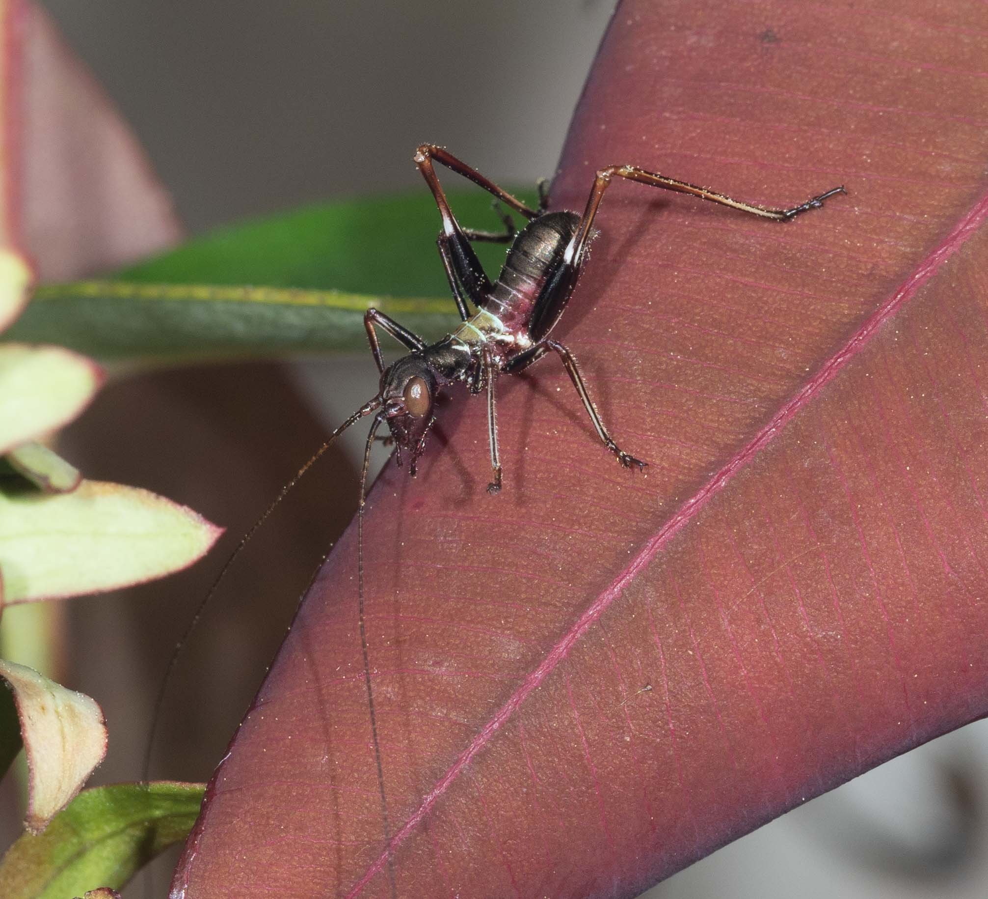
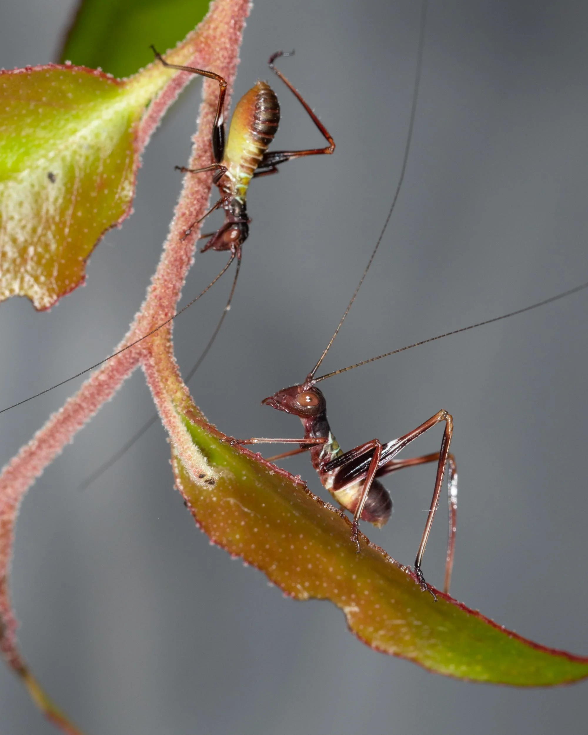
Soon after hatching, the nymphs were around 5-6mm in body length. The upper side of the body was chocolate brown, apart from the second and third thoracic segments (mesonotum and metanotum) which were a golden colour with a narrow white rear border. The underside was crimson red.
These first instar nymphs looked like ants. Ant mimicry is a well known feature of early Torbia nymphs (Rentz, 2010). It aids survival because most predators find ants distasteful.
Their oversized, staring eyes and dark bodies lend them a slightly menacing, alien appearance.


We first supplied the nymphs with Gahnia inflorescences and soon saw them grazing on the pollen grains. A couple of days later we gave them sprouting eucalypt vegetation which they readily accepted. The holes in the leaves in the images above bear testament to that fact!
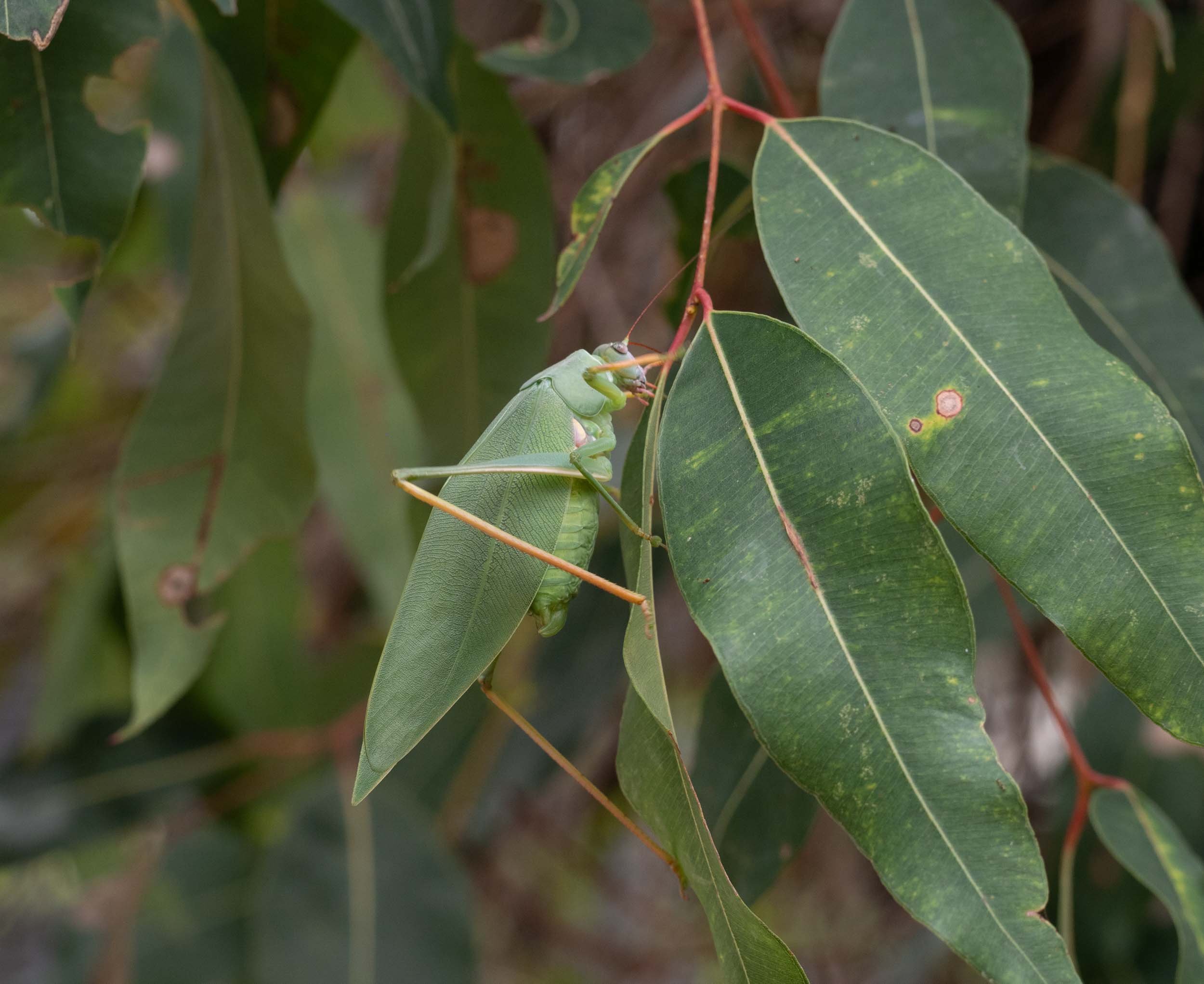
The nymphs preferred to be at the top of the branches and at the least disturbance would climb upwards. This was a totally stereotypic behaviour. In their natural environment this would place them amongst the young, reddish juvenile leaves to which they were well matched in colour. Indeed, it was difficult to sight them amongst these upper leaves in their enclosure.
2nd instar - growth and changes in body proportions
In the days that followed, the nymphs fed hungrily on the eucalypt leaves – as seen in this video – demanding a fresh supply of foliage every few days.
There was little change in their appearance, apart from an obvious swelling of the abdomen, which took on a pale yellow-green hue below. This expansion is possible even without moulting because the cuticular plates of the abdomen are joined together with flexible strips.


Nonetheless, a careful comparison of photos of nymphs taken on 29th October (first instar panel of photos) and 6th November (above panel) shows that the relative eye/head size and the head/thorax size is greater at the earlier date.
This strongly suggests that a moult occurred between these times. So a few precocious developers were now second instar nymphs.
On 19th November, we caught nymphs in the act of this first moult. Nymphs lay motionless alongside their discarded skin (exuvia). After a few minutes they proceeded to eat the exuvia, leaving no trace. Insect cuticle is made of the protein chitin, so is highly nutritious.


The measured body length of nymphs around this time was 10mm. So they had doubled their size in a month.


3rd instar - first sign of wing buds
On 2nd December, two weeks after witnessing that first moulting event, we saw other nymphs consuming their freshly exuvia - a second moult! The freshly moulted insects showed the first signs of wing buds - small yellow flaps on the sides of the second and third thoracic segments.


The body length of the largest nymphs had increased to 14mm.
4th instar - ears appear
The largest nymphs seen on 12th December were significantly longer than 3rd instar nymphs - 17mm vs. 14mm. Their pronotum/head length was greater than at the earlier stage, while their eye/head size was smaller – compare the two images below of nymphs from 4th and 12th December.
4th December. 3rd instar nymph
12 December - 4th instar nymph
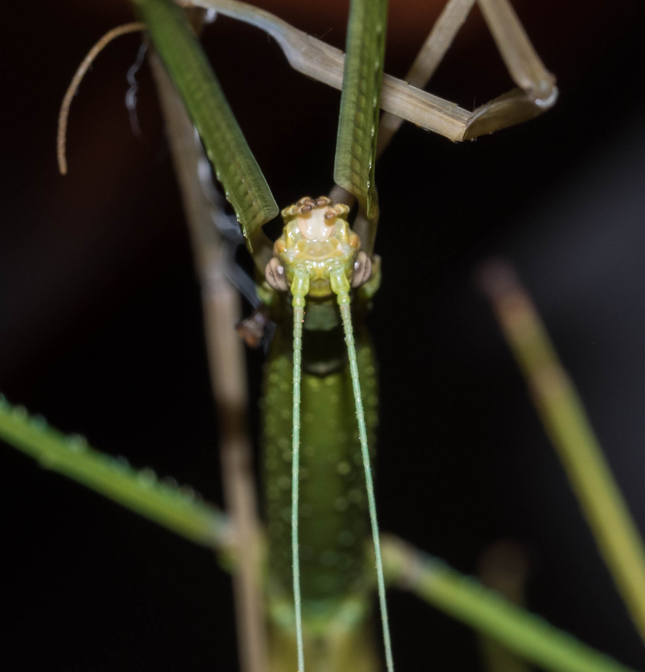
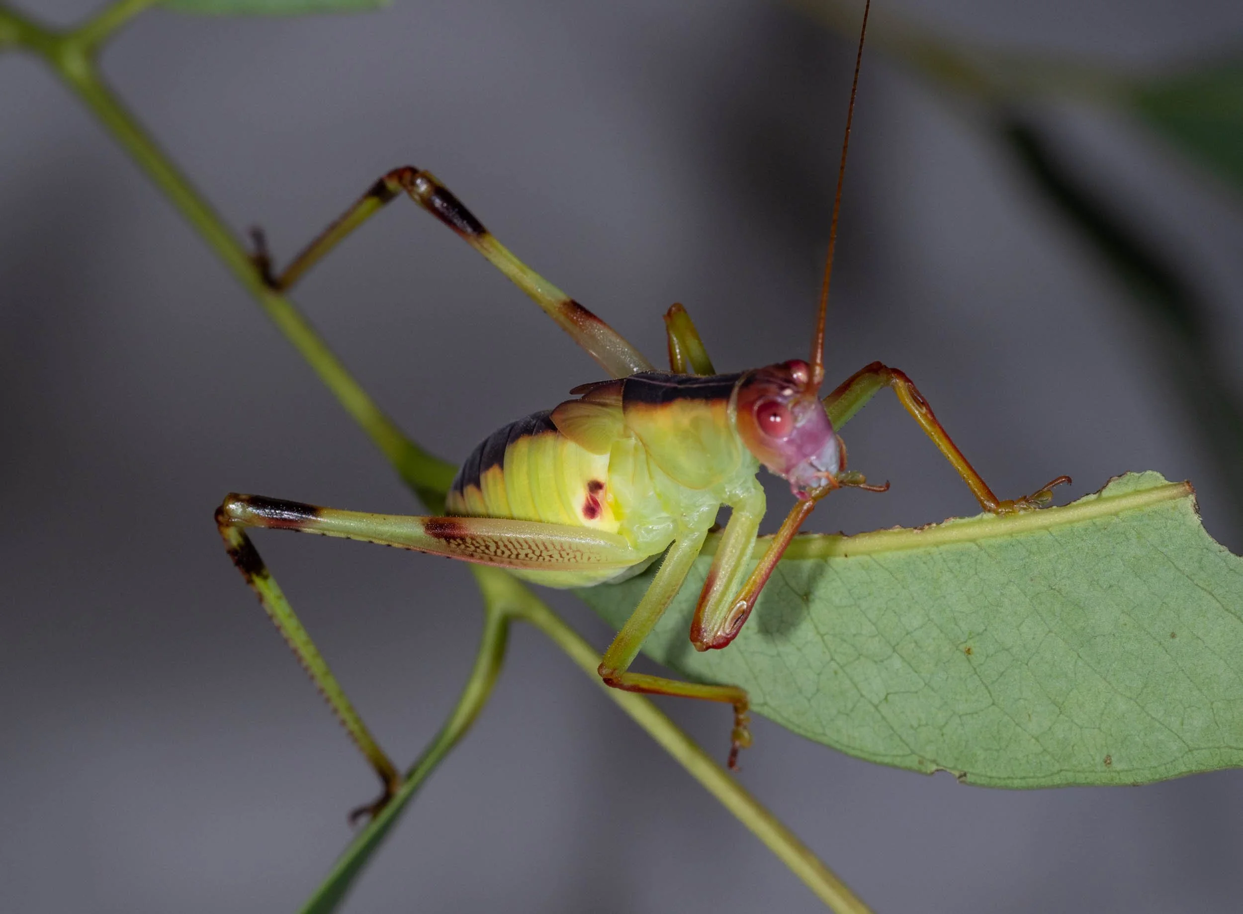
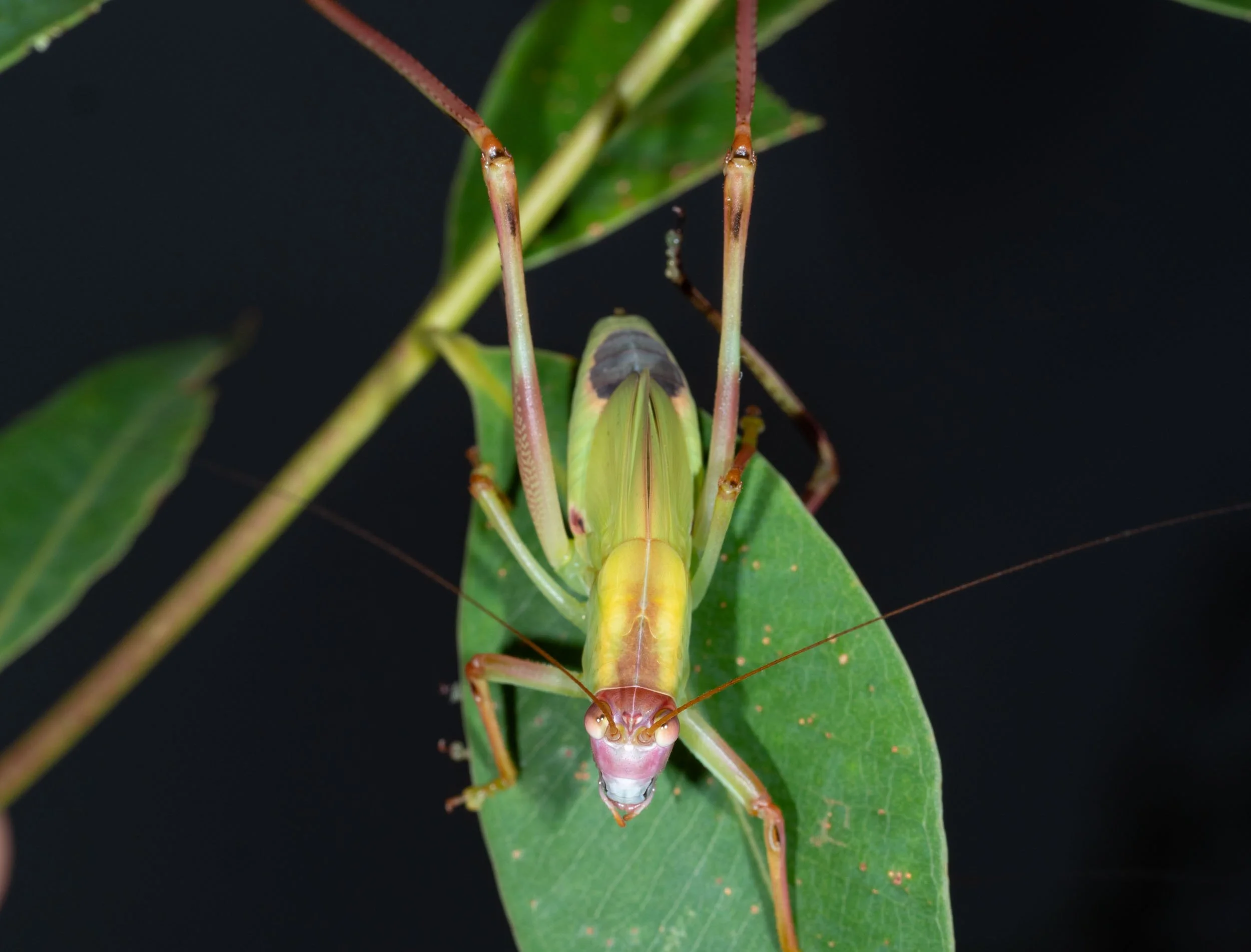
These changes were relatively subtle. But a major new addition was apparent in some nymphs - they sported a new organ, an ear!
The opening to the katydid’s hearing organ lies on the front and back of each fore tibia. The front opening is indicated with an arrow in the RH photo above of the nymph from 12th December. So it is clear that the nymphs in this pair of photos must be separated by a moult.
A thin membrane, the tympanum, sits beneath these oval openings. A sense organ connected to the tympanum converts air vibrations entering those openings into electrical signals. These travel along a leg nerve to the katydid’s central nervous system.
A slightly depressed, darkened area - the anlage of the tympanal opening - can actually be seen on the fore tibiae of 3rd instar nymphs (arrowed in the 4th December nymph).
From 15th December onwards the nymphs became progressively more active, suggesting that they had entered a dispersal phase.
We had planned to leave on a 2 week journey north on 21st December - with our katydid palace in tow. To simplify the logistics of katydid care during the trip, we released 9 of the nymphs into a eucalypt tree in our home forest on the day we left.
They appeared to quickly adapt to their new, natural home, grazing on leaves on the lower branches. The nymphs did not attempt to jump away after release. They simply clambered around or paused to chew on the vegetation. Eventually they began quietly climbing upwards towards the crown of the tall eucalypt.
This behaviour is consistent with what we observed at earlier and later developmental stages. They would seldom jump within their cage, although they are capable of powerful leaps to escape a threat. Rather they rely on their superb camouflage to avoid predators. We often struggled to find them on the eucalypt leaves, even within their small cage!


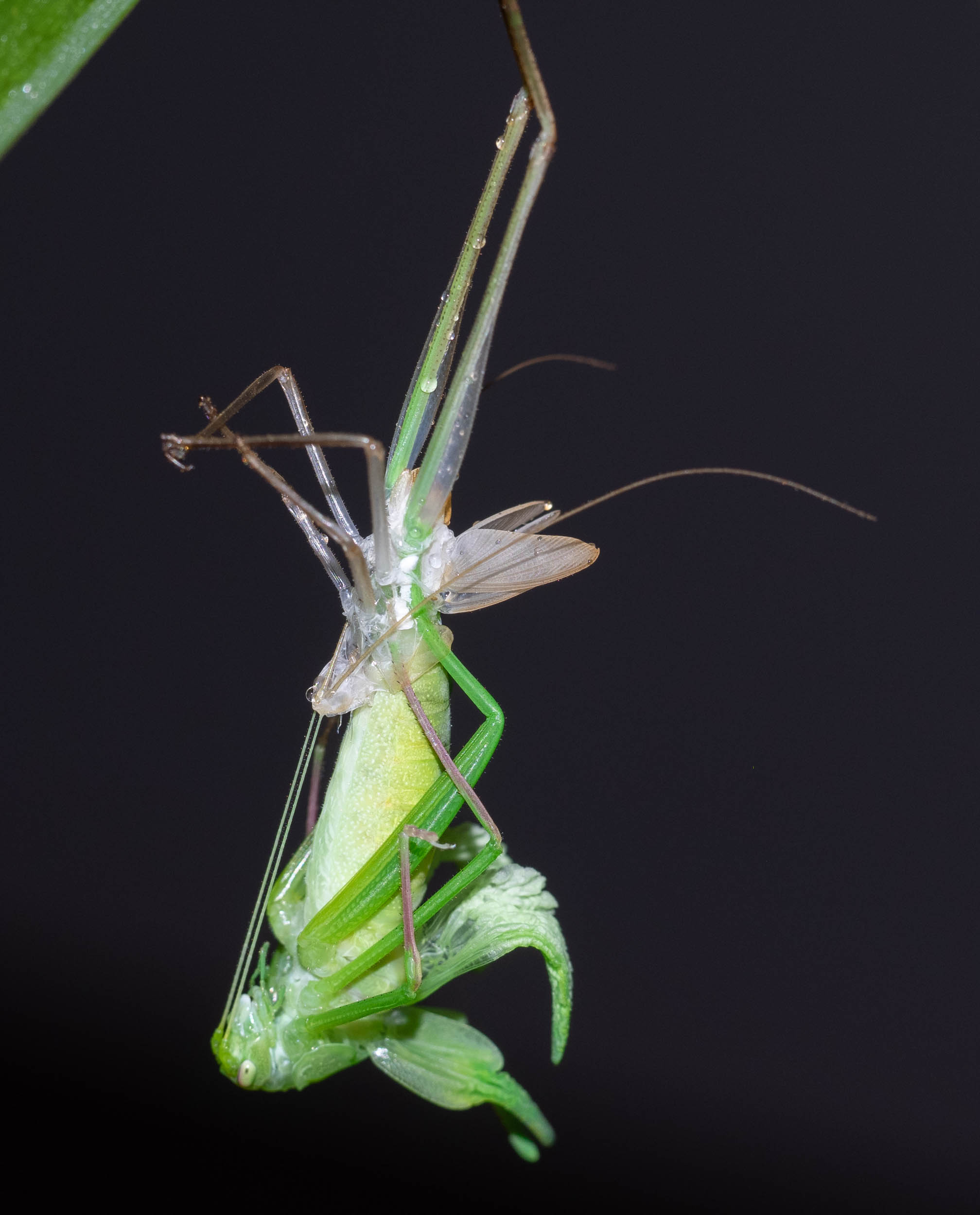
5th instar - reoriented wing buds
It was 4 days later, on 25th December, before we were next able to examine the nymphs closely. They had changed significantly. Both fore wing and hind wing buds projected directly towards the rear and lay parallel and close to the dorsal midline.
A moult to the 5th instar must have occurred to bring about these changes.


6th instar - enlarged wing buds
After our return home on 3rd January we found that one of the nymphs was substantially larger than its siblings - with a body length of 30mm. Its wing buds were also much larger and had a different form, so it must have advanced to the 6th instar.
Furthermore it clearly possessed an ovipositor - the structure used by female insects to deposit eggs during laying - or more correctly, an ovipositor anlage.


By 17th January, 8 of the nymphs had reached the 6th instar. All except one of these were females and had body lengths between 32-35mm. The other nymph, judged to be a male by the absence of an ovipositor, was much smaller with a body length of 25mm.
One of the two developmental “laggards” still at the 5th instar on 17th January was an animal that had torn off a leg during moulting to the 4th instar (RH photo below). It did successfully moult to the 6th instar the next day, but didn’t regenerate its missing leg parts. This nymph was also a male. We called him “Hoppy”.
To answer one of the questions posed at the outset - katydid sex can be determined from the structure of the terminal segments as early as the 6th instar.


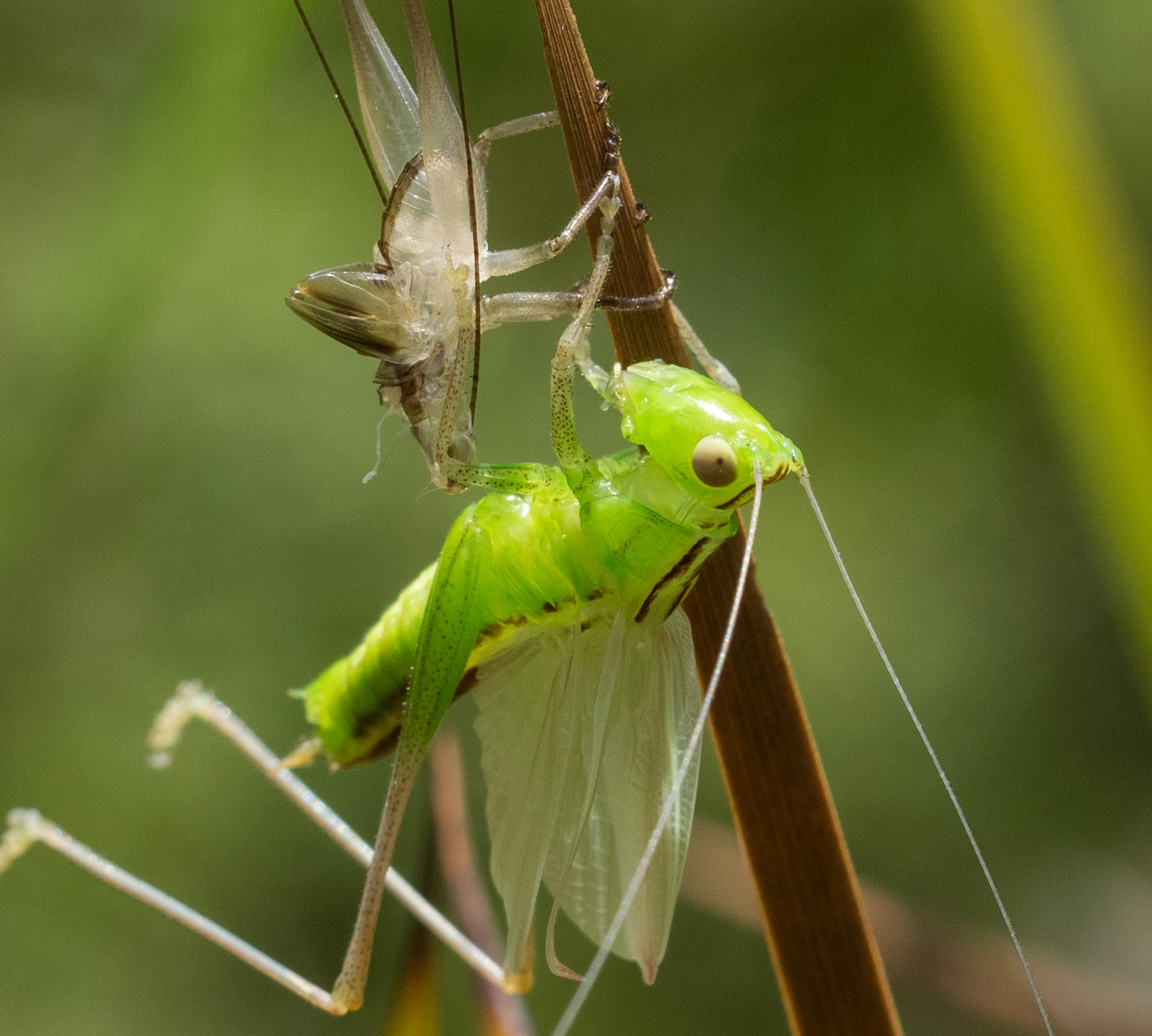
Adult stage - all grown up
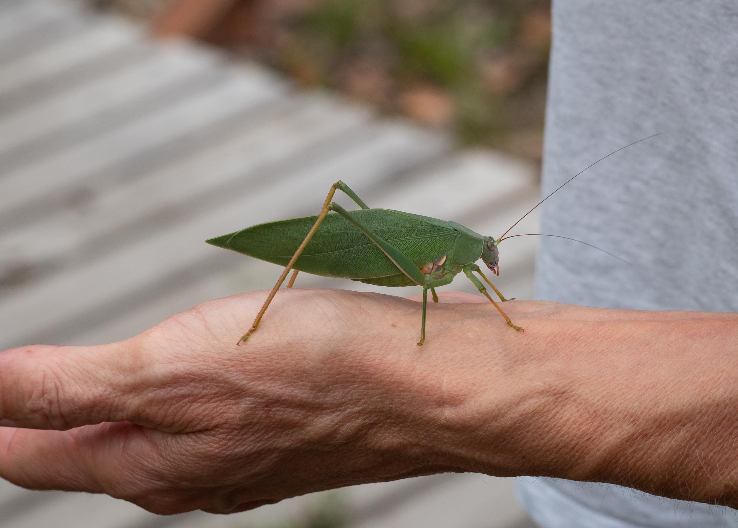
On 18th January we were excited to discover the first adult in the katydid cage. It is quite a magnificent creature, with its long wings, which are almost twice the size of the body. You can really only see the forewings, called tegmina, in a resting katydid. Just the tips of the hindwings, which lie beneath, are visible. The tough tegmina are just protective covers for the hindwings and are not used in flight. They are held out sideways when a katydid takes flight, allowing the hindwings to flap.
Over the next 19 days, all of the 6th instar nymphs turned into adults. So given that the clutch hatched between 20-23 October, we calculate that post-embryonic development takes between 87 and 109 days.
We never managed to catch a nymph in the process of carrying out its final moult - despite several late night attempts. However you can watch all of the action as I recorded it in a different home katydid species a few years ago.


Seven of these adults were females as shown by the presence of an ovipositor. Their short ovipositor is rather unusual for katydids. Most katydids possess a long, pointed ovipositor for pushing eggs into the ground. Remember, however, that our species deposits its eggs in a line on the surface of a plant stem. A short ovipositor is clearly admirably suited for this task.
The other 3 adults were males. Two of these were the last to undergo the final moult, on 5th and 6th February. The former was Hoppy, which we know entered the 6th instar on 18th January. The other male entered the 6th instar on 17th January. So the 6th instar took 18 and 20 days, respectively, for these male nymphs.
For a number of reasons, it wasn’t possible to determine precisely the lengths of the other instars. First, apart from Hoppy, we couldn’t recognise nymphs as individuals through development. Second, there was a high degree of asynchrony in development of the clutch. Finally, we didn’t make observations of the clutch on every day throughout development.


The adult females were noticeably larger than the males. The average tegmen length for the 3 males was 43.7mm, that of the 7 females was 53.8mm. And of course, their terminalia were different. Males lack an ovipositor. They have a pair of gonostyles in the equivalent position.
There is another clear difference between male and female katydids - males make a calling sound to attract females. We heard this soon after the first adult male appeared. It was a rather soft sound, quite unlike the raucous katydid calls we often hear on a warm summer’s night in our forest. I made a recording some years ago of such loud katydid calls, which you can hear below.
Male katydids call by rubbing their tegmina together. The base of the tegmina has a complex pattern of veins, which you can see in the third photo in the panel above. A row of teeth on the underside of the left tegmen functions like a file. They rub against a raised vein on upper side of the right tegmen, producing the calling sound. The pattern of file teeth and the resulting call is species specific.
What is this species?
Our adult katydids clearly display the defining features of genus Torbia, as described in Rentz (2010):
a flat dorsal surface of the pronotum (Fig. A, white arrow)
a broad lateral lobe…nearly as long as the dorsal length of the pronotum itself (Fig. A, black arrow)
narrow, laterally compressed thoracic sterna (Fig. B)
meso- and metathoracic lobes longer than broad (Fig. B, black arrows)


I am also confident that this is Torbia perficita, the Giant Torbia, based on the following defining characters (Rentz, 2010):
large size
veins of tegmina strongly indicated both in structure and emphasised by colour (Fig. A)
posterior margin of pronotum well-produced (Fig. A)
pinkish-purple, prominent mark on thorax (Fig. B)
Most convincingly, the venation at the base of the male’s left tegmen is identical to the type specimen of Torbia perficita in the British Museum of Natural History. Nothing has changed in 150 years!

Left mirror area of tegmen of male Torbia perficita Walker, 1869. Lectotype Natural History Museum, London. Image downloaded from Orthoptera Species File.

5 Feb 2020 Base of left tegmen of adult male (Hoppy)

Drawings of Torbia perficita Plate 119 from McCoy’s Zoology of Victoria Prodromus.
Torbia perficita is one of the species illustrated in Frederick McCoy’s Prodromus of the Zoology of Victoria 1890. At the time it was called Phaneroptera valida.
The illustrator did a wonderful job of depicting the anatomy of this katydid. Drawings in the top half of the page show the male and his various body parts, while the female is displayed in the bottom half (apart from her terminalia which are included with the male body parts).
Both a work of art and a valuable taxonomic record!
You can view a copy of these drawings together with the accompanying description by McCoy in this pdf file, which I have downloaded from the Biodiversity Heritage Library.
Freedom Day: a happy ending (hopefully)
We released all of the adults into the wild, placing them on the lower, epicormic growth of tall eucalypts in our forest. Watching them quietly crawl away on the leaves, we were once again struck by their superb camouflage.
We’re secretly hoping that potential predators have as much trouble finding the katydids amongst the eucalypt foliage as we did.