Breaking out
A secure refuge can also be a prison unless you have a way of getting out. Insects have found diverse ways of addressing this challenge. We recently witnessed the Houdini-esque struggles of katydid nymphs breaking free from the confines of their embryonic home.
The insect egg - a (relatively) safe place to develop
Many insects produce eggs with a hard shell (a chorion) to protect their offspring during embryonic development - the most vulnerable stage of any animal’s life. The mother chooses a suitable spot to lay her eggs and then generally leaves them to their own devices. Safely encased within, the embryo develops new cells and tissues which are assembled into organs and body parts.


Insect eggs are not always an impregnable bunker. Some species of wasps can penetrate the chorion of an insect egg with their ovipositor and deposit their own egg inside. The wasp embryo then develops at the expense of the host.


Mystery eggs
On 16th September, I found a clutch of 29 insect eggs neatly arranged on an upper twig of a 1m high eucalypt seedling. Each had a dark seam running along its length. They looked like tiny purses.
Their shape and arrangement was somehow familiar. A search in David Rentz’s field guide “A Guide to the Katydids of Australia” confirmed our suspicion that they were katydid eggs, although his photo (page 12) did not identify the species.
The clutch of 29 mystery eggs found on 16th September
These eggs must have been there for some time. We hadn’t seen an adult katydid for many months.
It’s not uncommon for insects to overwinter as eggs. Embryonic development in insects usually takes only a few weeks. So what does the embryo do to bridge this time gap? It goes into a state of arrested development called diapause. David Rentz notes that some katydid species undergo a diapause.
How long would we have to wait to see our hatchlings? What species of katydid were they? To get answers to these questions, we put the eggs into our “buddy tank” - our repository for rearing developing creatures, be they eggs, nymphs, larvae or pupae.
Hatchlings!
Late on the morning of 20th October, just over a month after we found the eggs, Kerri spotted 6 tiny, crimson insects clambering over the clutch. Their long antennae confirmed their identity - if there was any doubt - as katydid nymphs. Very elegant creatures.
The six eggs from which the nymphs had emerged had a white object attached to one end of the opened seam. This looked like some sort of membrane. It turned out to be the cuticle layer that covers the embryo inside the egg, which is shed during hatching. More about that later.


No more nymphs appeared on that day. Following a tip in David Rentz’s book that katydids often hatch at dawn, we decided to arise very early the next morning. Our sleep sacrifice was handsomely rewarded.
Hatching - from start to finish
An early start was warranted. My first glance of the eggs at 4:00am revealed a nymph already starting to hatch. The egg had split along its seam giving a first glimpse of the creature within.
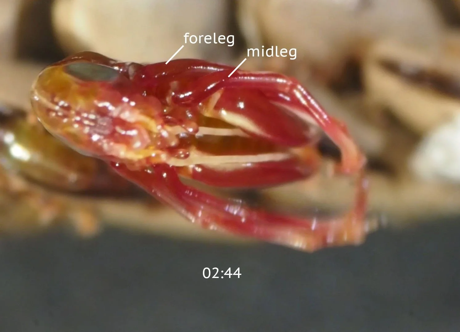
Over the next 5 minutes, shown in the following image sequence and video, the nymph thrust its body out of the egg case, starting with its head. The forelegs and middle legs were flung apart in dramatic fashion as if the nymph were announcing its arrival (2:46m).
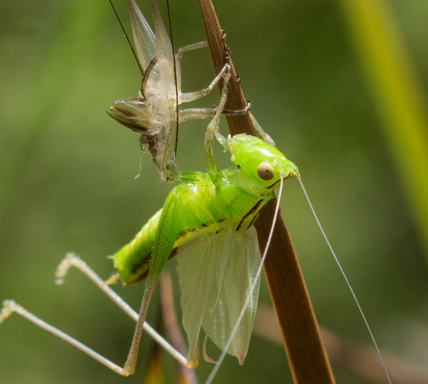
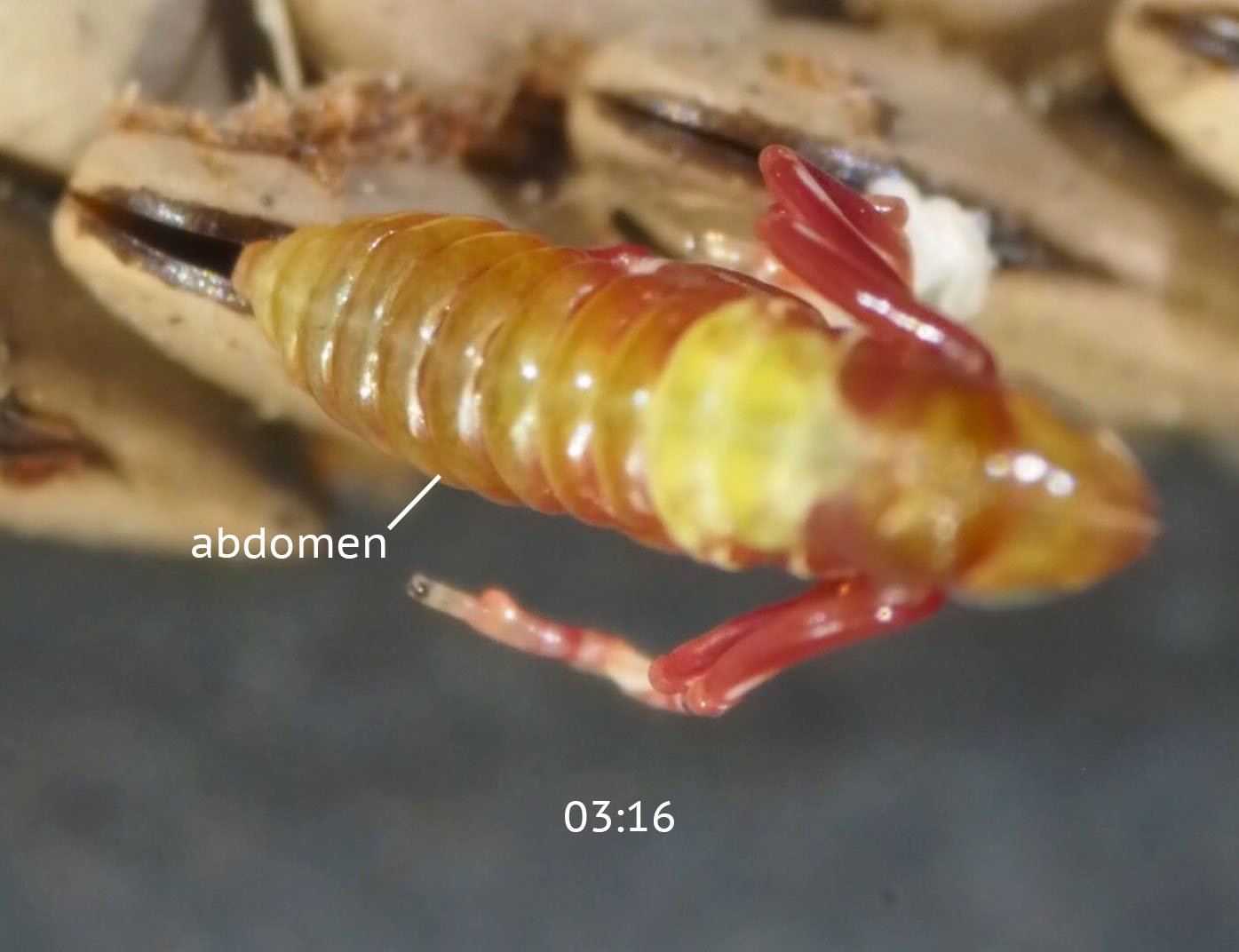
Once the abdomen had been withdrawn, the nymph paused with its hindlegs still trapped inside the egg case (3:16m). The transparent skin on its body started to wrinkle, marking the beginning of its first post-embryonic moult (3:59m). The cuticle coat it had developed inside the egg was being discarded.
This process, called ecdysis, will be repeated on several occasions throughout the the life of the insect. Its inflexible cuticle coat must be shed to allow continued growth of its body.


The movie of this process also shows the start of hatching in the neighbouring egg.
I reoriented the clutch to get a different view of this second insect. At the start of the movie below the nymph had already withdrawn its head and part of its forelegs. This different viewing angle reveals the process of withdrawal of the limbs and antennae to good effect.
Quite reminiscent of a scene from “Alien”!
Final escape from the cuticle and egg case
In the next phase of hatching, shown in the sequence of stills below, the nymph completes its escape from the confines of its embryonic skin.
The cuticle wrinkles as the nymph withdraws its body - indicated in images 00:35 to 03:24. The hindlegs are the final parts to be freed. The discarded cuticle remains attached to the egg case - that white object previously referred to. The cuticle actually remains attached to the egg case throughout the process, providing grip as the nymph extracts its body.
The nymph pulls its hindlegs clear of both the cuticle and the egg case. The tibia bends alarmingly during this process. The new exoskeleton is quite elastic at this stage (4:41, 6:23) but hardens quickly after hatching is completed.


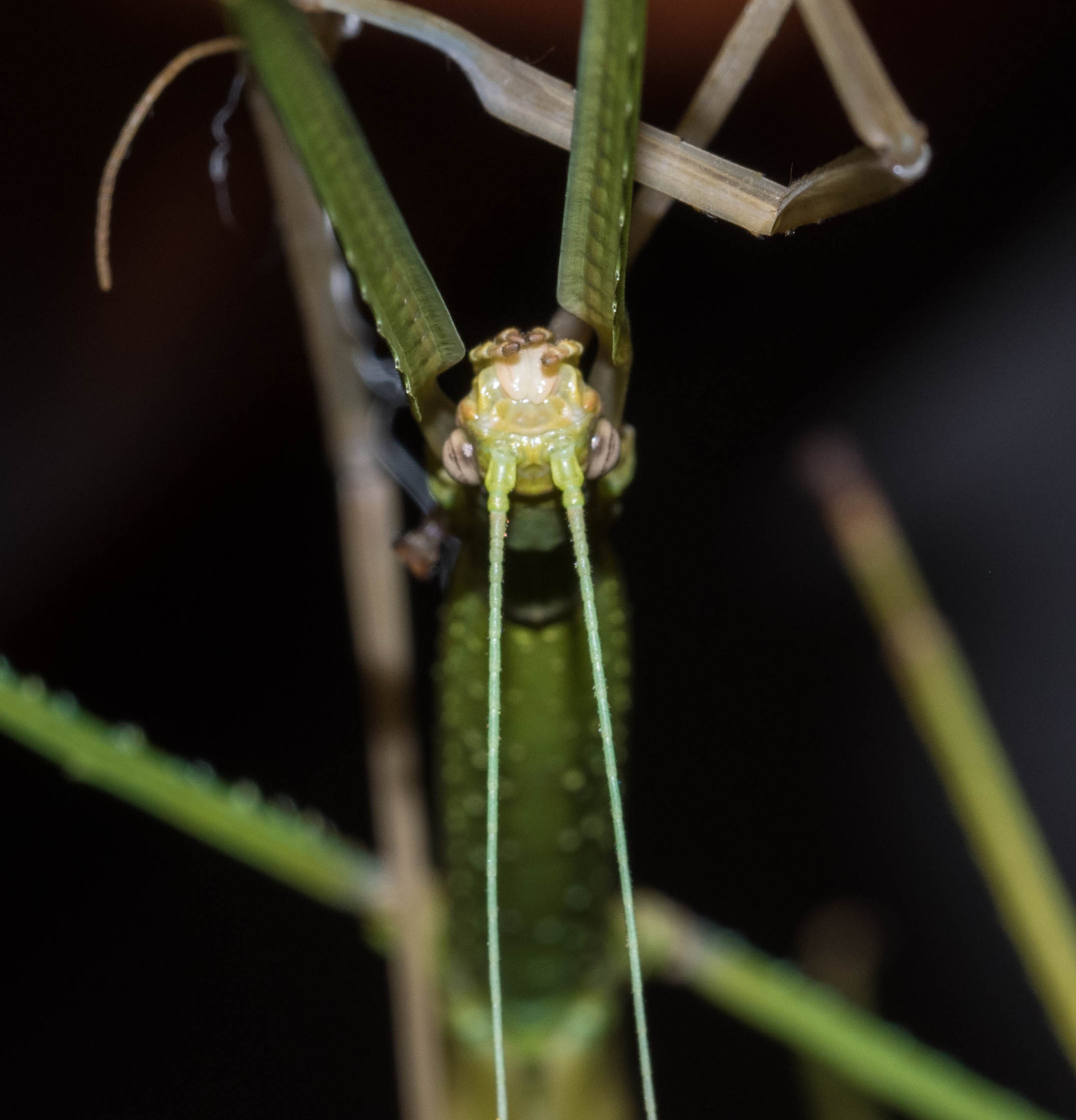
One final task remains - freeing the antennae from their embryonic cuticle coat. The next movie shows how this is accomplished. The nymph grasps the antennae between its mouthparts and pulls them forward in a ratchet-like fashion until they are released from the cuticle.
The whole process of ecdysis - shedding your former skin - is a particular challenge for an insect like a katydid that has long limbs and antennae. It’s much more straightforward for something like a caterpillar or a maggot, which has either short legs or lacks limbs entirely.
Ecdysis doesn’t get much easier when a katydid has become a nymph. The moults it undergoes on the way to becoming an adult also require Houdini-like skills.
I documented the moulting of a katydid nymph to the adult stage in my post from January 2019 - A katydid gets too big for its skin.
The final count
11 nymphs hatched on the second day of observation and 8 more on the day after that. Our buddy tank was becoming quite a menagerie!
In total, 25 of the clutch of 29 eggs hatched successfully. Not a bad outcome!


Rearing the nymphs
We placed a variety of seeding grasses and other vegetation into the buddy tank with the hatchlings as we weren’t sure what their food requirements were.
They accepted pollen grains from Gahnia flowers in the first few days but later fed avidly on new growth eucalypt foliage.
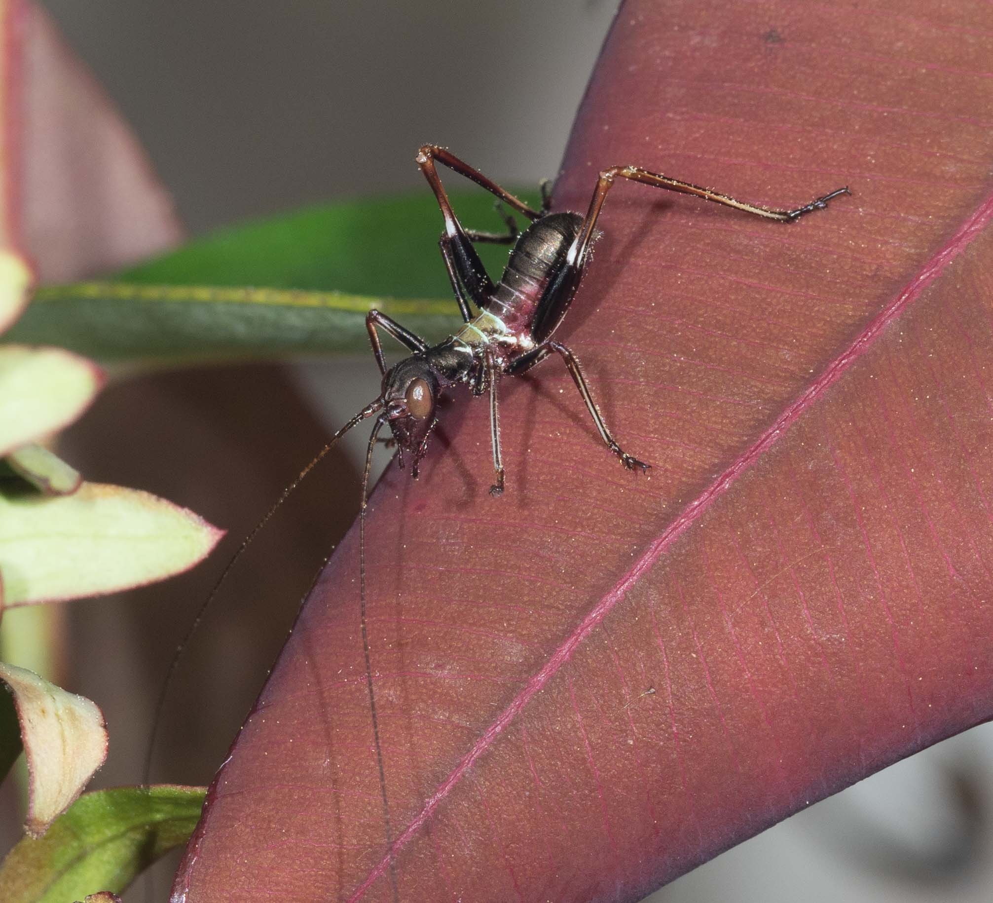
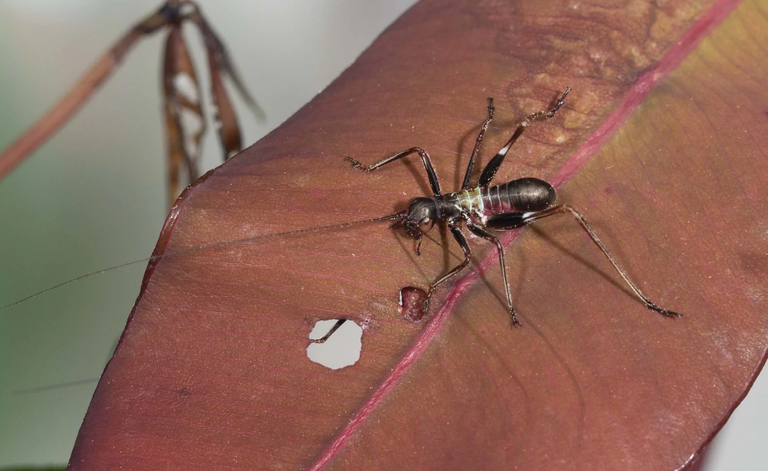
The nymphs quickly adopted the same dark crimson colour we saw in the hatchlings on the very first day. They look a lot like ants. This is no coincidence. It serves to protect them from vertebrate predators that avoid ants.
Our hatchlings appear to be doing well, although a couple have succumbed to what appears to be a fungal infection.


We’re hoping to raise some of them through to adulthood to positively identify the species. Comparison with images on iNaturalist strongly suggest they’re the Giant Gumleaf Torbia, Torbia perficita. Time will tell.
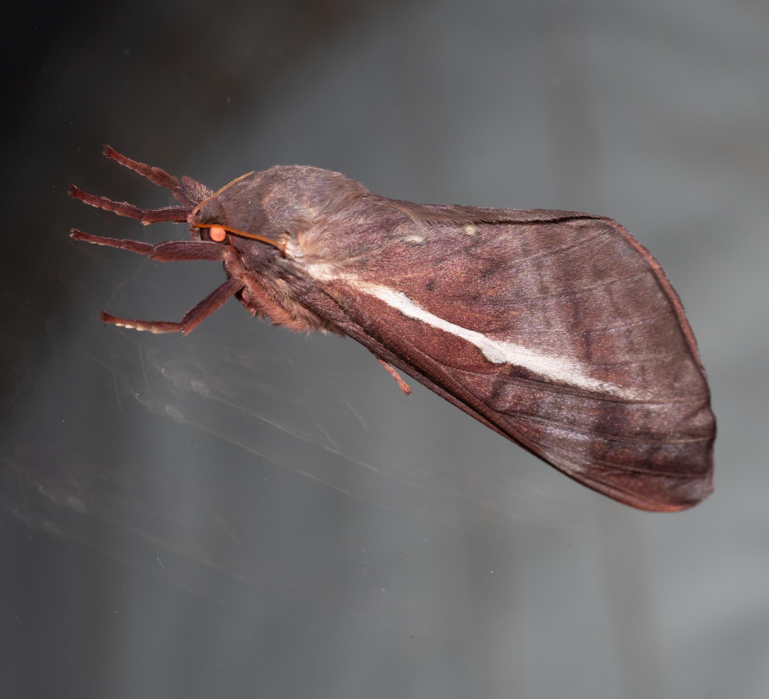
If they are Torbia perficita, they’ve got a lot of growing to do. The newly hatched nymphs are 5mm long while the adults we observed on a night time moth viewing session are 35-40mm.
In the meantime, they make great pets!
Update: As you can see in my later blog “Growing Up”, I was able to rear these nymphs through to adulthood. And indeed they turned out to be Torbia perficita.
The first nymph to moult to the adult stage did so on 18th January, the last on 6th February - between 3-3.5 months (87-109 days) after they hatched from the egg.