
Sawfly anatomy
Of necessity, taxonomic papers contain many technical terms for describing the details of various body parts. Few people, apart from specialists in the field of study will be familiar with this jargon, making species descriptions difficult to interpret.
The aim of this page is to help the non-specialist reader become familiar with names of sawfly body parts. The following pair of drawings shows some of these. The sawfly illustrated, Aglaostigma quattuordecimpunctatum is a North American species, but the same naming system is used for Australian sawflies.


from Goulet & Huber 1993 “Hymenoptera of the World: An Identification Guide to Families”.
The Sawflies of Britain and Ireland website has an excellent illustrated glossary to sawfly body parts available for download here. These anatomical terms apply equally well to Australian sawflies.
The labelled photos which follow aim to help you recognise these and other body parts in the actual insect. I have used four different representative Australian sawfly species for this purpose – Lophyrotoma interrupta, a member of the subfamily Pterygophorinae, Pseudoperga ferruginea and Perga dorsalis, both members of the subfamily Perginae and Clarissa sp., a member of the Euryinae.
All of these images, with the exception of Figs. 4D, 6, 7 & 8 are either from this Workbook page on our blog or our own or other people’s iNaturalist sawfly observations (see attributions with photos).
Overall body structure & leg segments
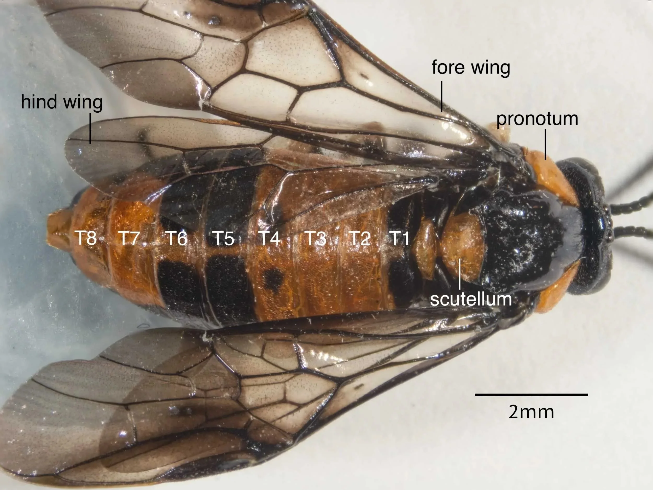
Fig. 1 - Dorsal view of the female of Lophyrotoma interrupta. The terga (dorsal cuticular plates) T1-T8 of the abdomen, the thorax, the base of the head the fore and hind wings are visible.
Fig. 2 - Ventral view of same specimen showing names of leg segments.
Fig. 3 - segments of the hind tarsus in the same specimen
Wings - veins and cells


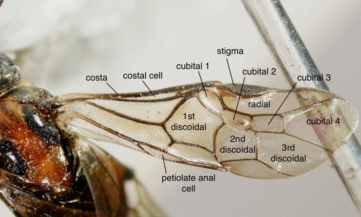
Fig. 4 - Names of veins and cells in fore wing of Lophyrotoma interrupta (A, B) and cells of Pseudoperga ferruginea (C) and Clarissa sp. (D). The wing venation is similar in these Pergidae species.
However, like all members of the subfamily Pterygophorinae, Lophyrotoma interrupta has 3 cubital cells, whereas Pseudoperga ferruginea, like all members of the subfamily Perginae, has 4.
The fore wing of Clarissa, like other members of the subfamily Euryinae, has an additional cell - anal cell - at the bottom edge of the wing.
Different naming systems - some of which are shown in this table - have been used for the wing veins and cells of sawflies over the years.
The wing terminology section of the glossary on the Sawflies of Britain and Ireland website has excellent diagrams showing wing cell and venation labelling (Figs. 90-94).
Antennae
Fig. 5A. Lophyrotoma interrupta female
Fig. 5B. Lophyrotoma interrupta male
Fig. 5C. Pseudoperga ferruginea female
Fig. 5 - Sex and species differences in antennal structure. Lophyrotoma interrupta like other species in the subfamily Pterygophorinae has pectinated antennae. The pectens are much longer in the male than the female (compare A and B). Members of the subfamily Perginae, such as the female Pseudoperga ferruginea in C, have simple antennae, lacking pectens and with fewer segments. The last segment is often expanded to form a club in these sawflies. The total number of antennal segments - the number of repeating units in the flagellum plus the pedicel and the scape/funicle - is often given in species descriptions.
Parts of the head, thorax and abdomen
Fig. 6A. male Perga sp. Photo kindly provided by cgrant64 (CC BY-NC) from this iNaturalist observation.
Fig. 6B. same specimen labelled
Fig. 6 - The side view of this male Perga sp. sawfly (either Perga dorsalis or Perga affinis) shows a number of structures which are often used in sawfly species descriptions.
The asterisks in Fig. 6B show a spine on the mid and hind tibiae, a feature of all subfamilies in the family Pergidae, except for Pterygophorinae and Euryinae.
Fig. 7A. female Perga sp. Photo kindly provided (CC-BY) by Reiner Richter from this iNaturalist observation.
Fig. 7B.
Fig. 7C. Photo kindly provided (CC-BY) by Reiner Richter from this iNaturalist observation.
Fig. 7D.
Fig. 7 - dorsal (A, B) and dorso-lateral (C, D) views of a female Perga sp. (probably Perga affinis) providing views of key thoracic structures.
Fig. 8A. Pergagrapta latreillii. Photo kindly provided (CC-BY) by Reiner Richter from this iNaturalist observation
Fig. 8B. Pergagrapta latreillii. Photo kindly provided (CC-BY) by Reiner Richter from this iNaturalist observation
Fig. 8C. Pergagrapta polita. Photo kindly provided (CC-BY) by Reiner Richter from this iNaturalist observation
Fig. 8D. Xyloperga lalage. Photo kindly provided (CC-BY) by Reiner Richter from this iNaturalist observation
Fig. 8 - lateral (A) and anterior (B) views of Pergagrapta latreillii, dorso-lateral view (C) of Pergagrapta polita and dorso-lateral view (D) of Xyloperga lalage showing further parts of the head and thorax.
Saw anatomy
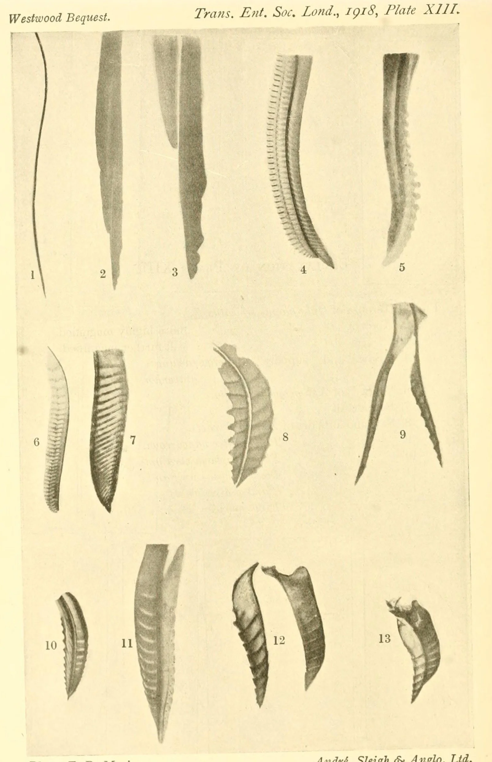
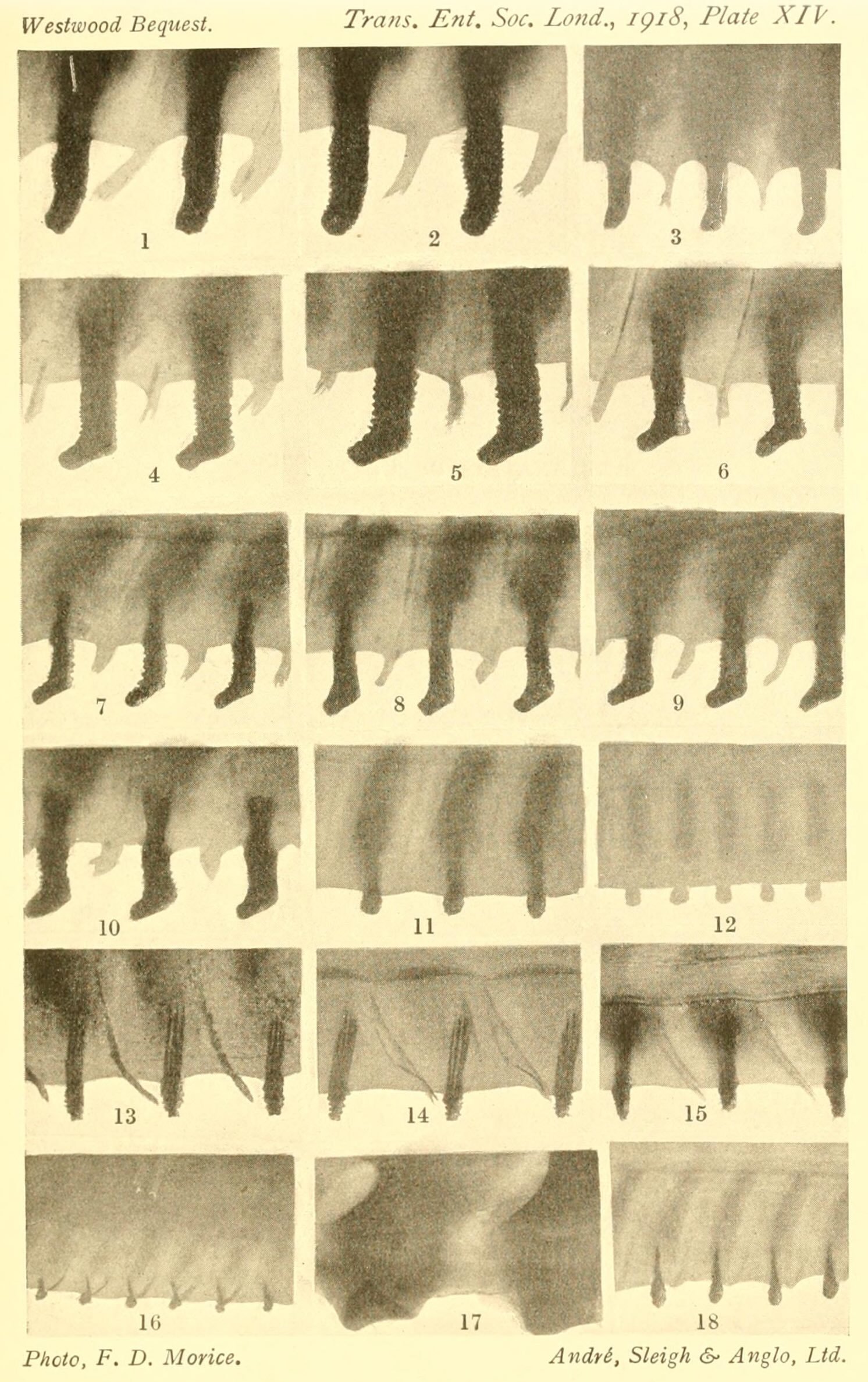
The morphology of the saw is an important diagnostic character for most sawfly species.
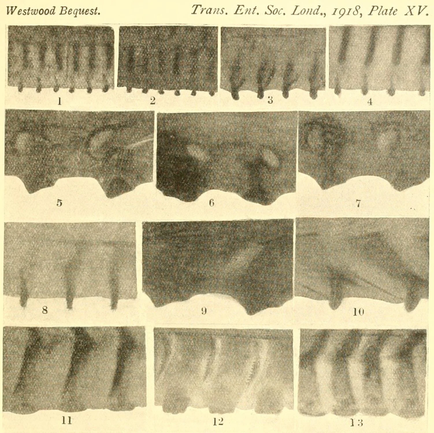
The following images from Morice (1919) show the range of morphology of the lancet (saw, first valvula) by itself or the lancet with the lance (saw support, second valvula) in a range of species.
Click on each plate and roll cursor over the photo to see species names. Some names were changed following Benson’s 1939 revision of the Perginae. The previous name is given in brackets after the current name.
References:
Benson, R.B. (1939) “A revision of the Australian sawflies of the genus Perga Leach, sens. lat. (Hymenoptera, Symphyta)”. The Australian Zoologist 9: 324-357.
Morice, F.D. (1919) “Notes on Australian sawflies, especially the “Authors' Types” and other specimens in the British Museum of Natural History and the Hope Collections of the Oxford University Museum; with diagnostic synopses of the genera and species, and photographs illustrating their structural characters”. Transactions of the Entomological Society of London 66: 247-333
Goulet, H. & Huber, J. T. eds. (1993). “Hymenoptera of the world: an identification guide to families”. Research Branch, Agriculture Canada Publication 1894/E. freely available from a variety of sources including https://doi.org/10.1002/mmnd.19950420212
This is a workbook page … a part of our website where we record the observations and references used in making species identifications. The notes will not necessarily be complete. They are a record for our own use, but we are happy to share this information with others.