
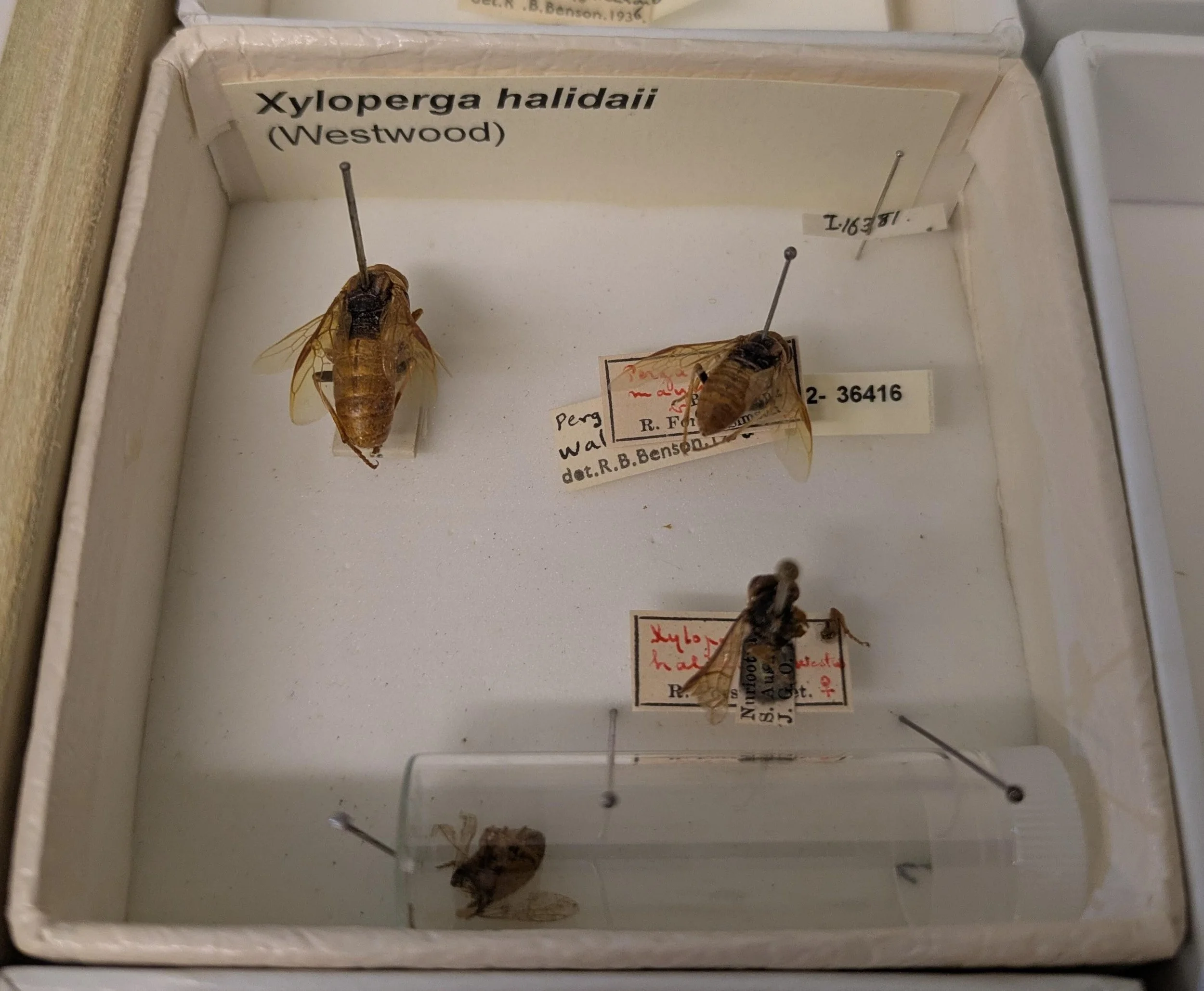
Xyloperga halidaii vs. Xyloperga amenaida
Xyloperga halidaii and Xyloperga amenaida are very similar in appearance.
The original descriptions of these species by Kirby (1882) and Westwood (1880) are brief, unclear and ambiguous, making it difficult to separate them confidently from field photographs.
On this page, I have detailed the morphology of 3 Xyloperga specimens collected in the field. My goal is to use this information to align features visible in field photographs with the original species descriptions of X. halidaii and X. amenaida.
Summary of findings to date
A comparison of specimens A, B and C with published descriptions of X. halidaii and X. amenaida strongly suggests that A and B are a female and male of X. halidaii while C is a female of X. amenaida.
This conclusion is supported by the strong similarity between specimen B and a BOLD specimen of a male identified as X. halidaii.
Discrepancies between features in these specimens and the published descriptions may stem from the brevity of the latter. I hope to obtain photos of the holotypes of both species to clarify the situation.
Observations of specimens
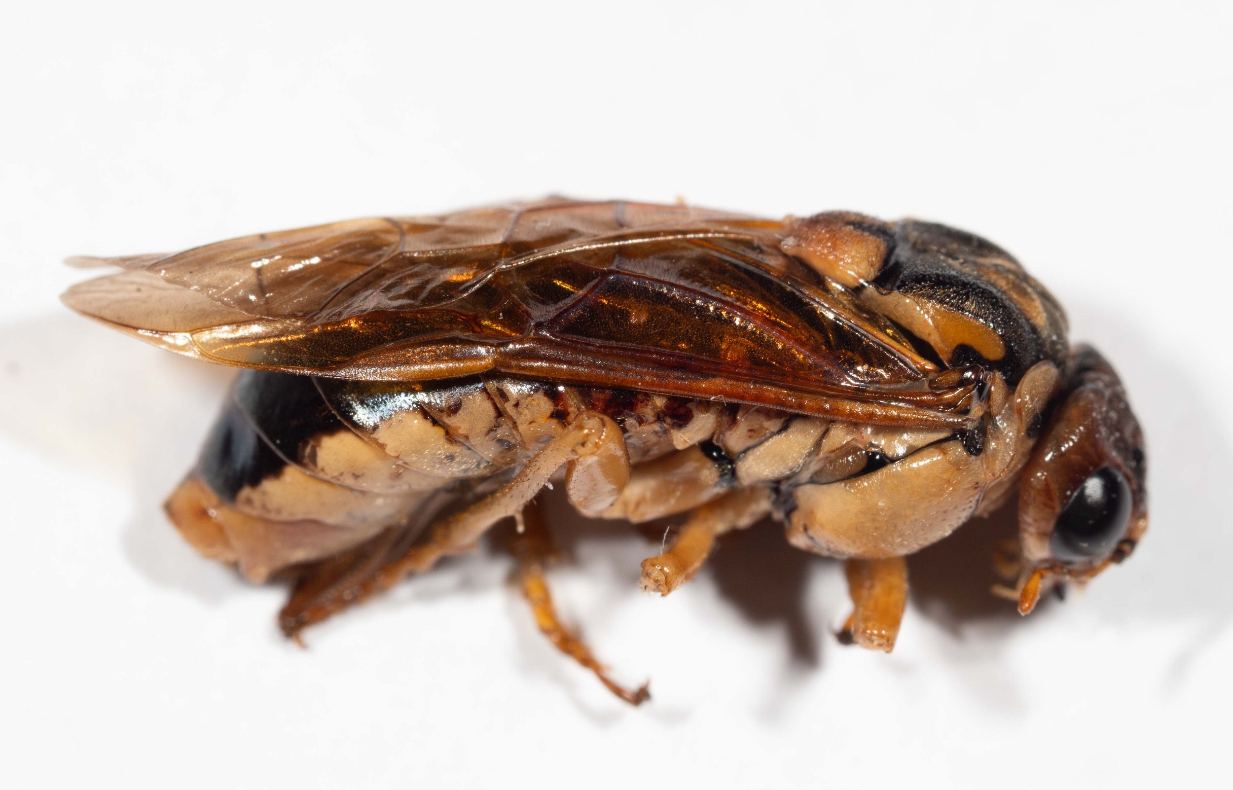
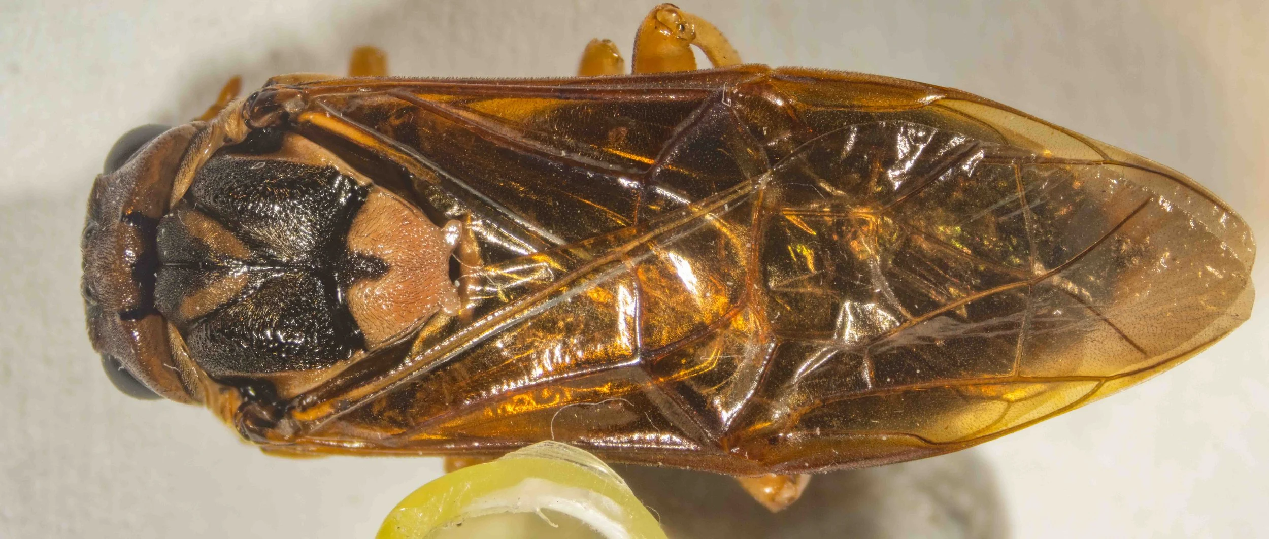
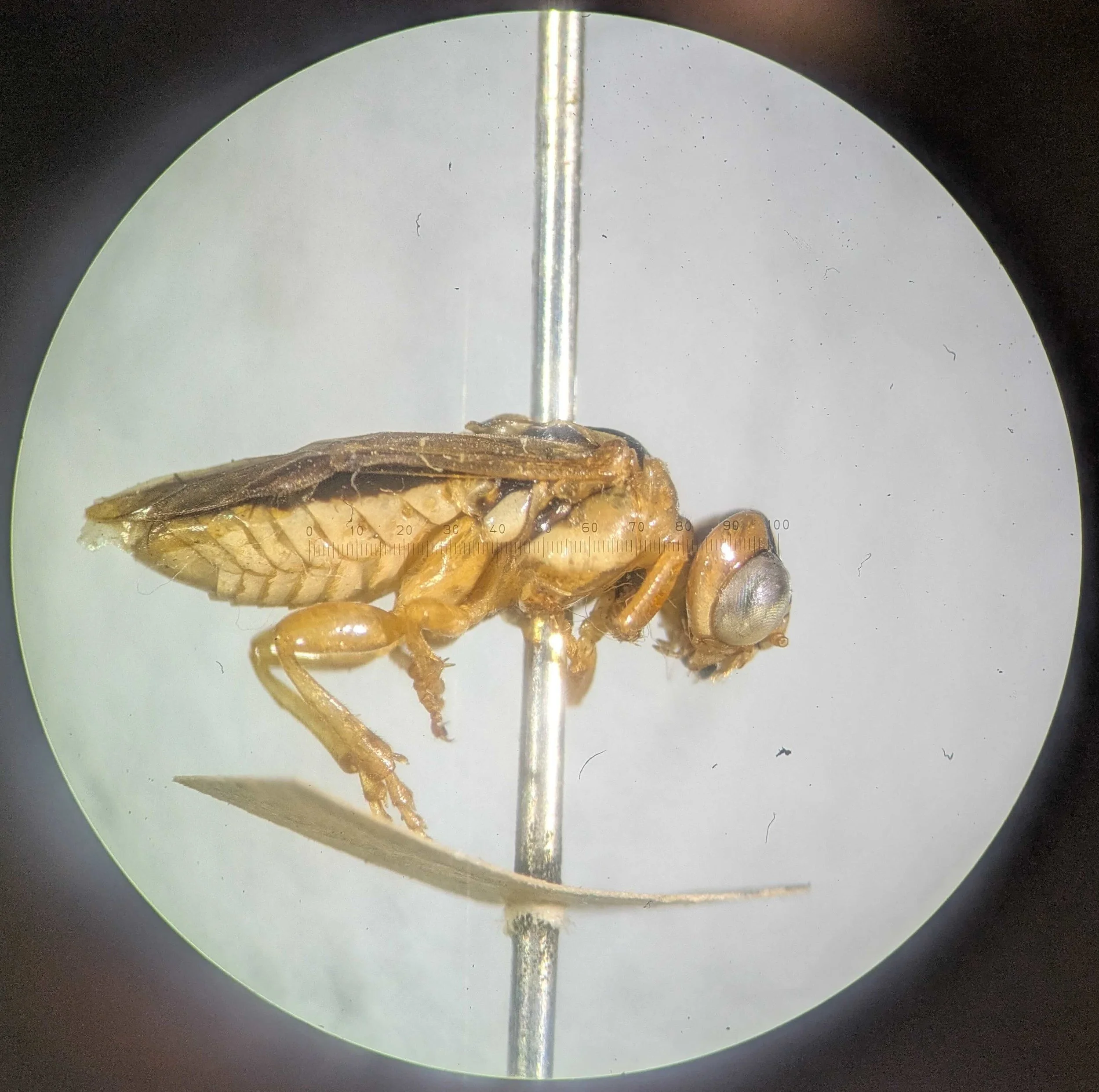
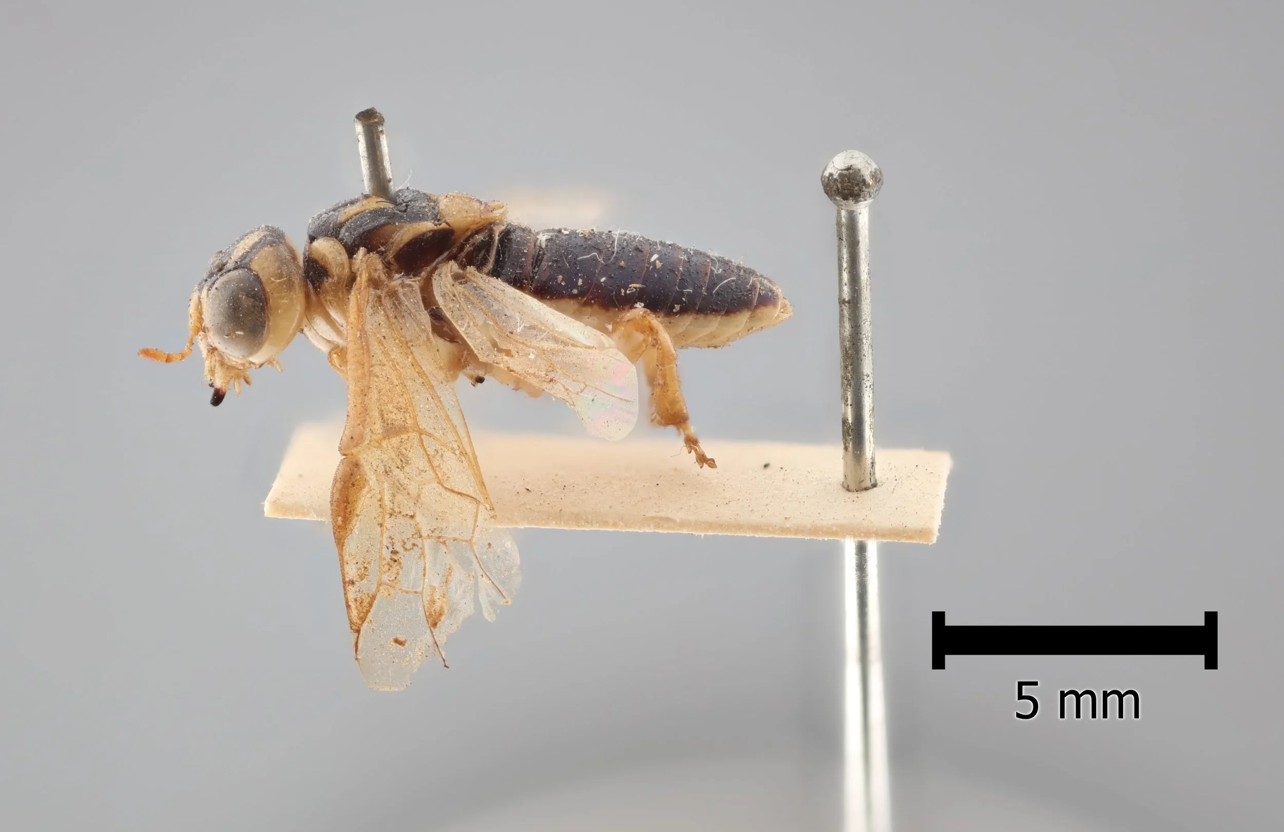
Specimen A
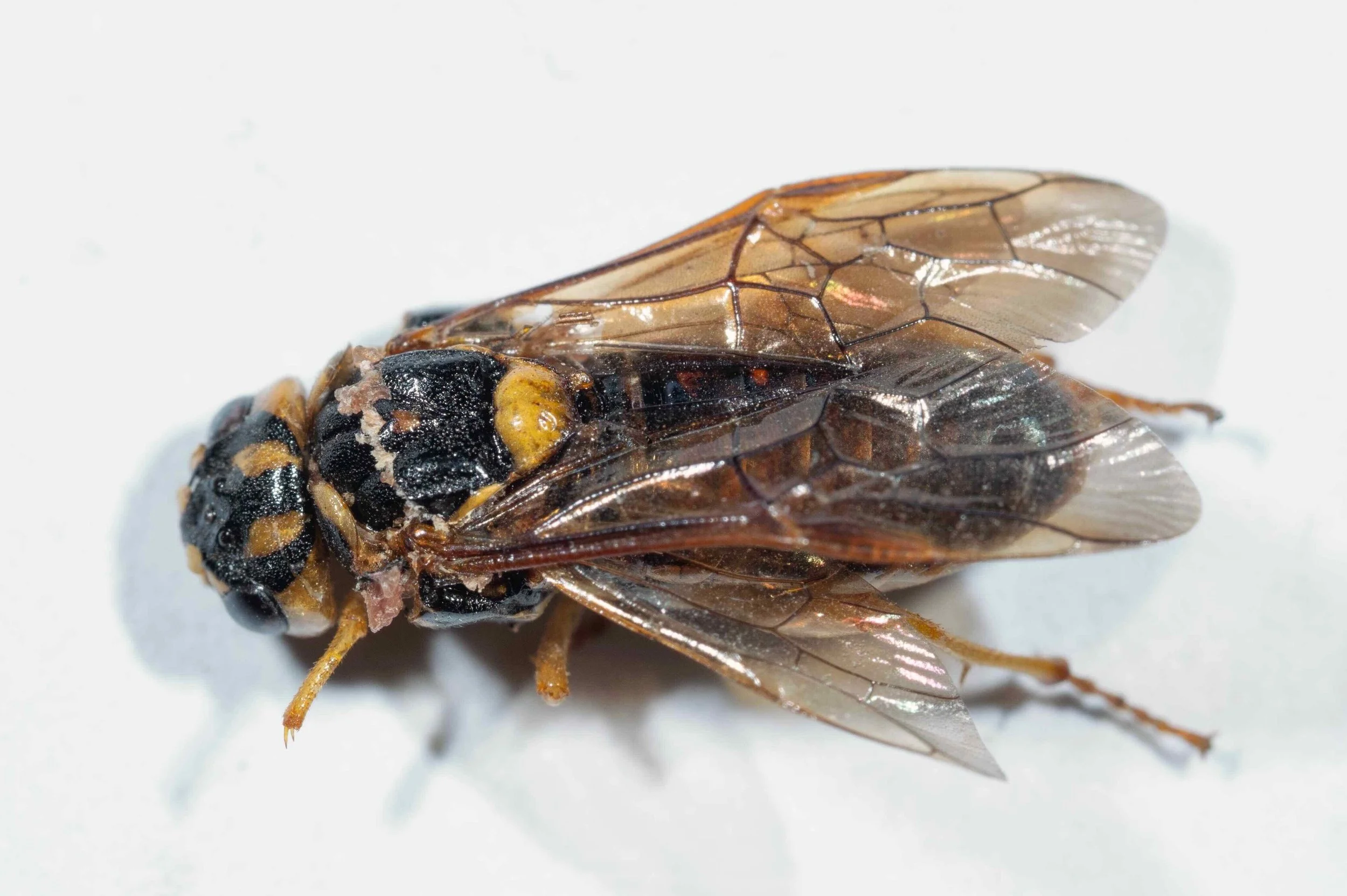
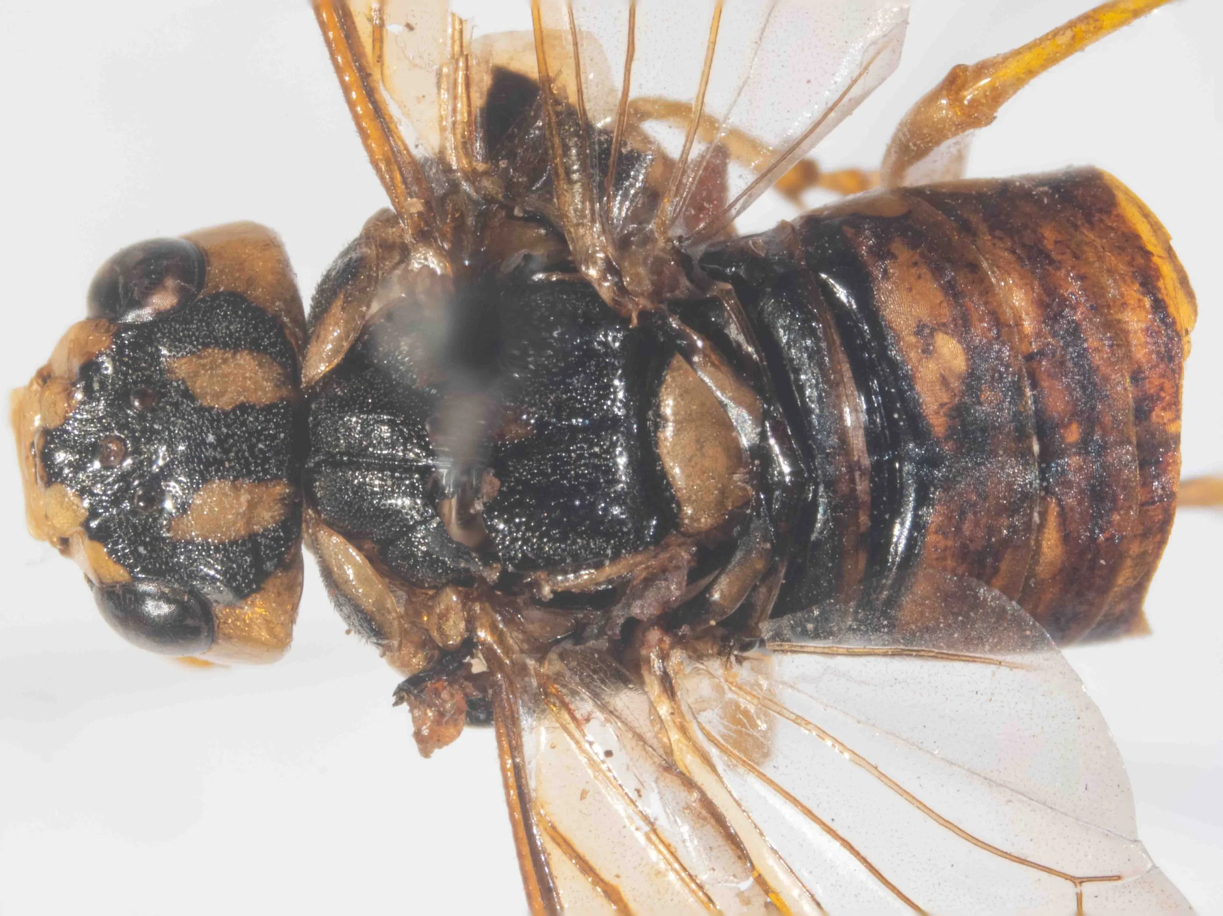
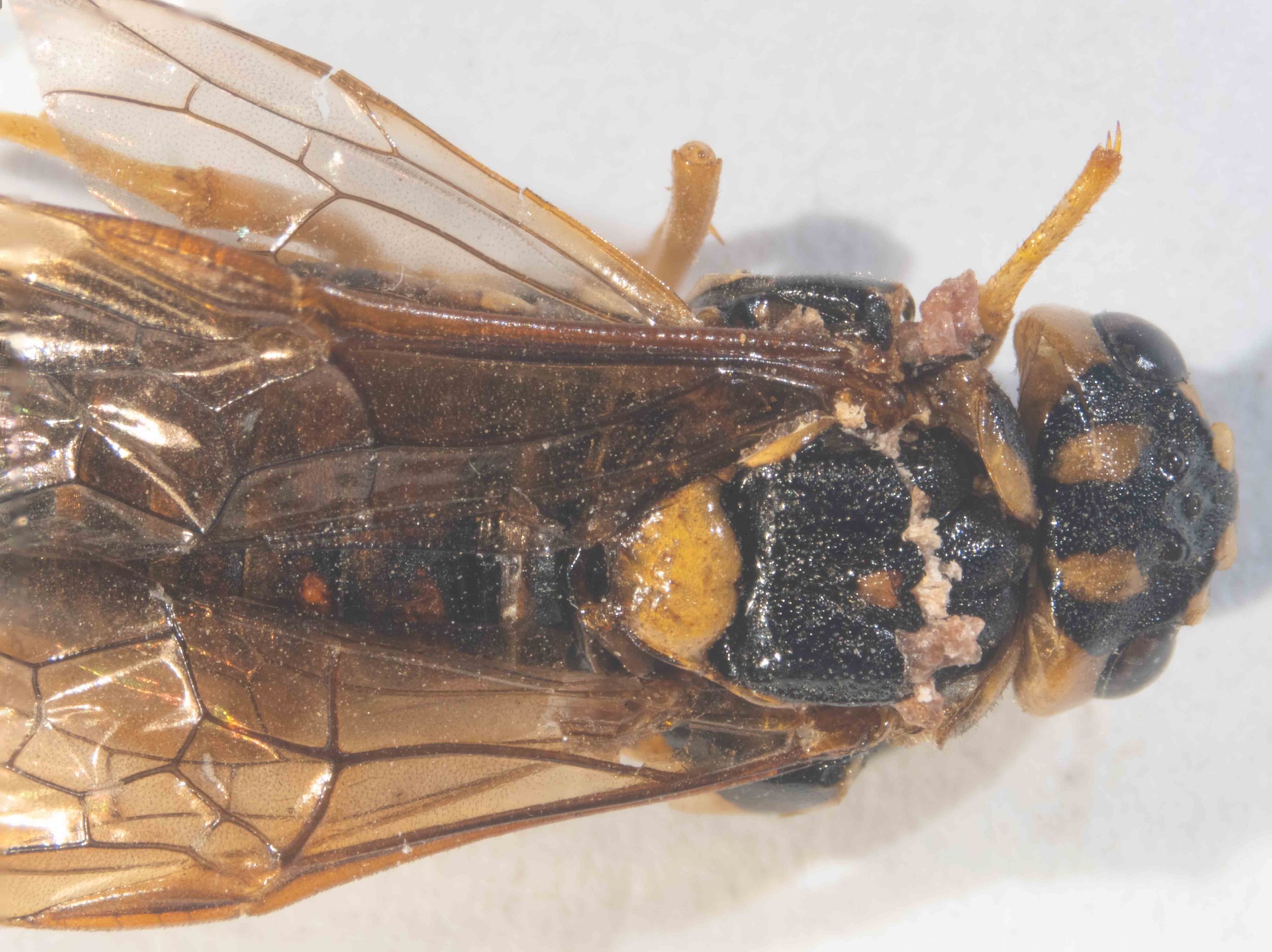
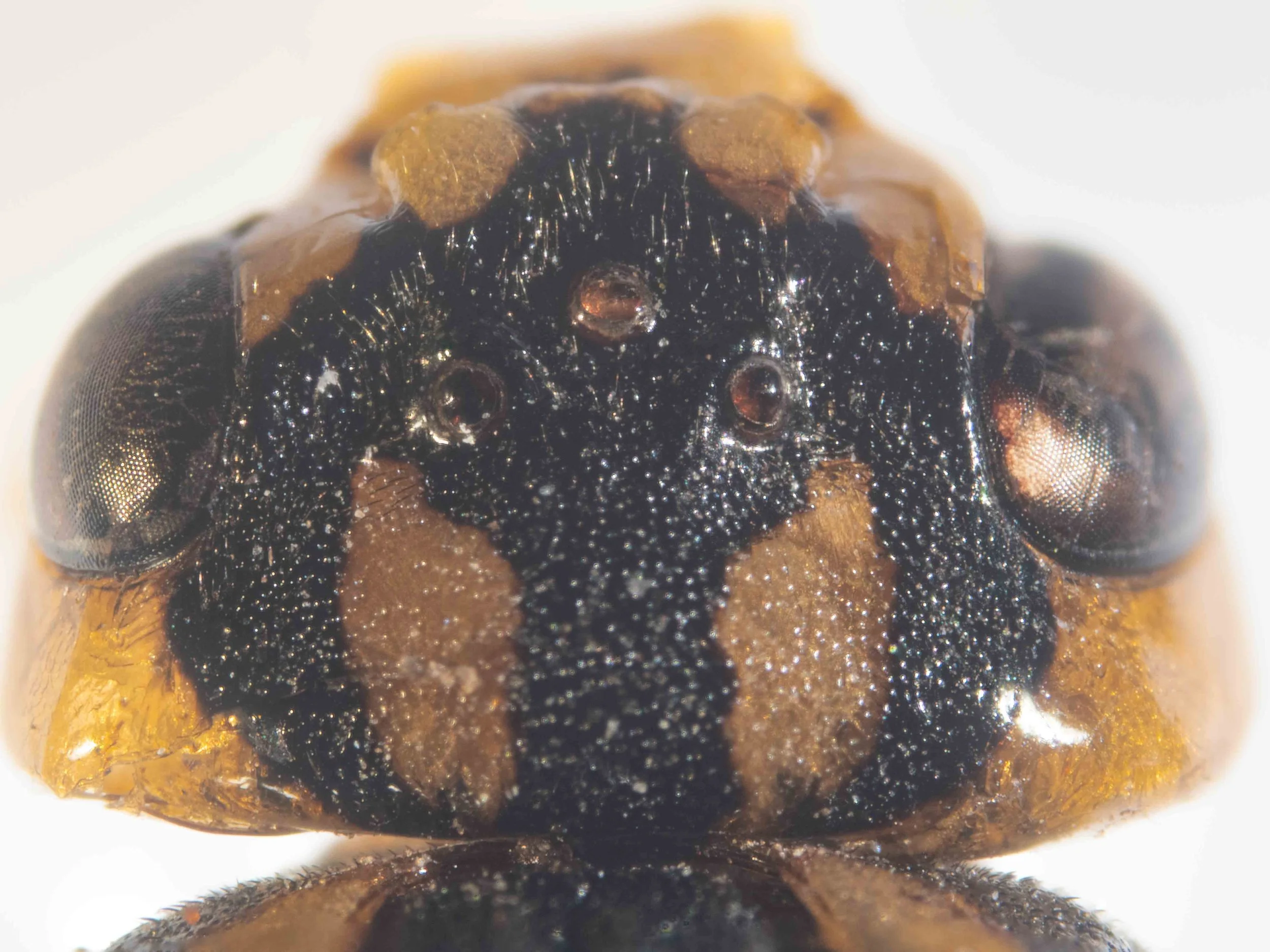
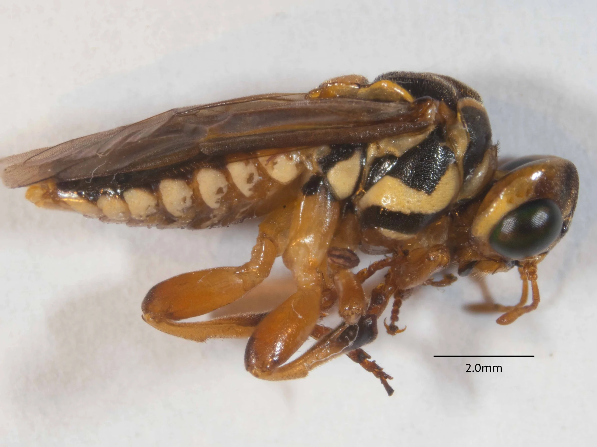
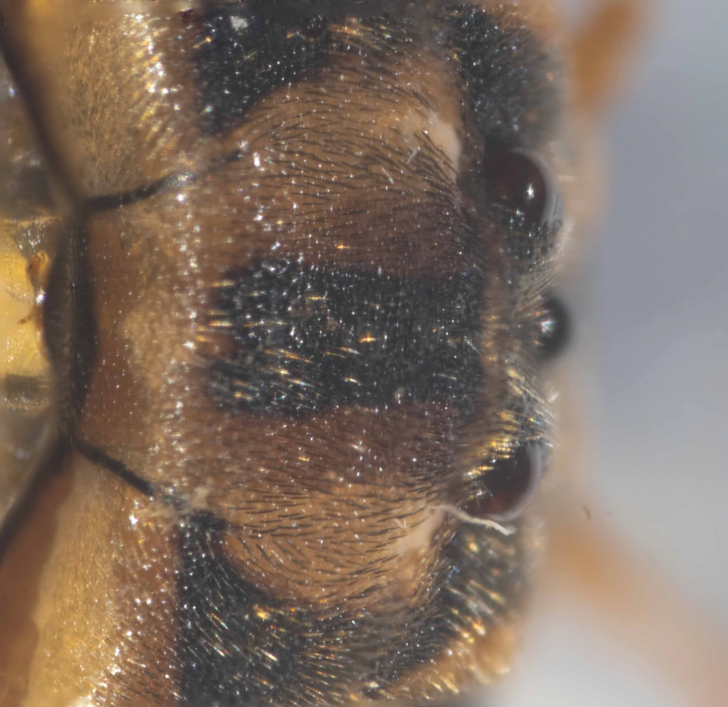
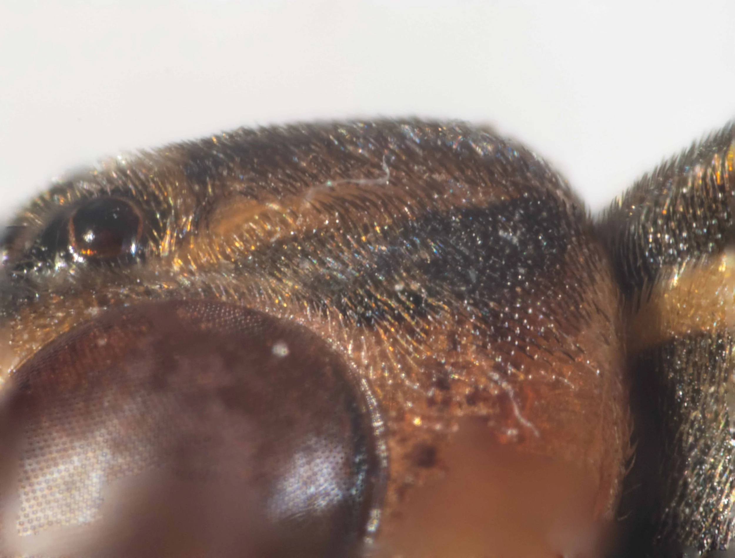
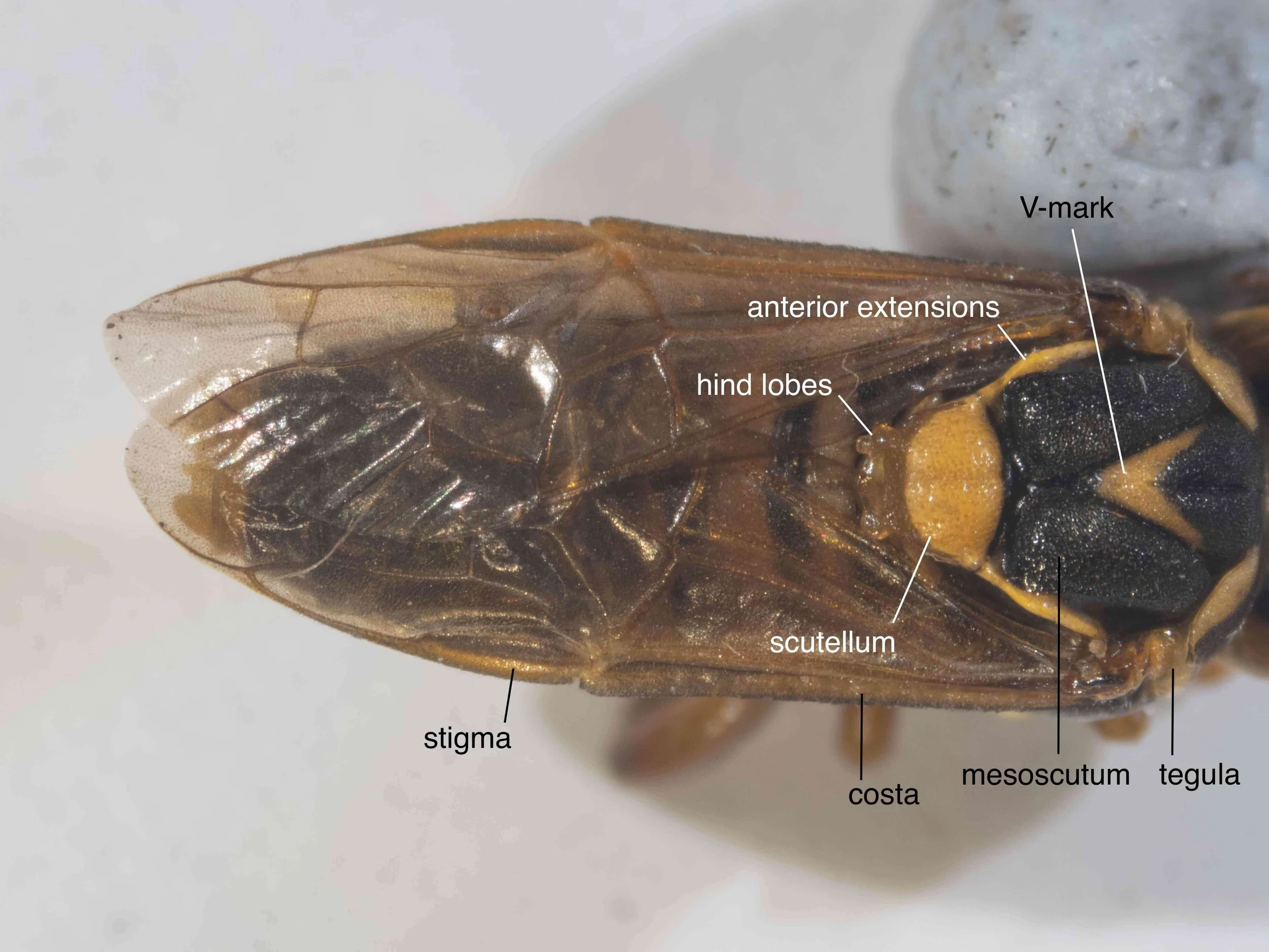
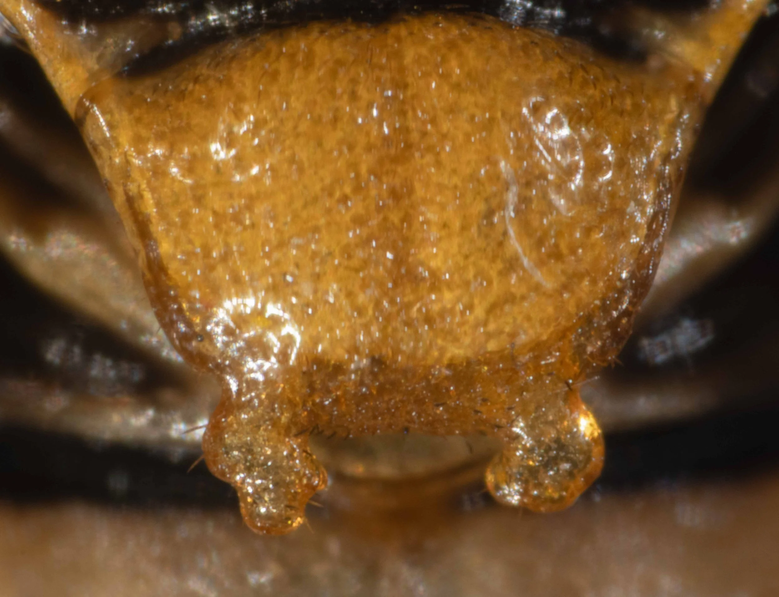
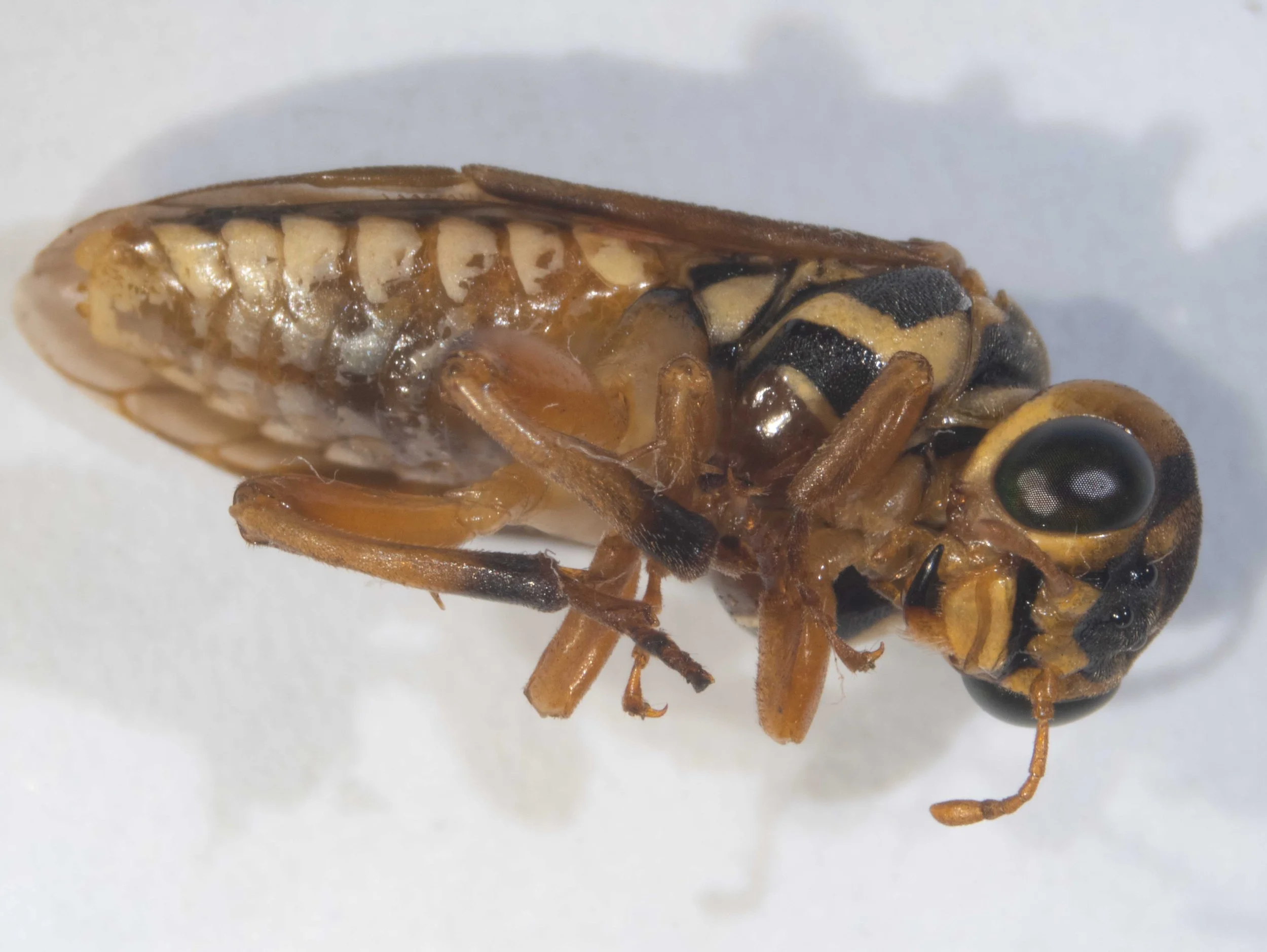
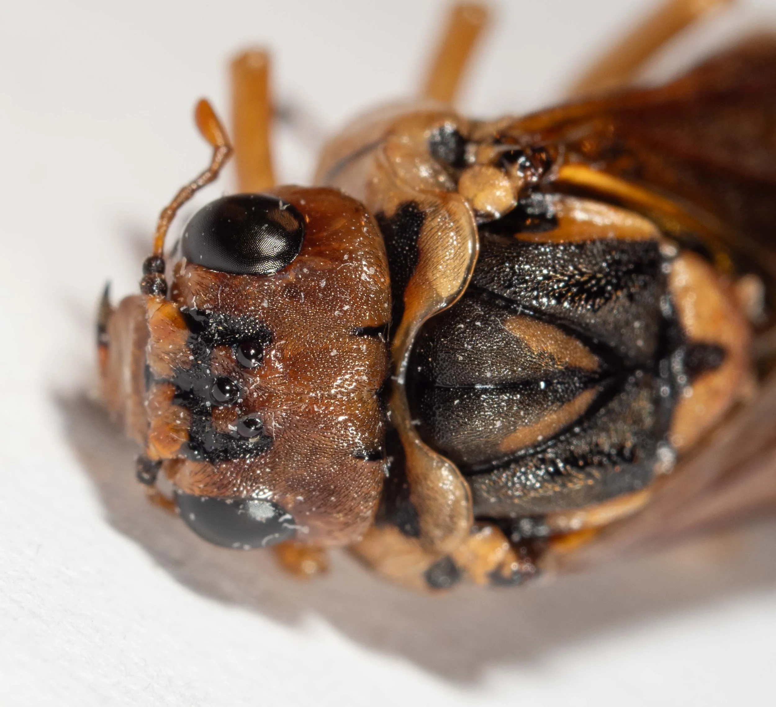
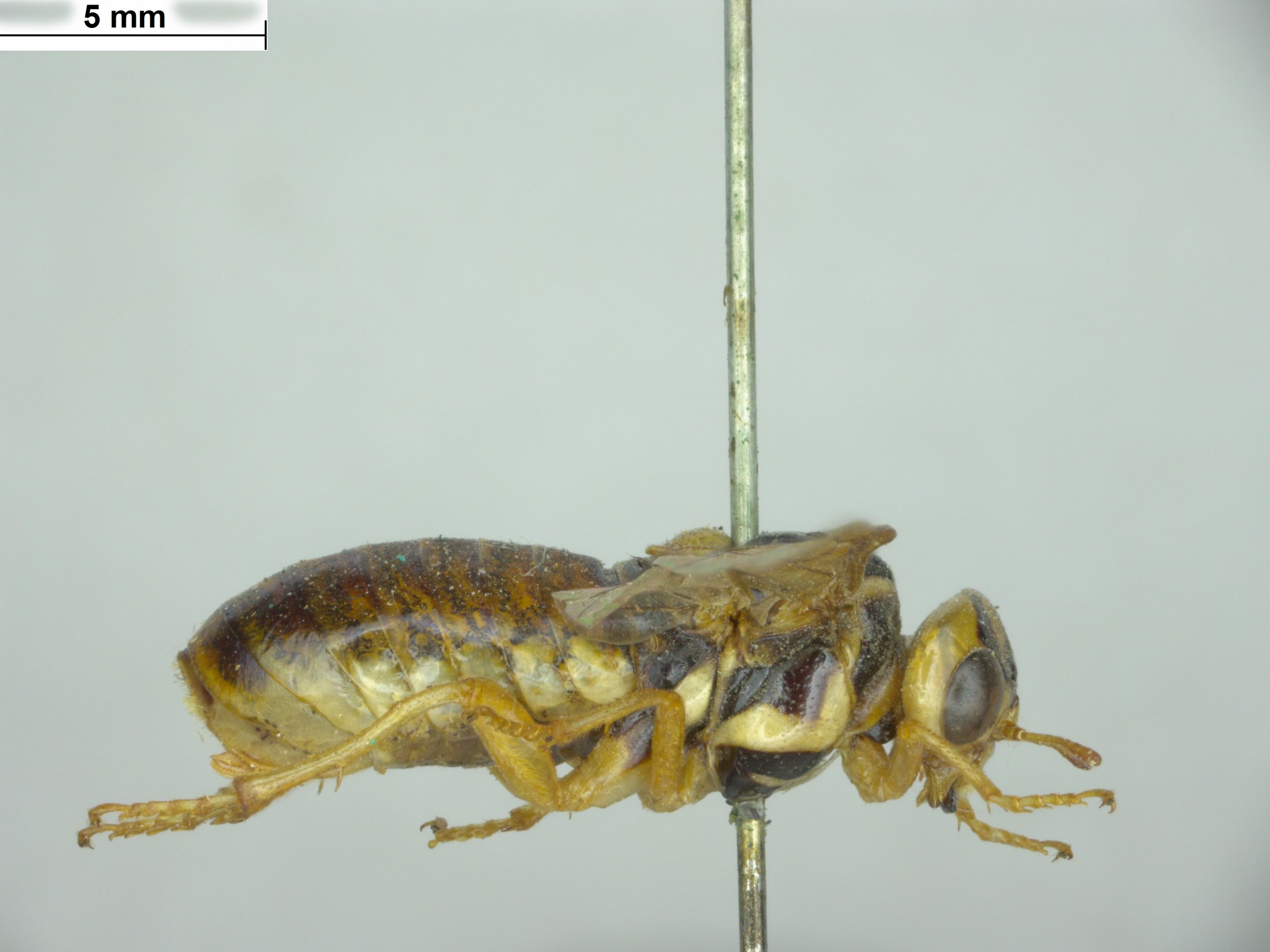
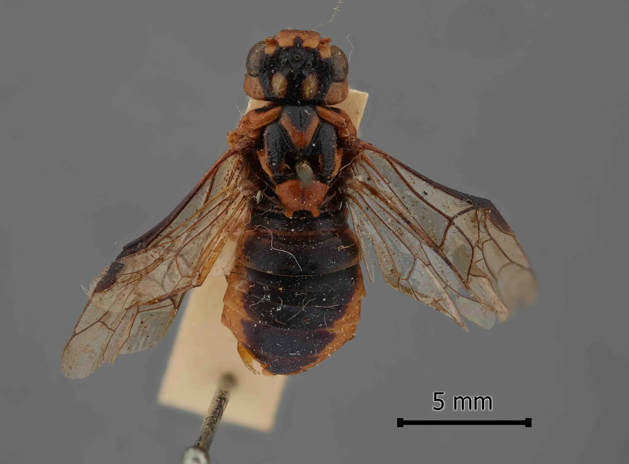
Specimen A is a female, collected by Marianne Broug (iNaturalist observation) at Belair SA in January 2026 (specimen #PW076). It was found dead on a road. Antennae and some leg segments are missing, the mesonotum split but otherwise the insect is intact. While lacking antennae, it shows the following characters which are diagnostic of genus Xyloperga: scutellum convex, with well developed hind lobes; head swollen behind the eyes; clypeus with a transverse fold in the middle.
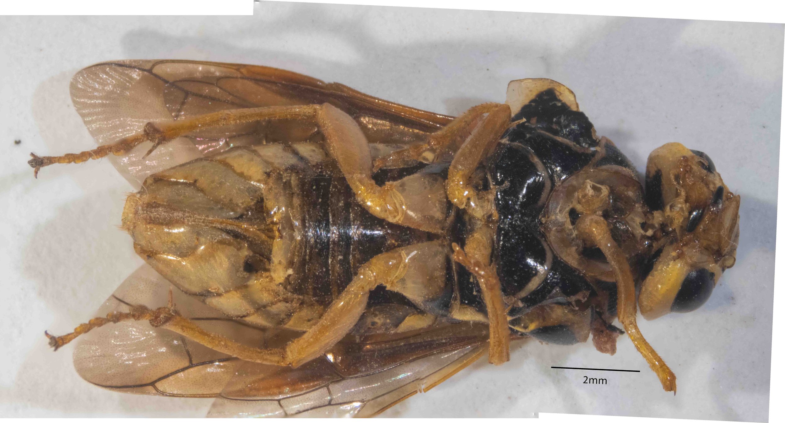

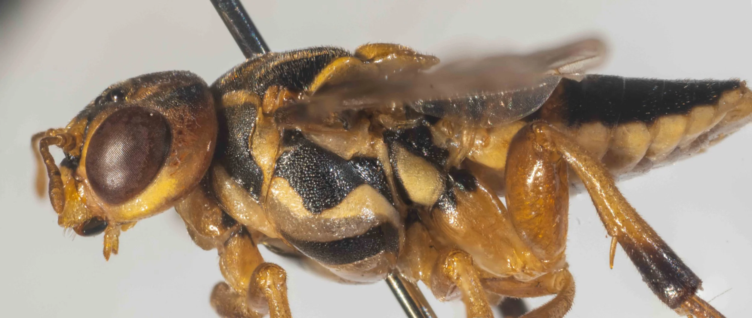
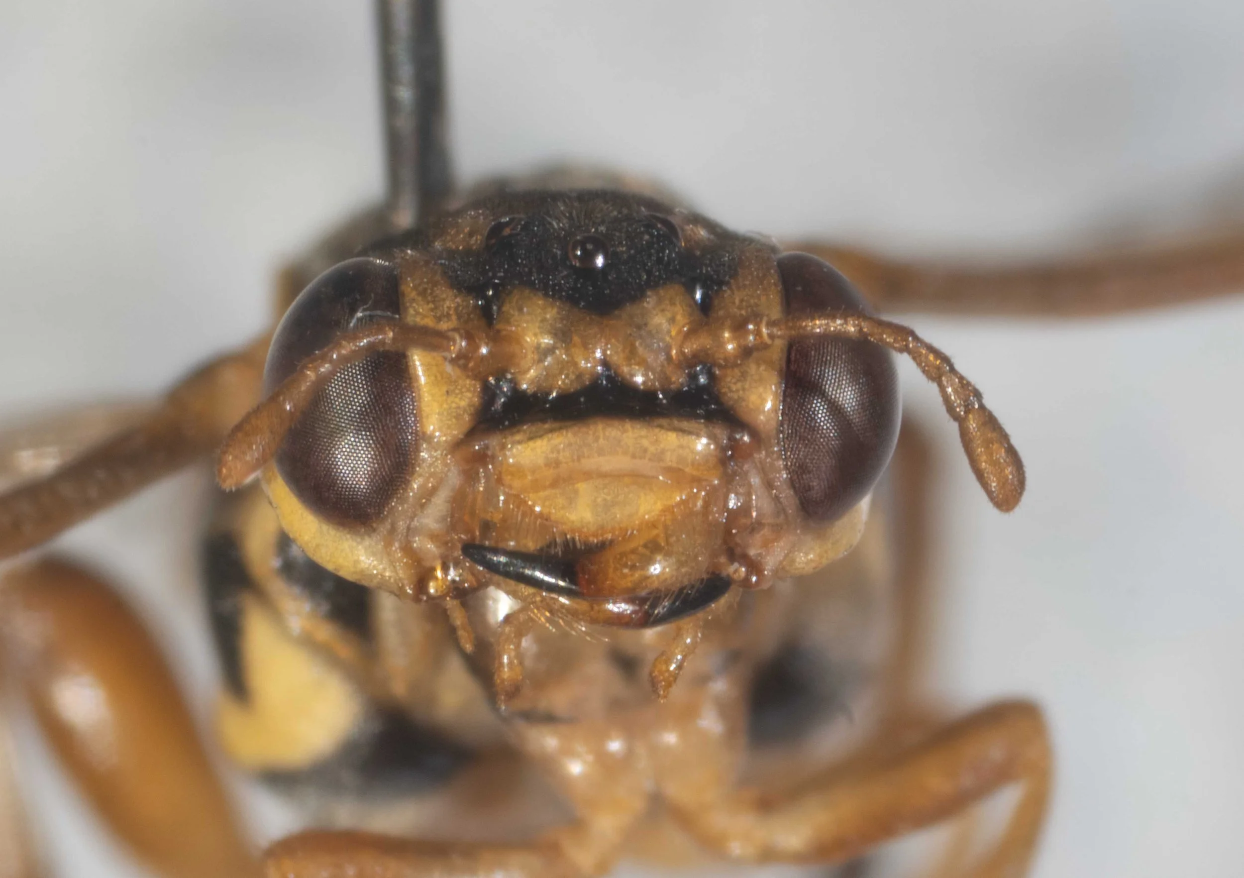
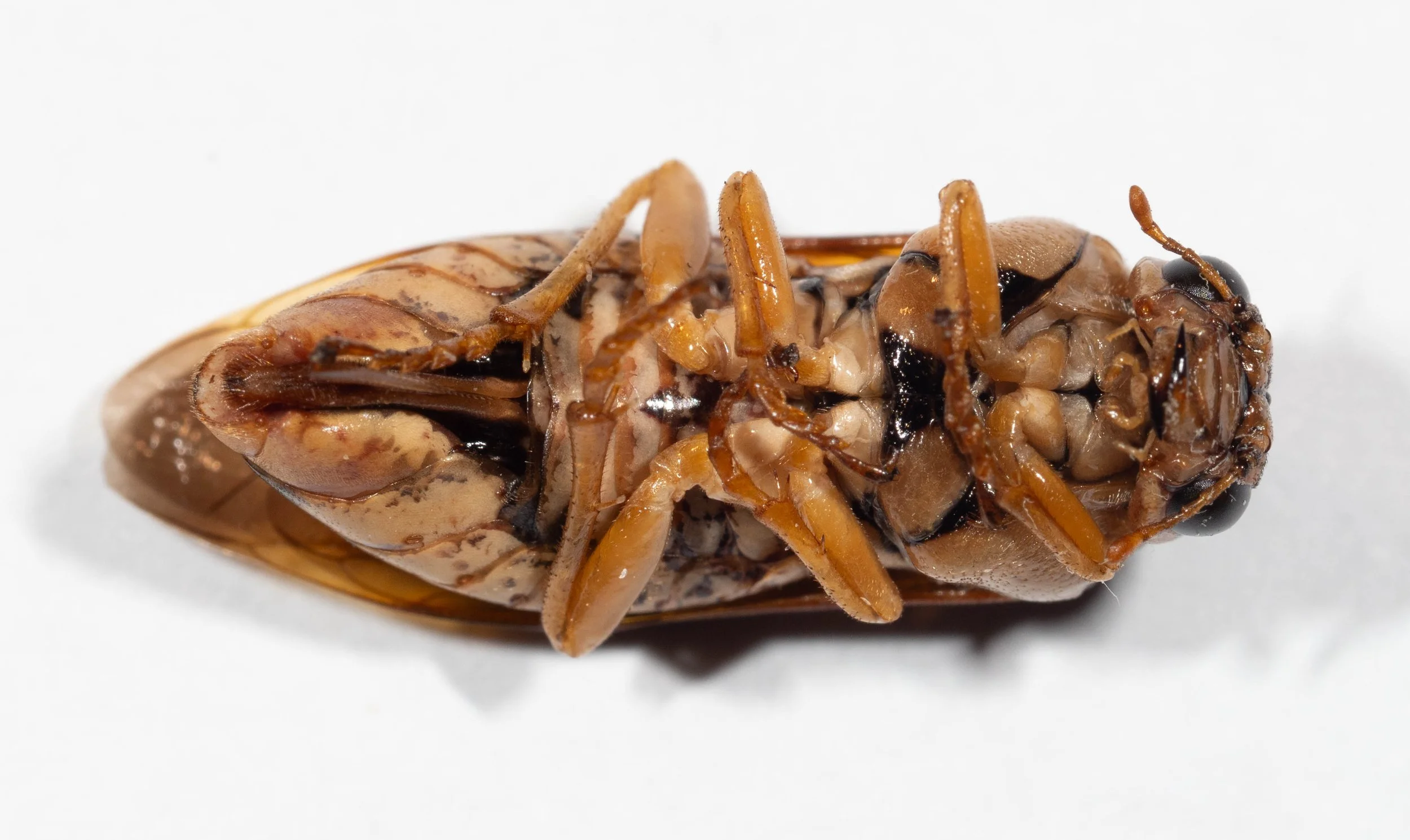
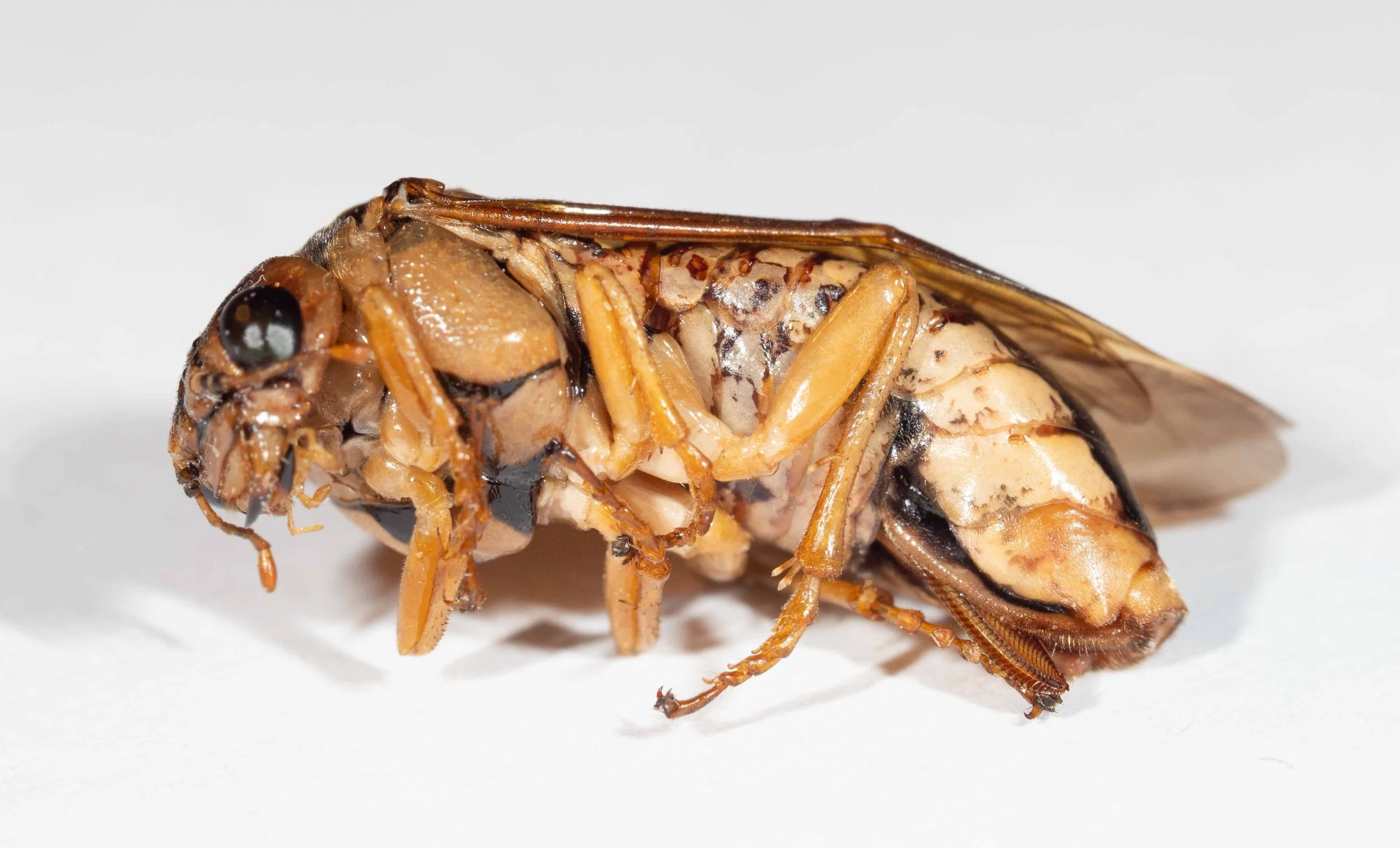
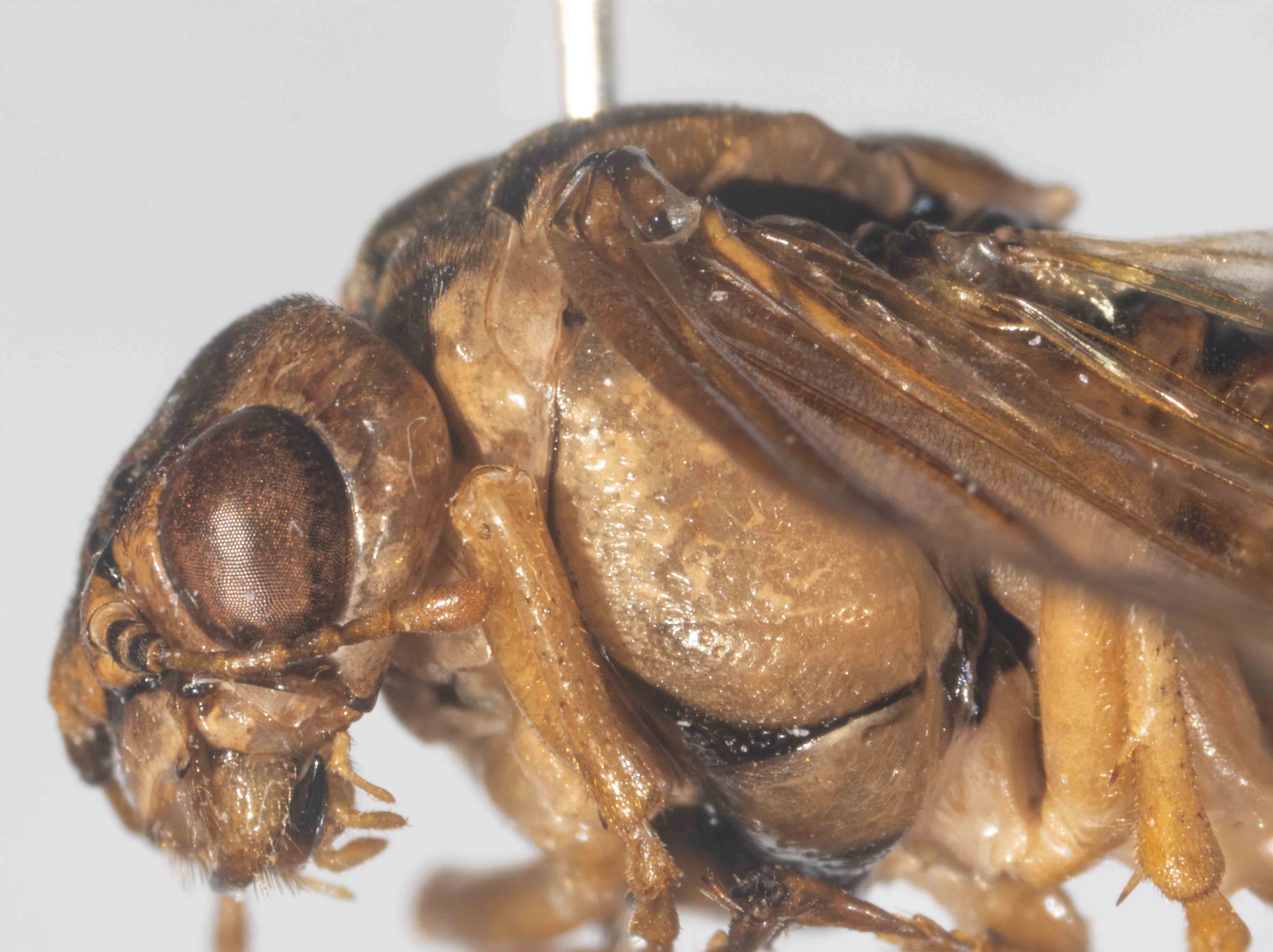
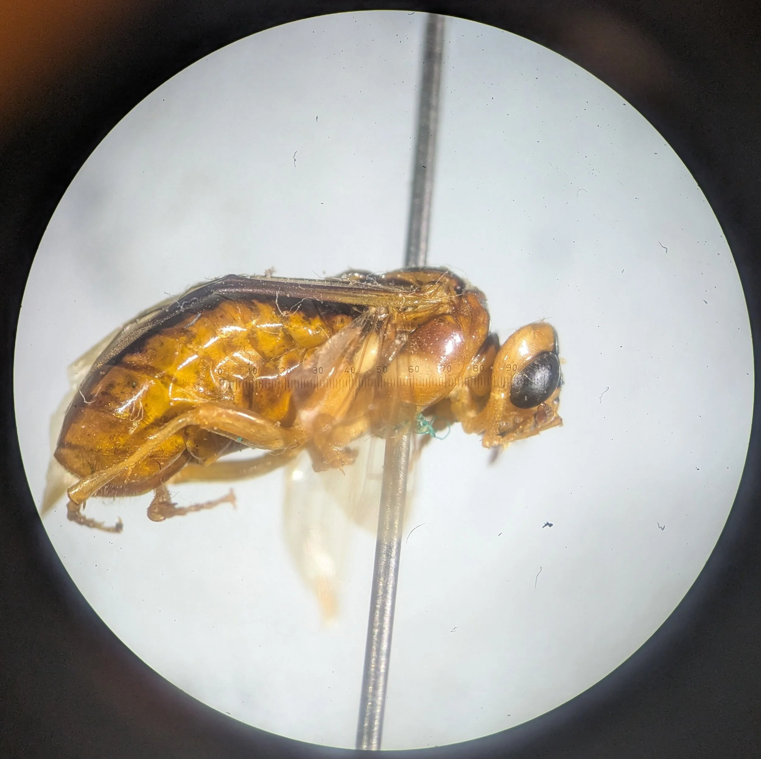
Specimen B
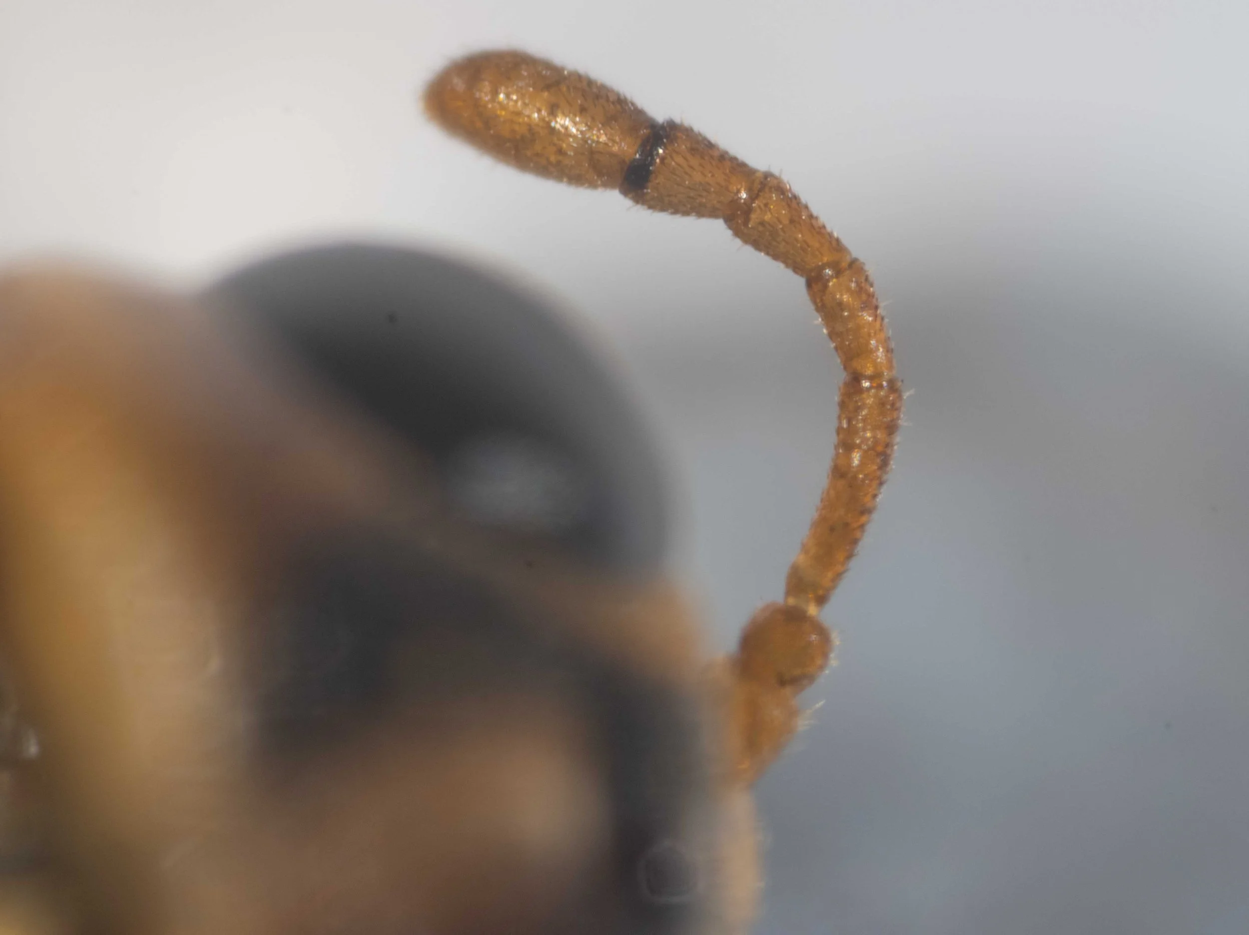
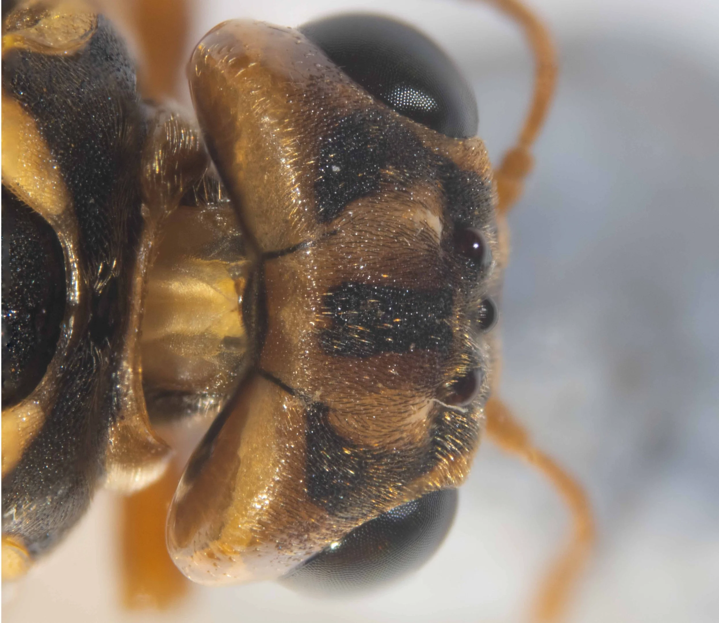
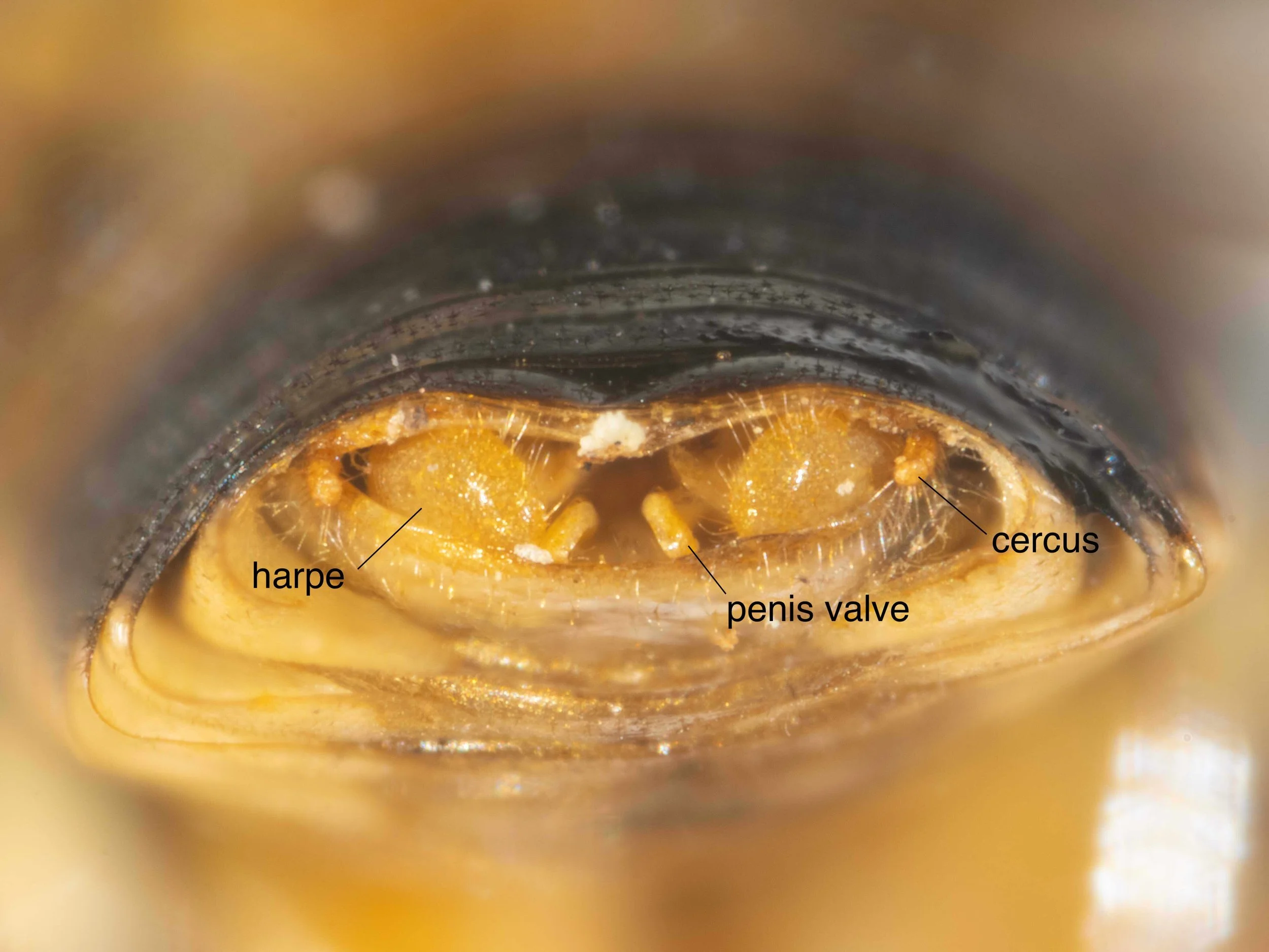
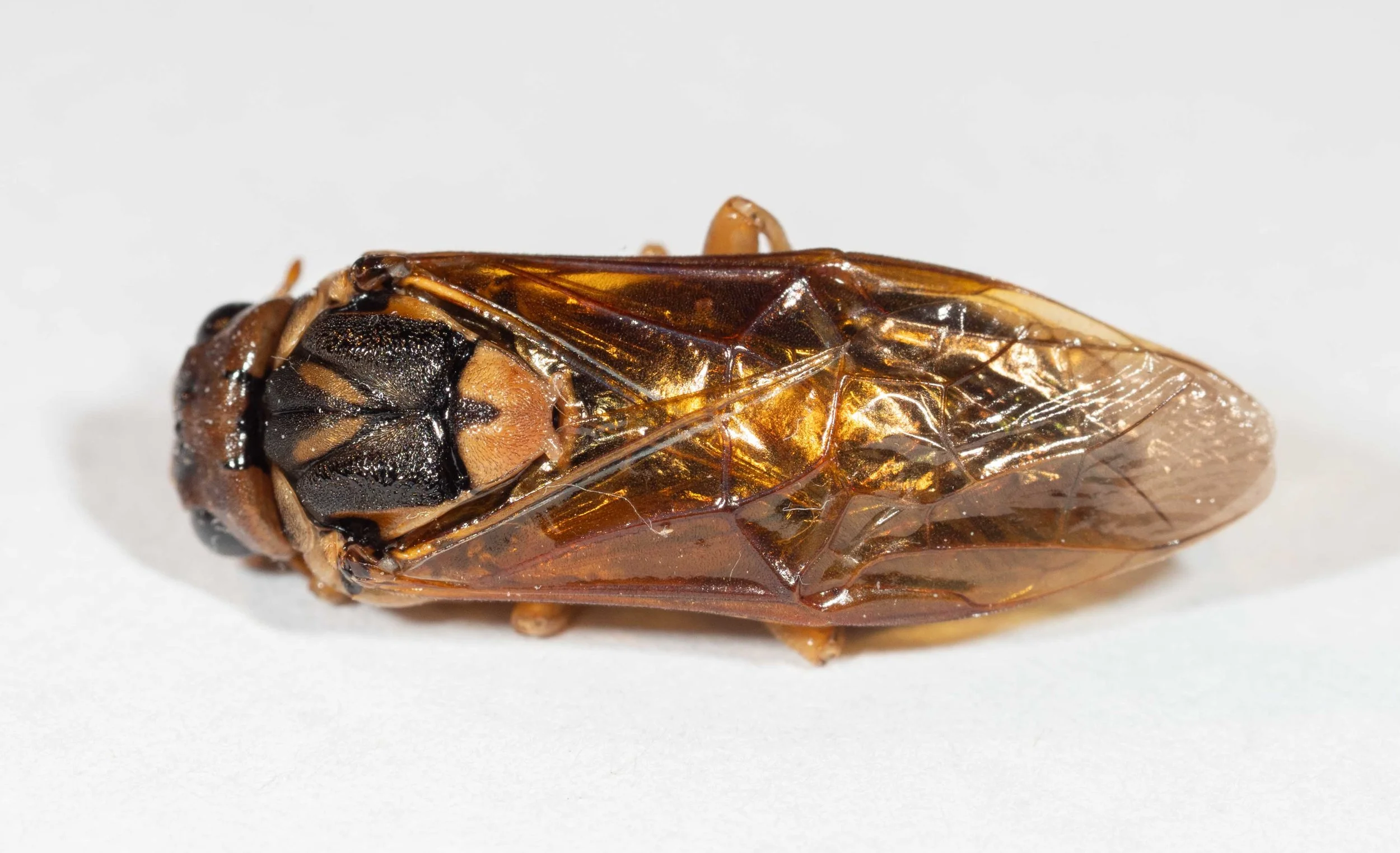
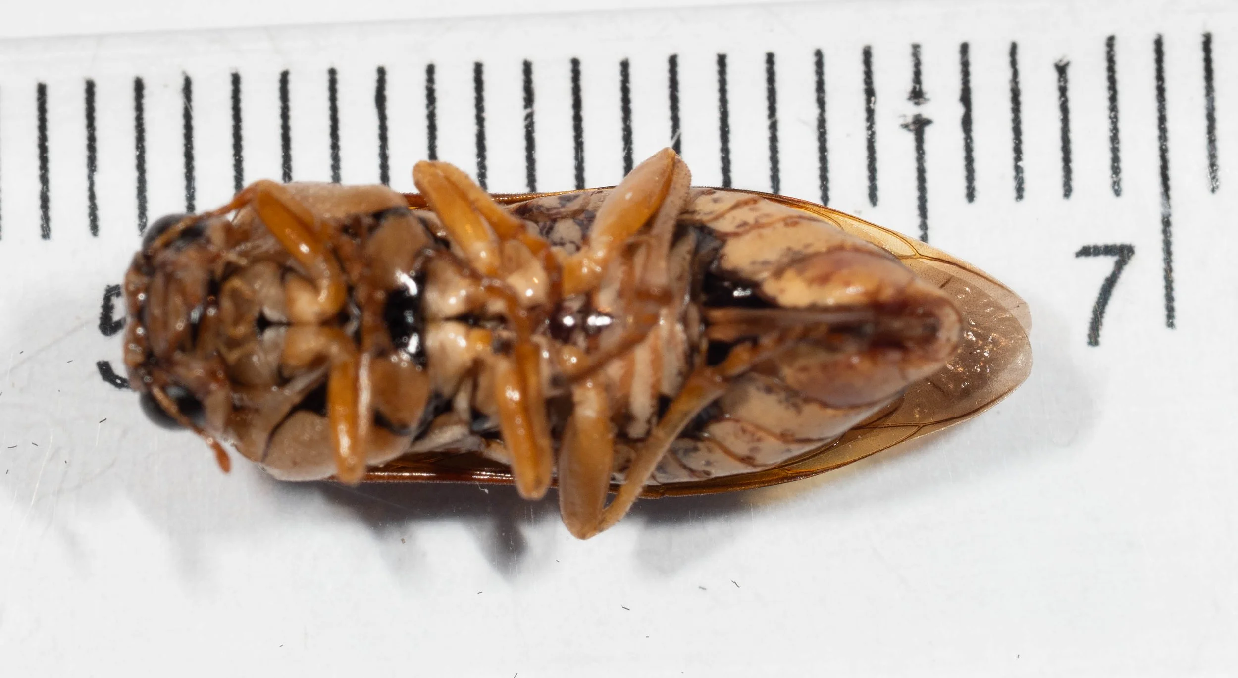
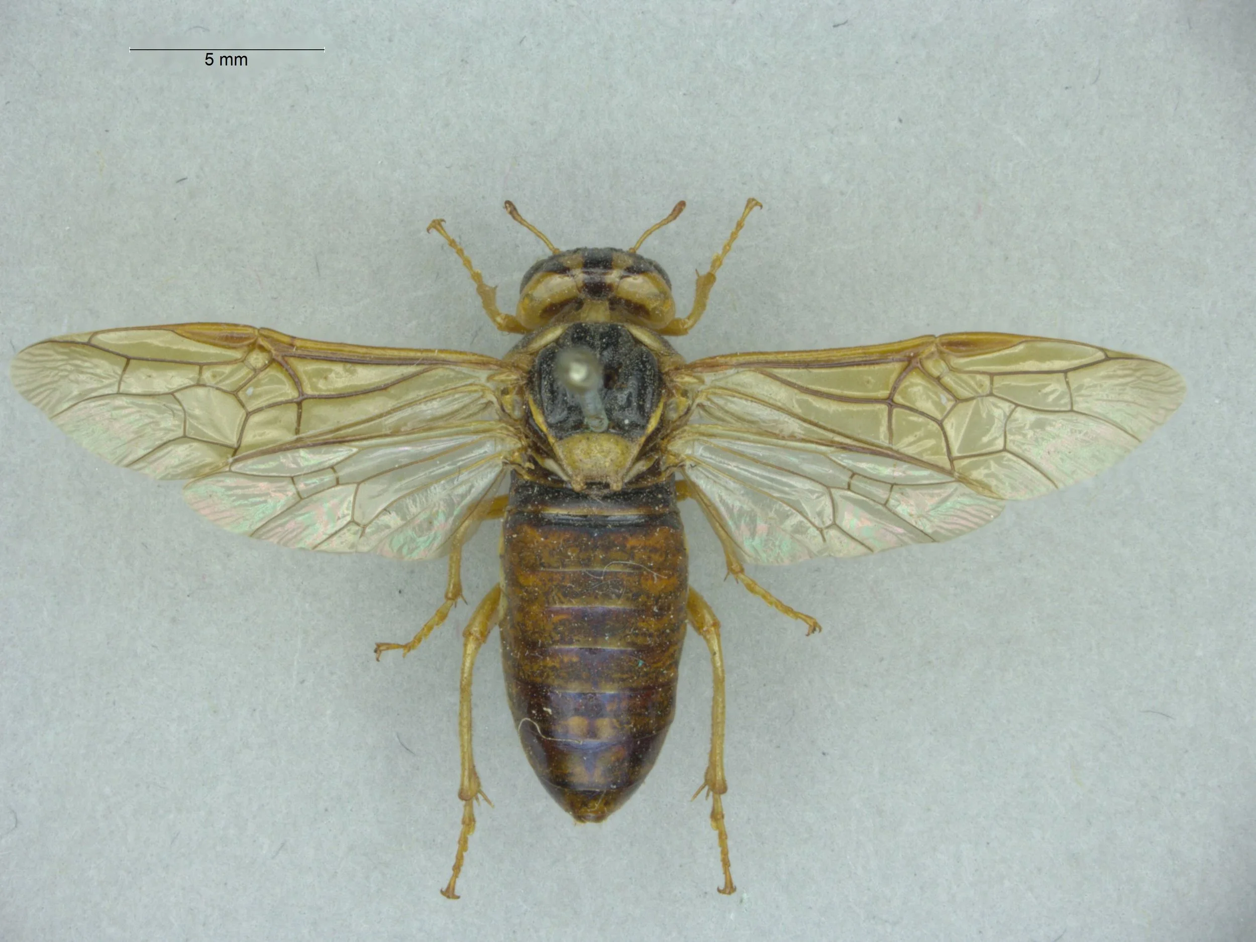
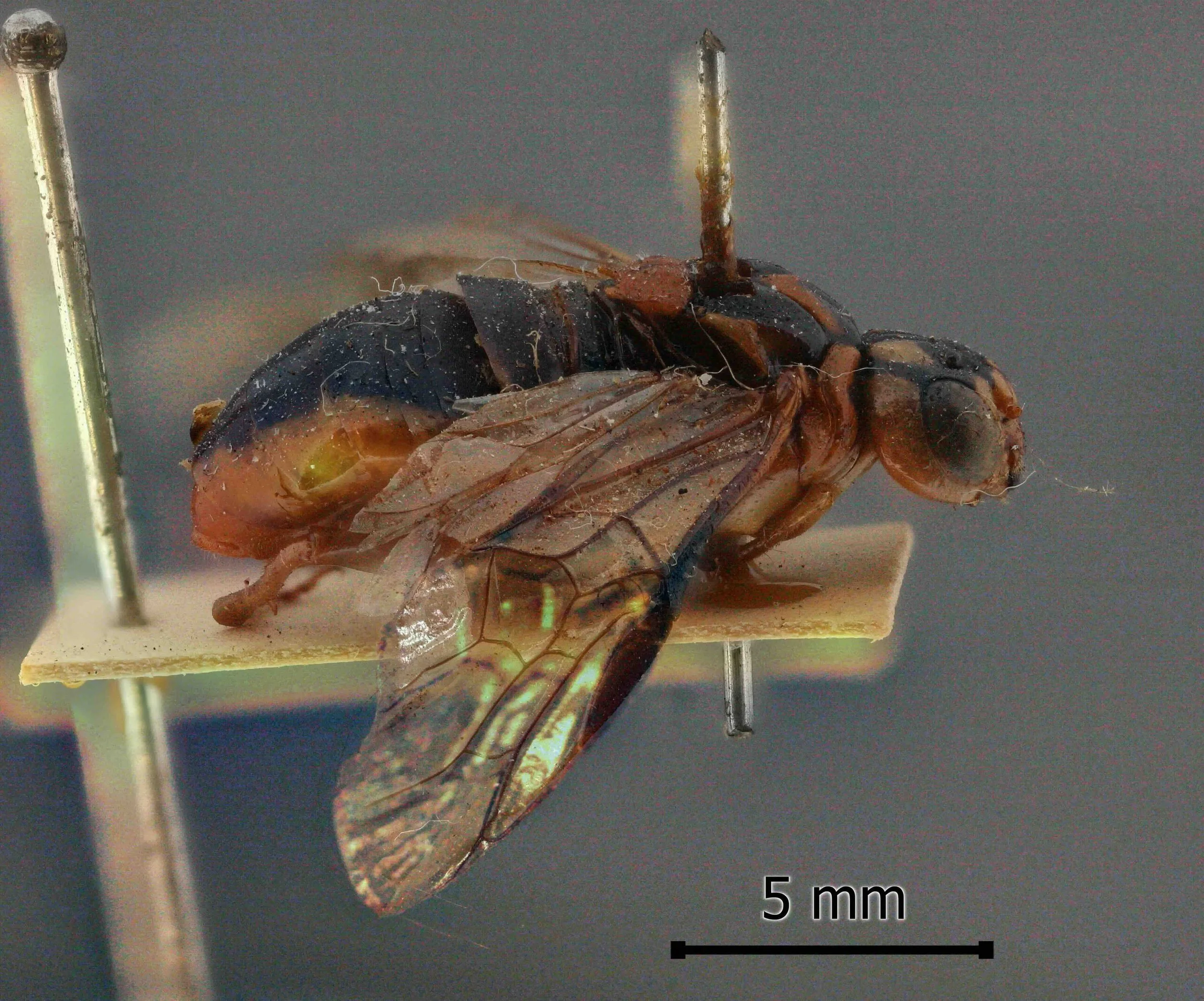
Specimen B is a male, collected by Chris Pease (@cole0ptera, iNaturalist observation) near Eumundi, Qld on 20/1/26 (specimen #PW085). It shows the following diagnostic characters of genus Xyloperga: 7-segmented antenna, shorter than the width of the head; scutellum convex, with well developed hind lobes; head swollen behind the eyes; clypeus with a transverse fold in the middle.
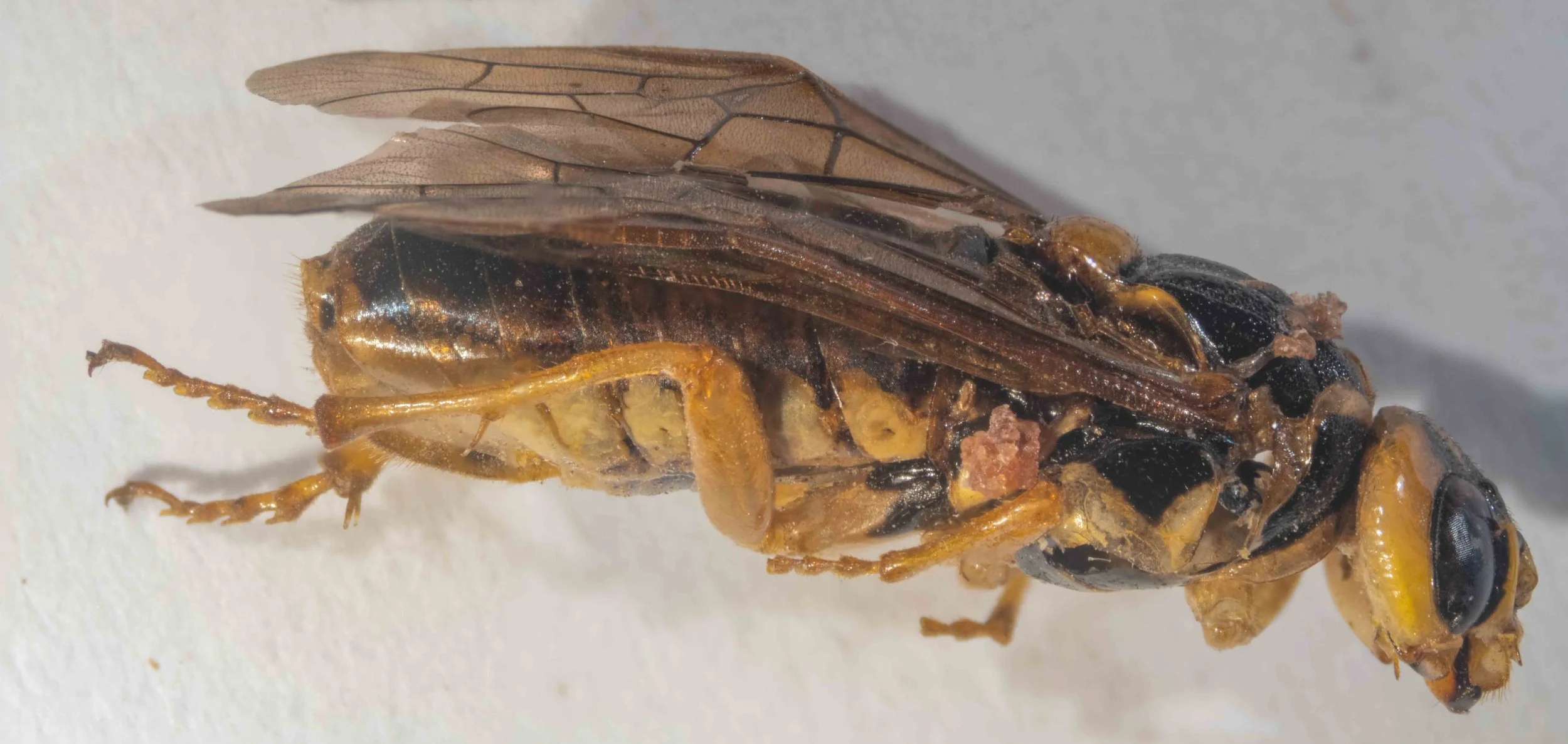
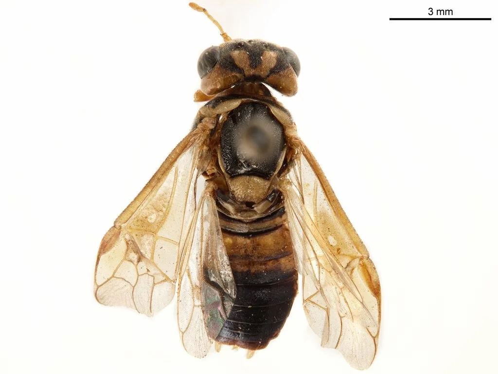
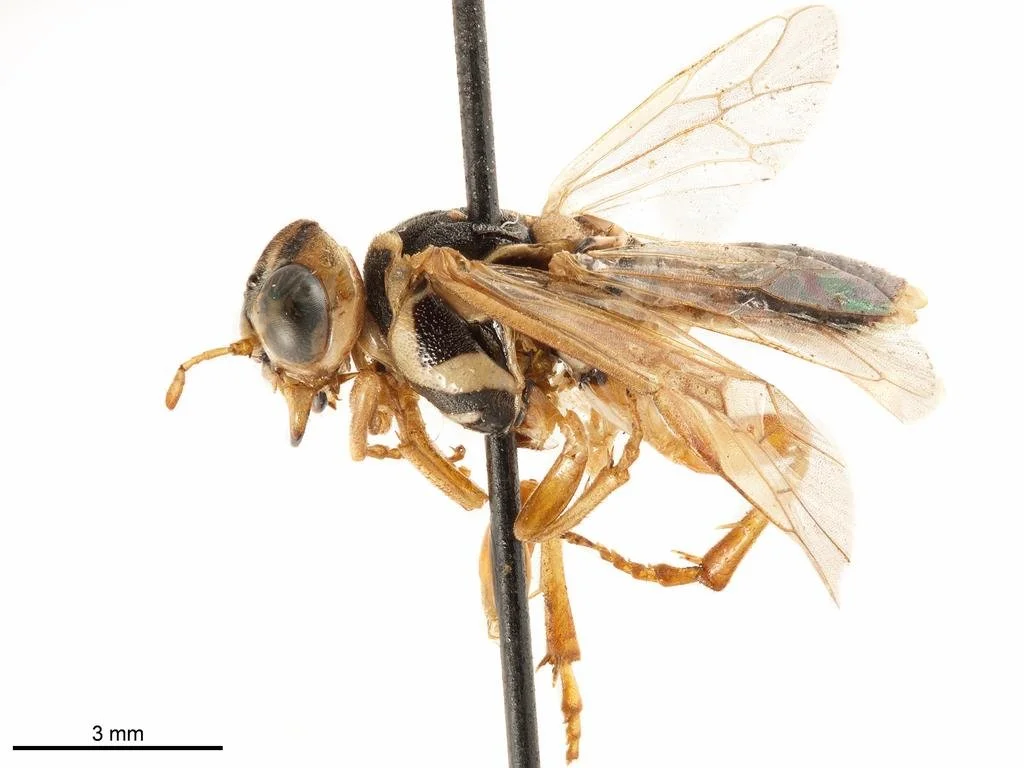
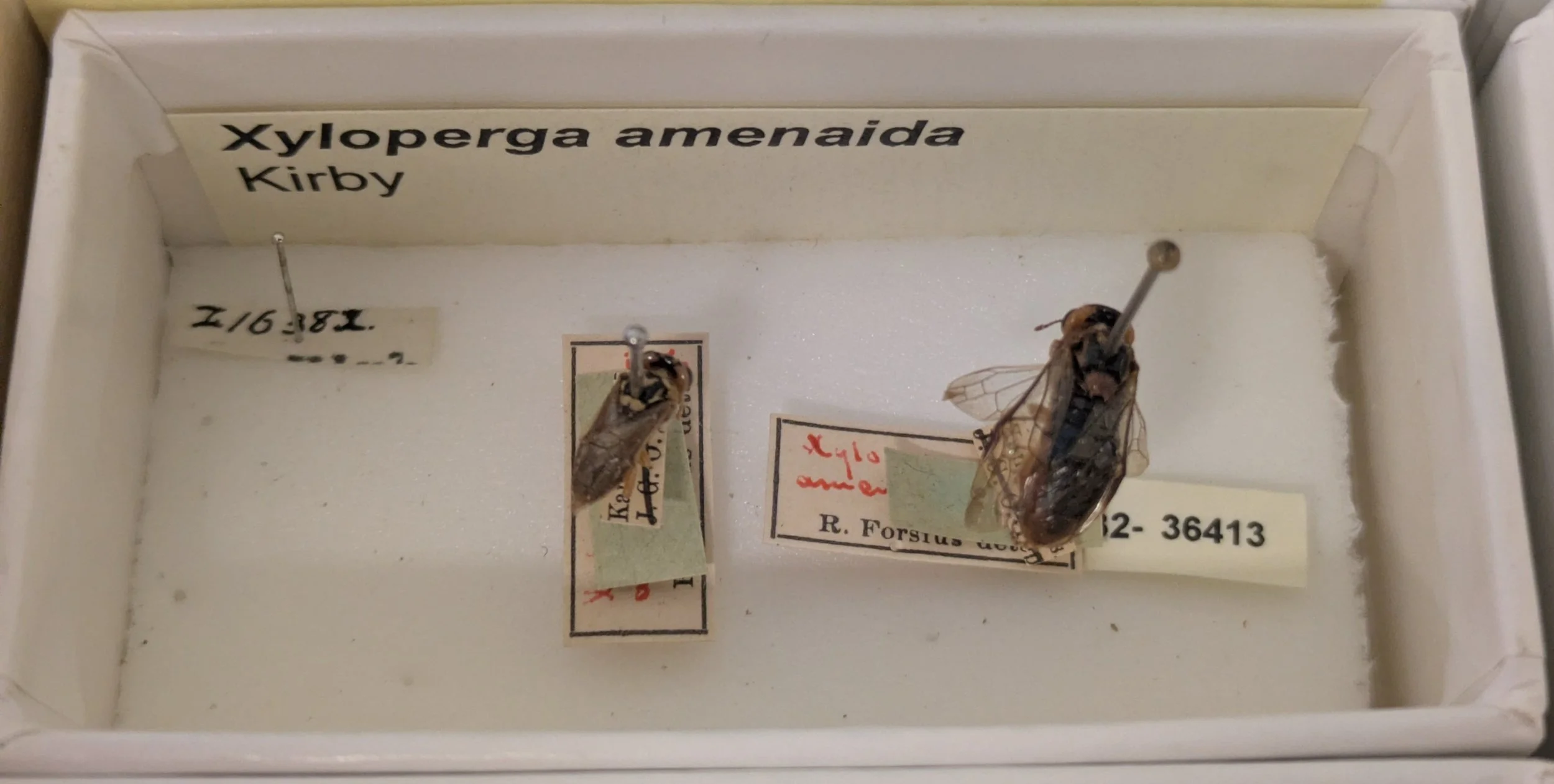
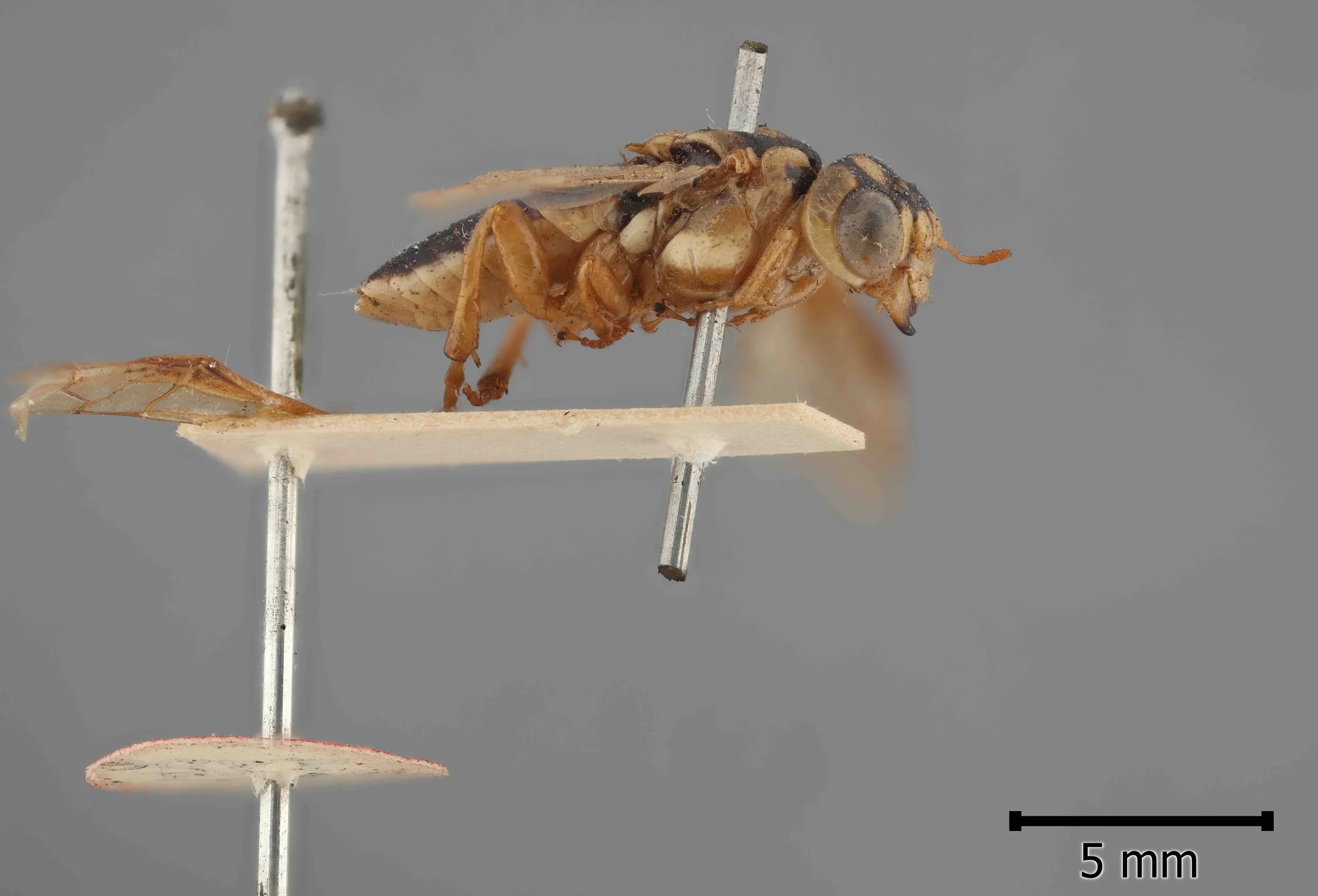
Specimen C
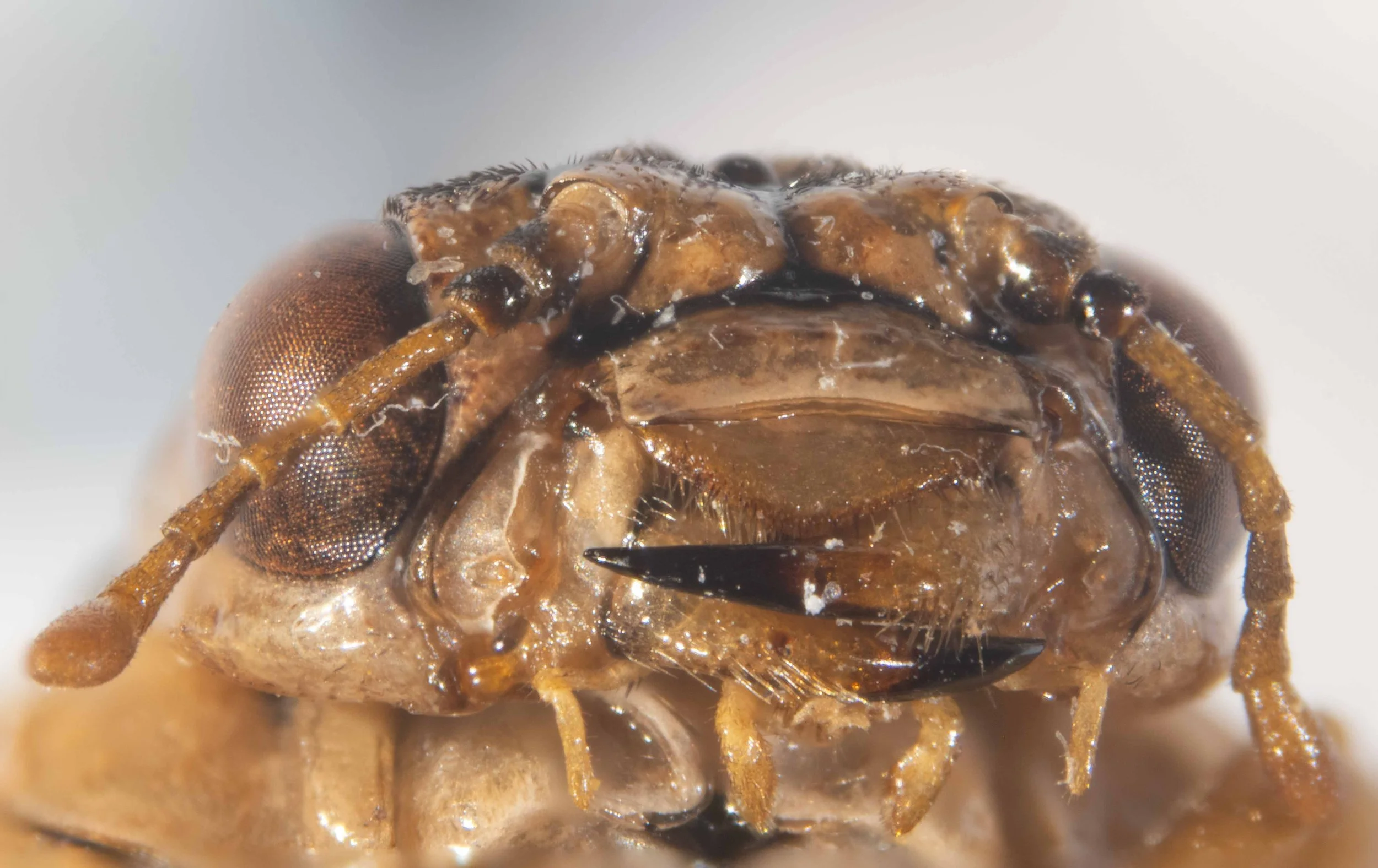
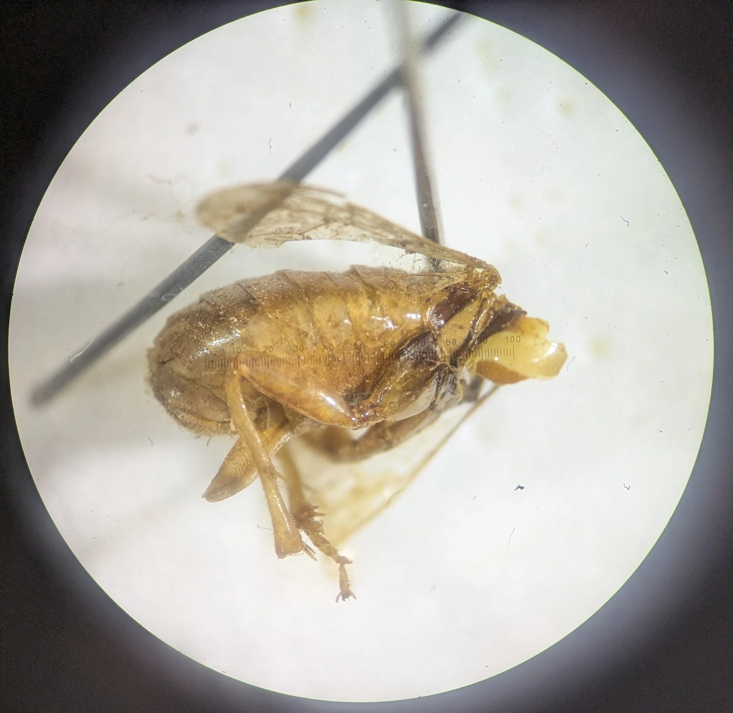
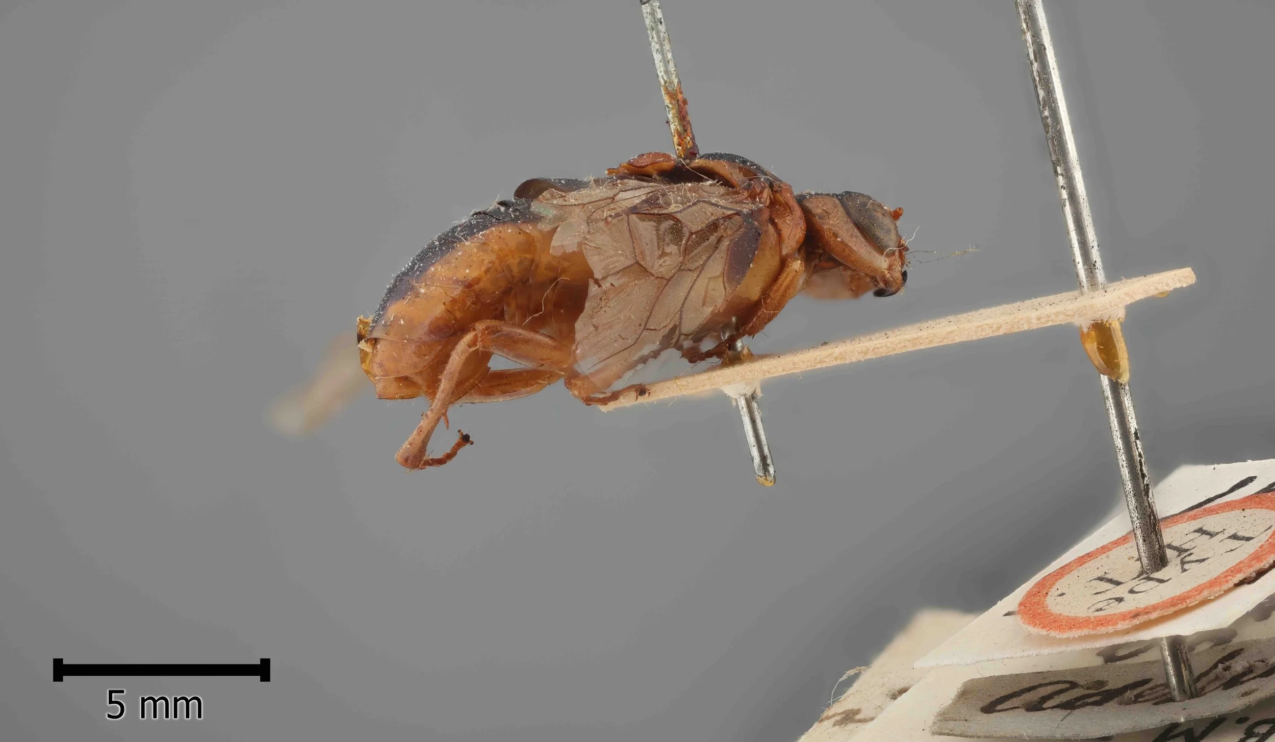
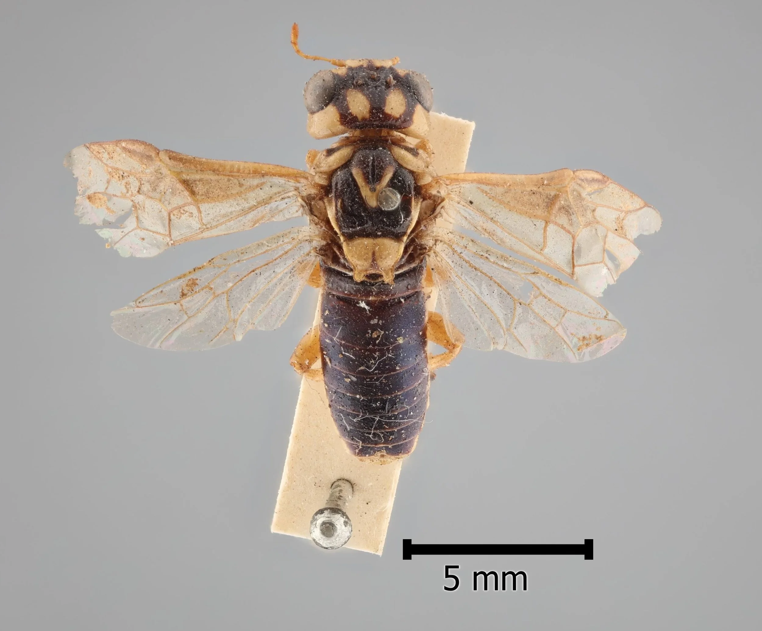
Specimen C is a female collected by James Peake, (iNaturalist observation) at Mt. Buller, Vic on 23/12/25 (specimen #PW077). It shows the following diagnostic characters of genus Xyloperga: 7-segmented antenna, shorter than the width of the head; scutellum convex, with well developed hind lobes; head swollen behind the eyes; clypeus with a transverse fold in the middle.

Comparison of specimens A, B, C from above observations
Click on the image below to download a high resolution pdf version (54KB).
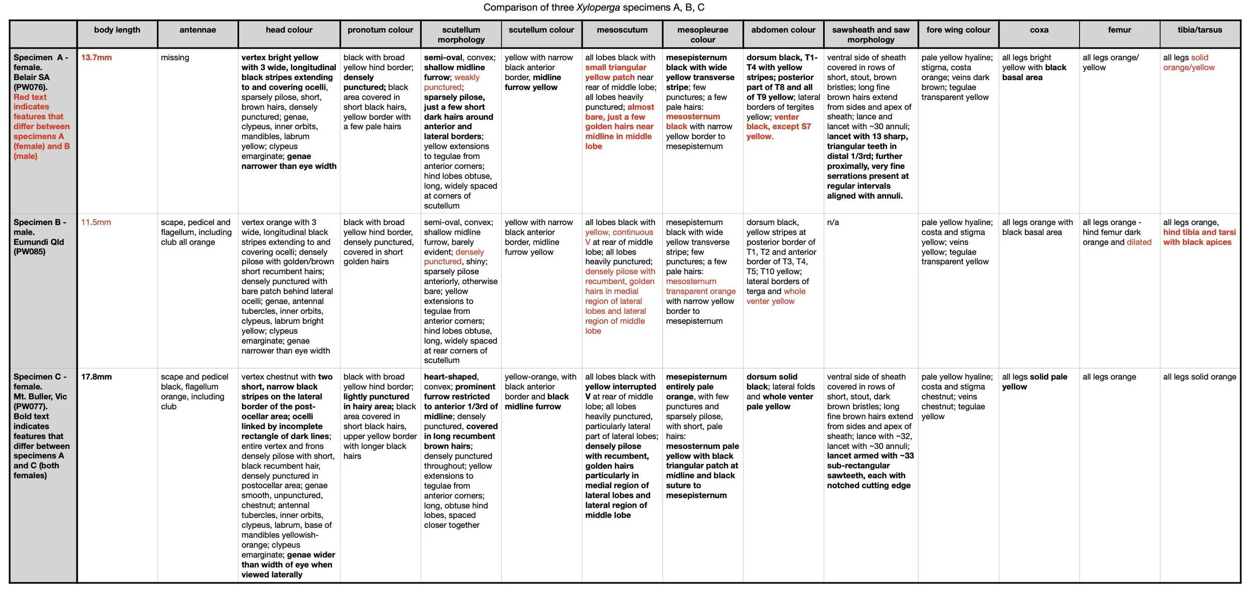
Summary:
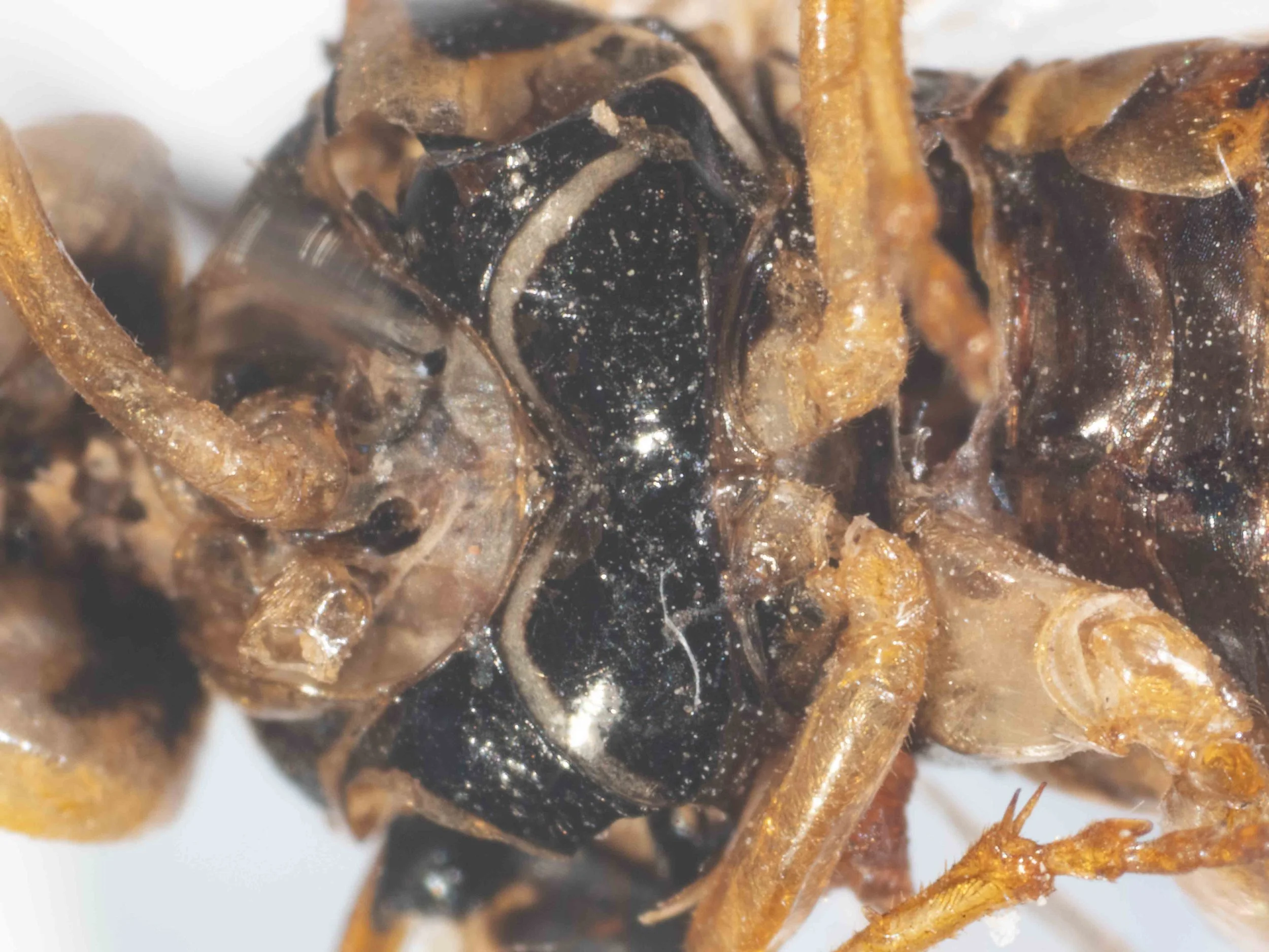
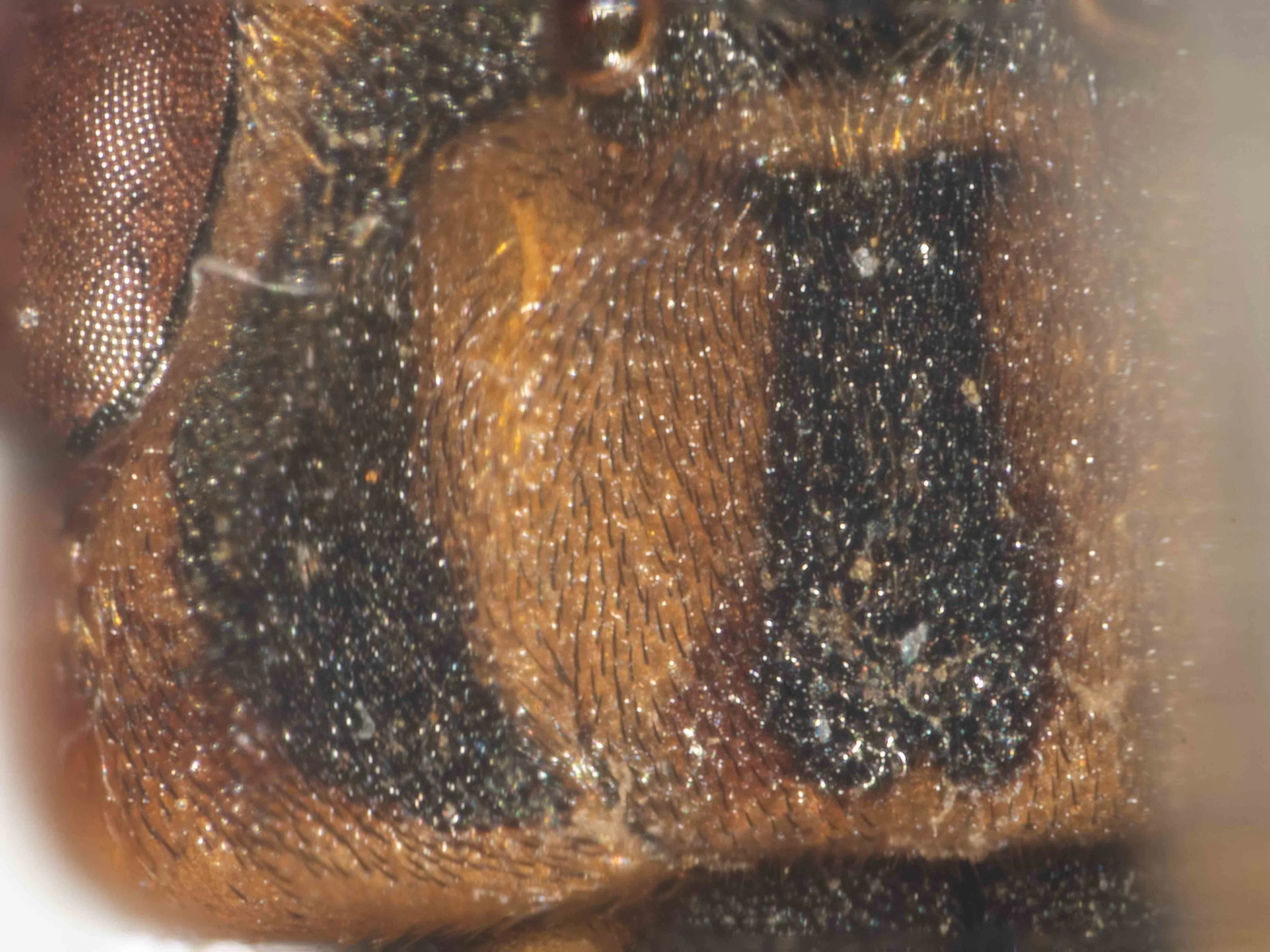
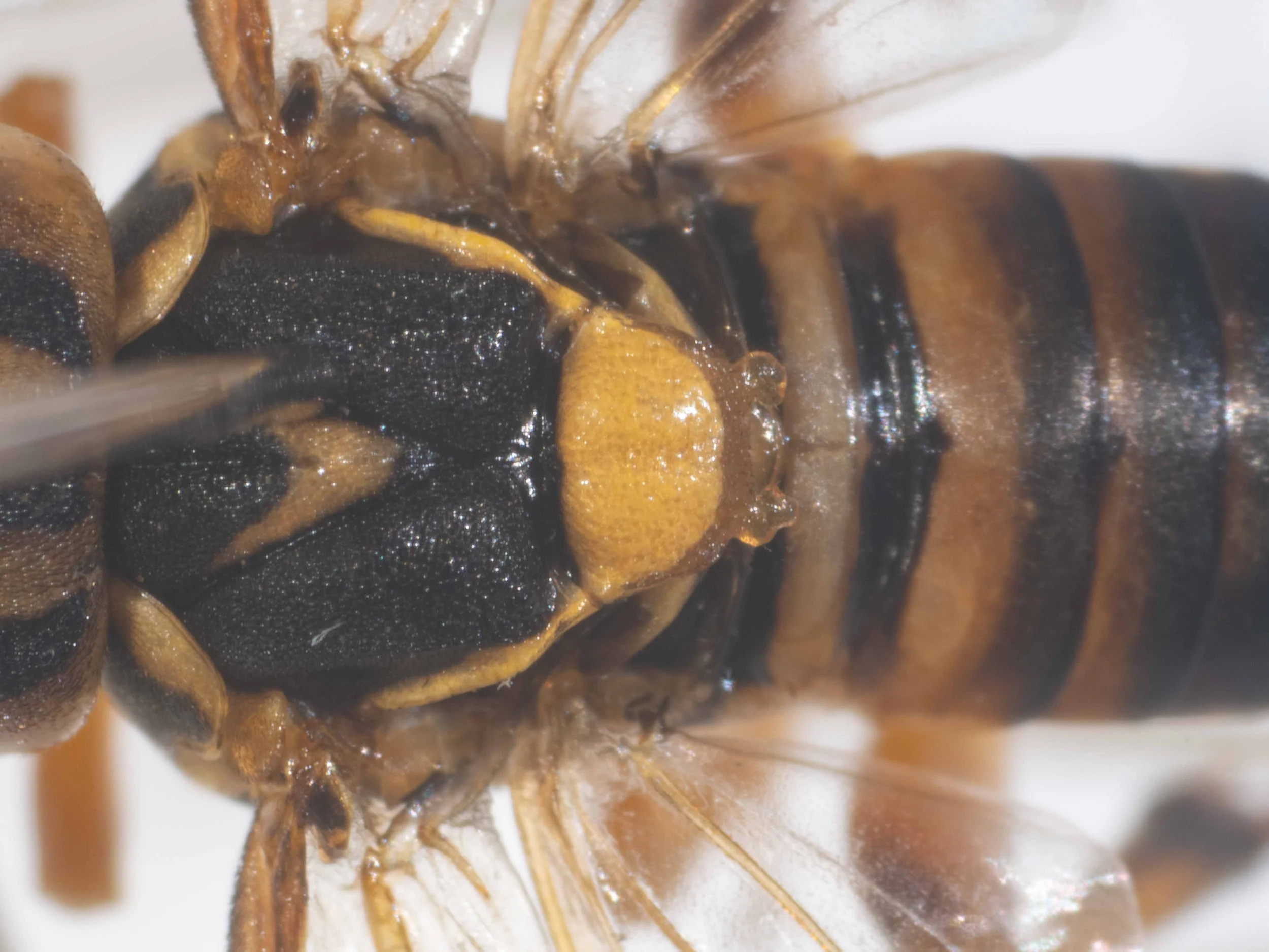
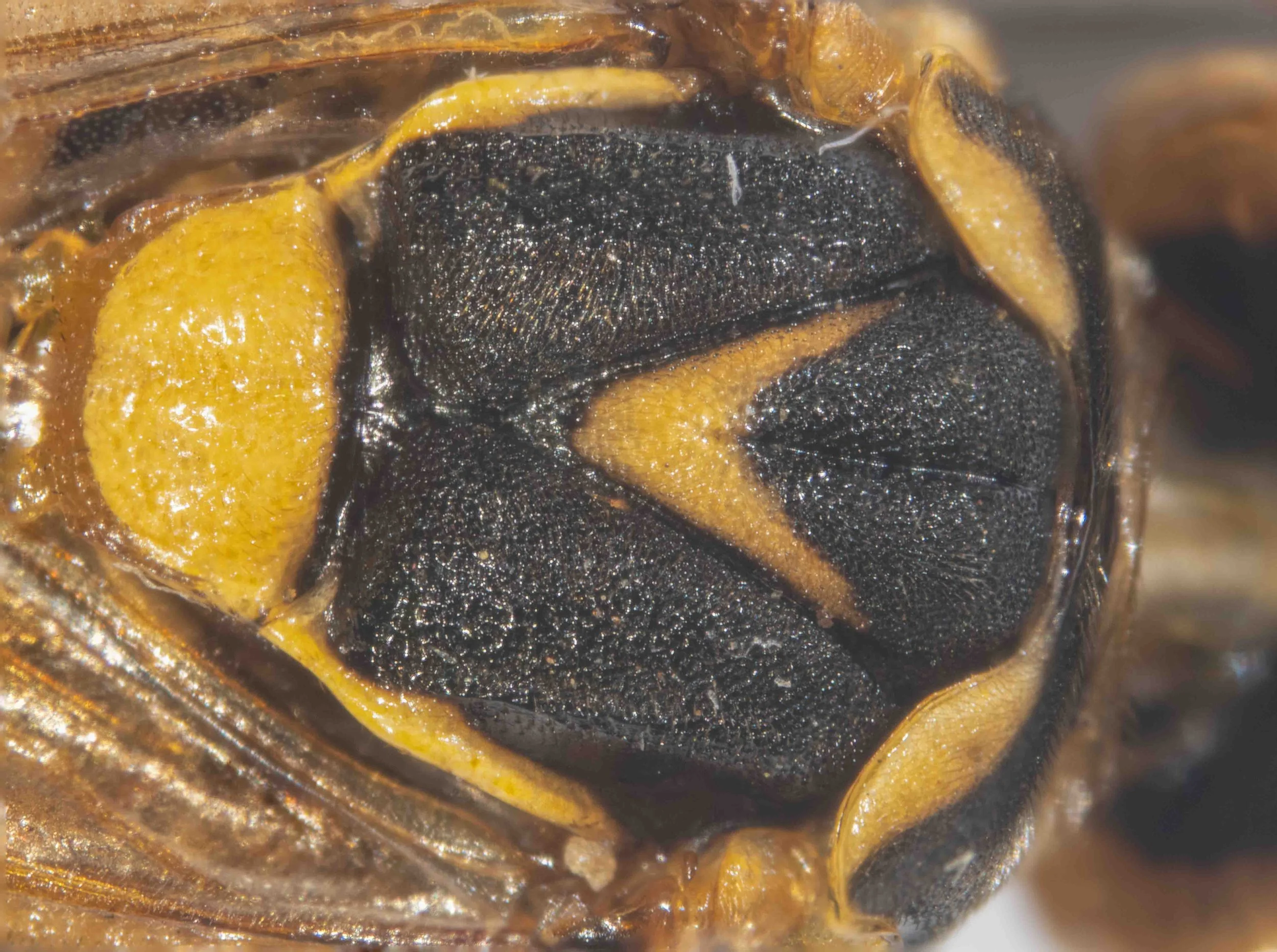
There are few differences between specimen A (a female) and specimen B (a male) - indicated by the red text in the Specimen A row. These include degree of punctation of the scutellum, size of the yellow patch on the middle lobe of the mesonotum, degree of pilosity of the mesoscutum and colour of mesosternum (black vs. transparent orange) and venter (black vs yellow).
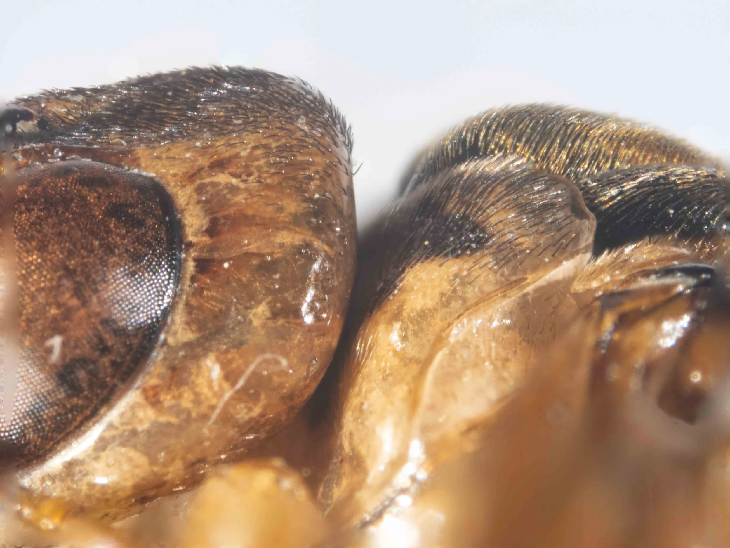
In contrast, there are many differences between specimens A and C - indicated by the bold text in the Specimen A and C rows. The most significant are head colour and morphology (pattern of stripes on vertex, relative genae vs. head width), scutellum colour and morphology, different V-mark on middle lobe of mesoscutum, mesepisternum colour and saw morphology.
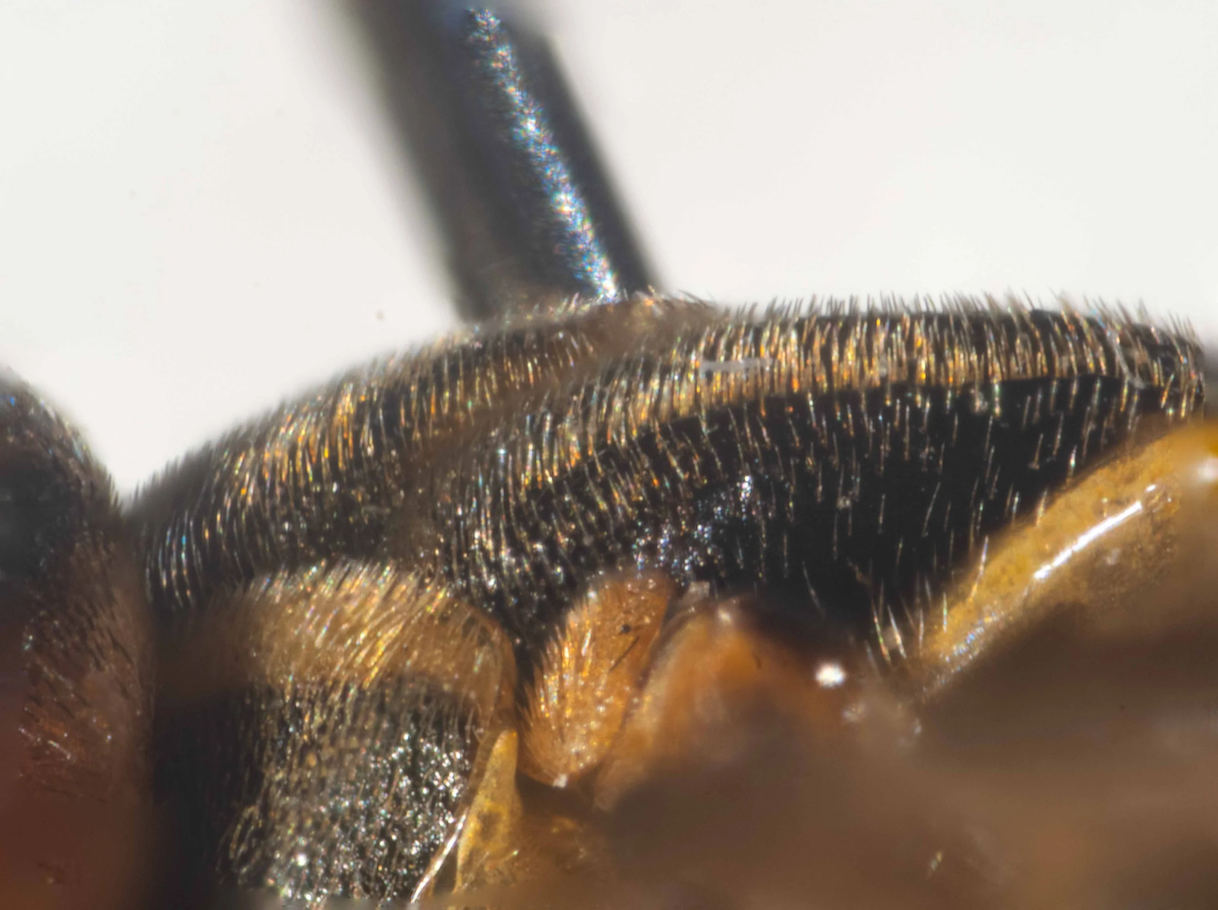
The last two characters - mesepisternum colour and saw morphology - are included in published descriptions of X. halidaii and X. amenaida and therefore play a key role in species delimitation. The other characters - head morphology and scutellum morphology/colour - were either not recorded or only superficially described in published descriptions.
How representative are Specimens A, B, C of other iNaturalist observations?
The descriptions of specimens A, B, C will be used to assist species identification of X. amenaida and X. halidaii in iNaturalist field photos. It is therefore important to know whether these specimens are representative of iNaturalist observations.
The following table shows whether the iNaturalist observation listed matches specimens A, B or C. Features that don’t match that specimen are noted.
Click on the thumbnail to download a full PDF version (59KB).
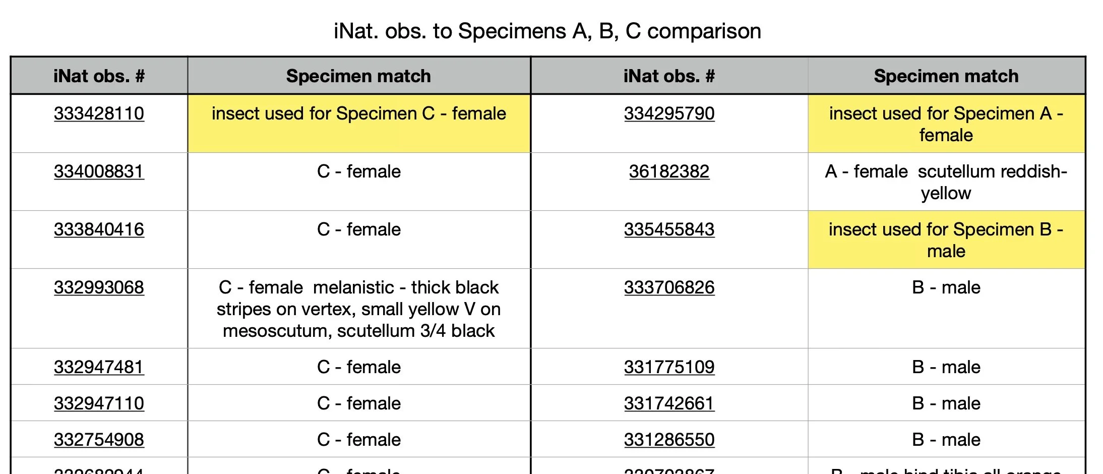
Summary:
iNaturalist observations of X. amenaida/halidaii fall clearly into 2 groups, corresponding to specimens A/B (female/male) and specimen C (female).
I conclude that the specimens are representative of iNat observations.
There are minor differences between individuals within each of these groups:
- some insects in group C are strongly melanistic, showing thick black stripes on the vertex reaching to the ocelli, rather than narrow, short stripes and thicker black stripes on the scutellum
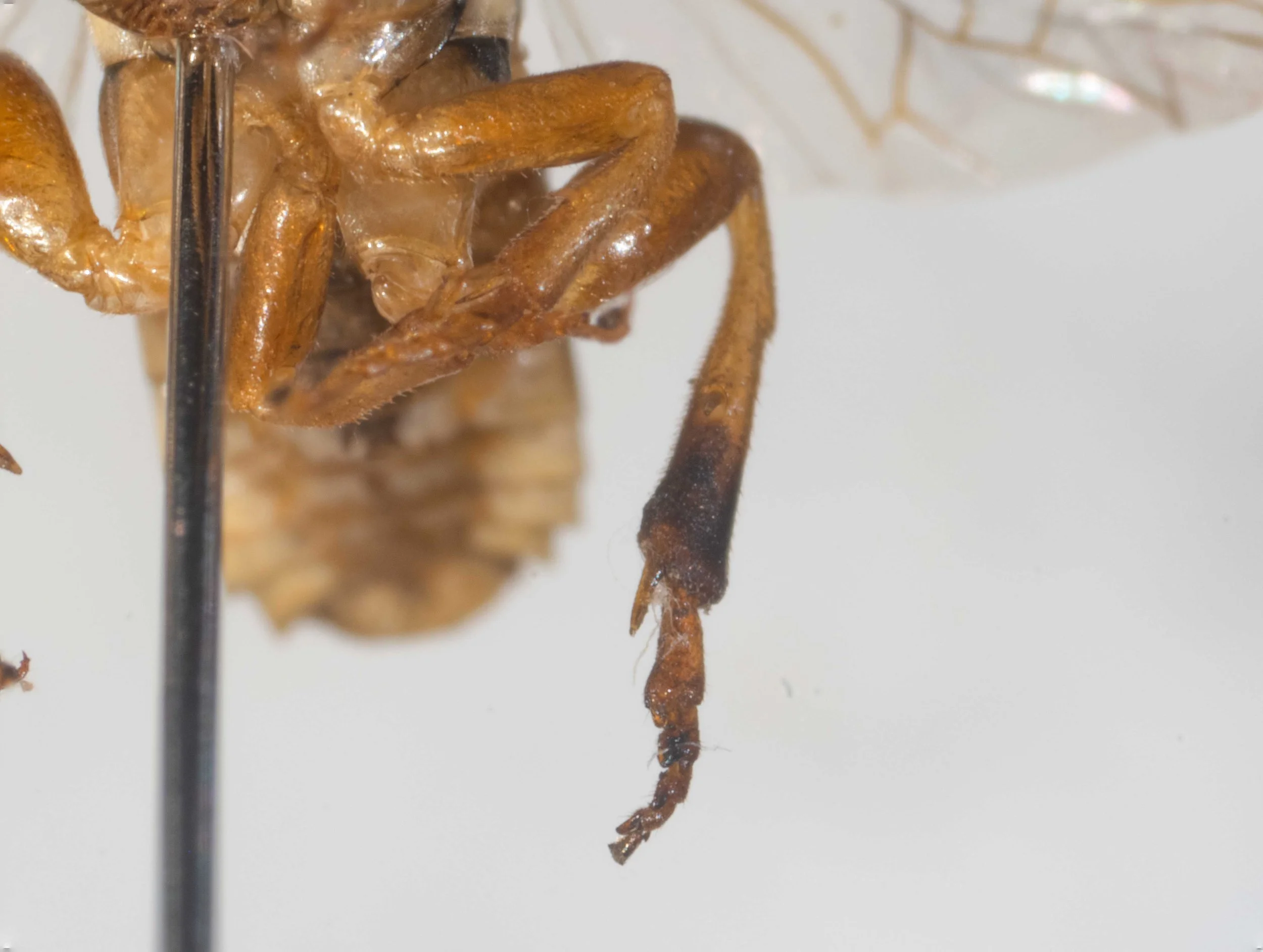
- the apex of the otherwise orange hind tibia is black in some individuals in group B
- the hind femur is clearly dilated in some individuals in group B but not in others
Differences between X. amenaida and X. halidaii in published descriptions
The matrix below compares features of X. amenaida and X. halidaii extracted from published descriptions of these species by a range of authors.
Red text highlights features that differ between these species.
Click on the image to download a pdf of the matrix (871KB).
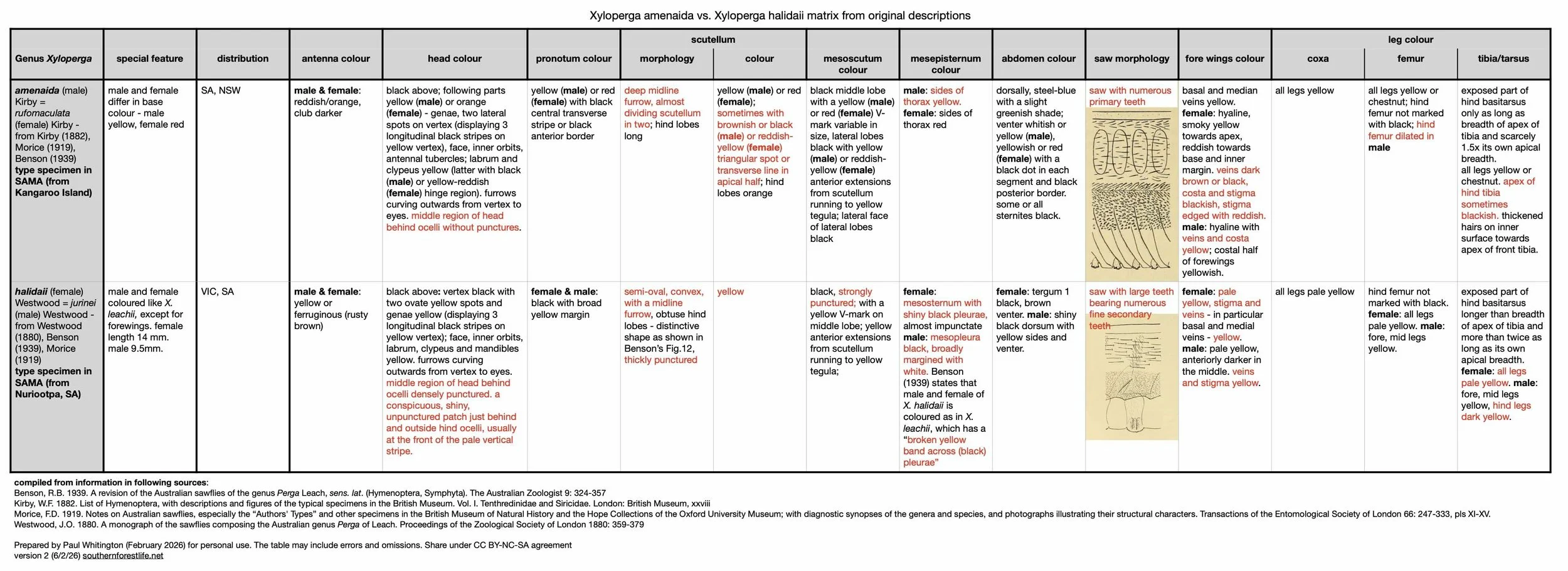
Summary of distinguishing features in published descriptions:
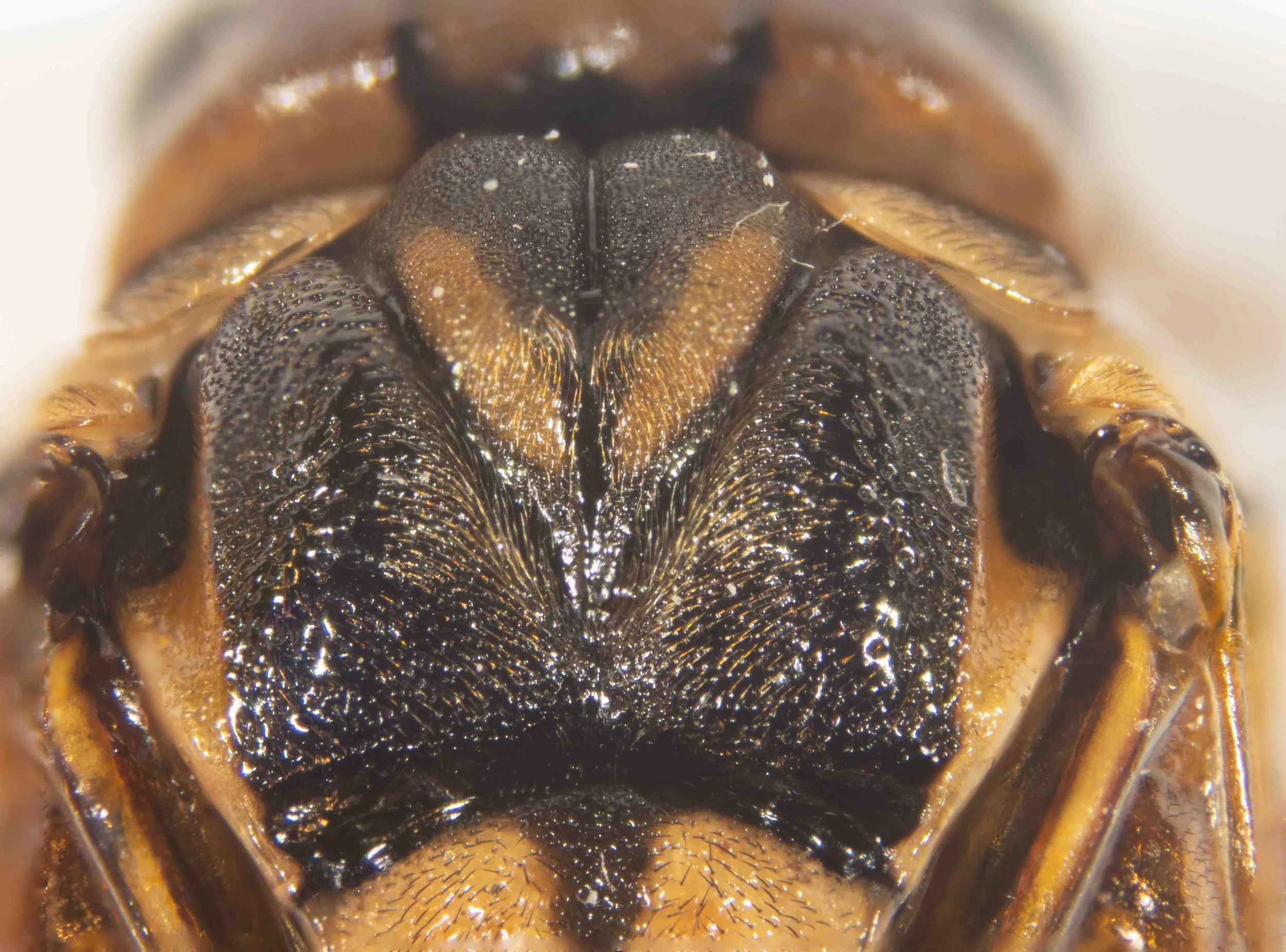
Post-ocellar region of X. halidaii is densely punctured, apart from a patch behind the hind ocelli, whereas it is unpunctured in X. amenaida
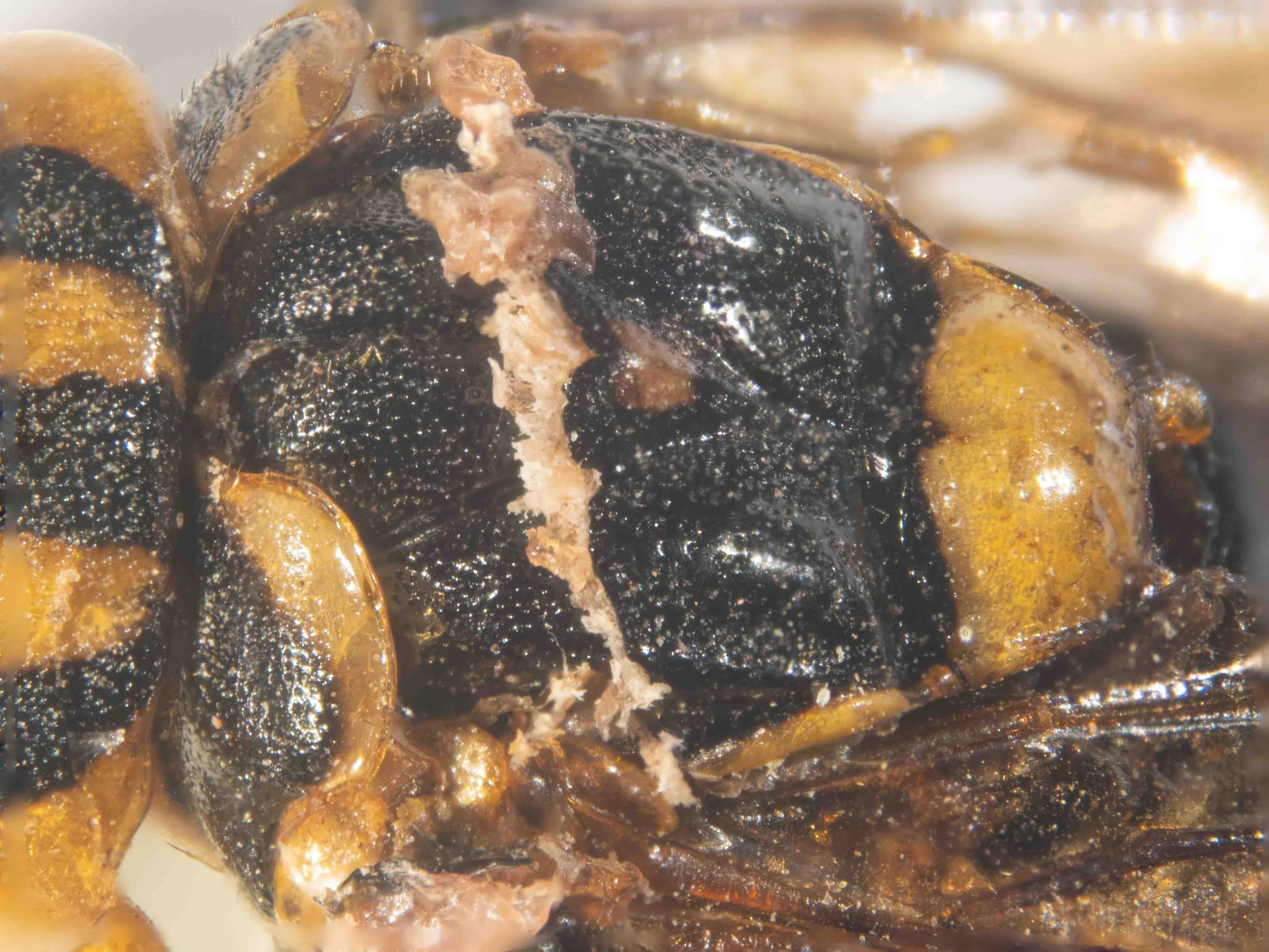
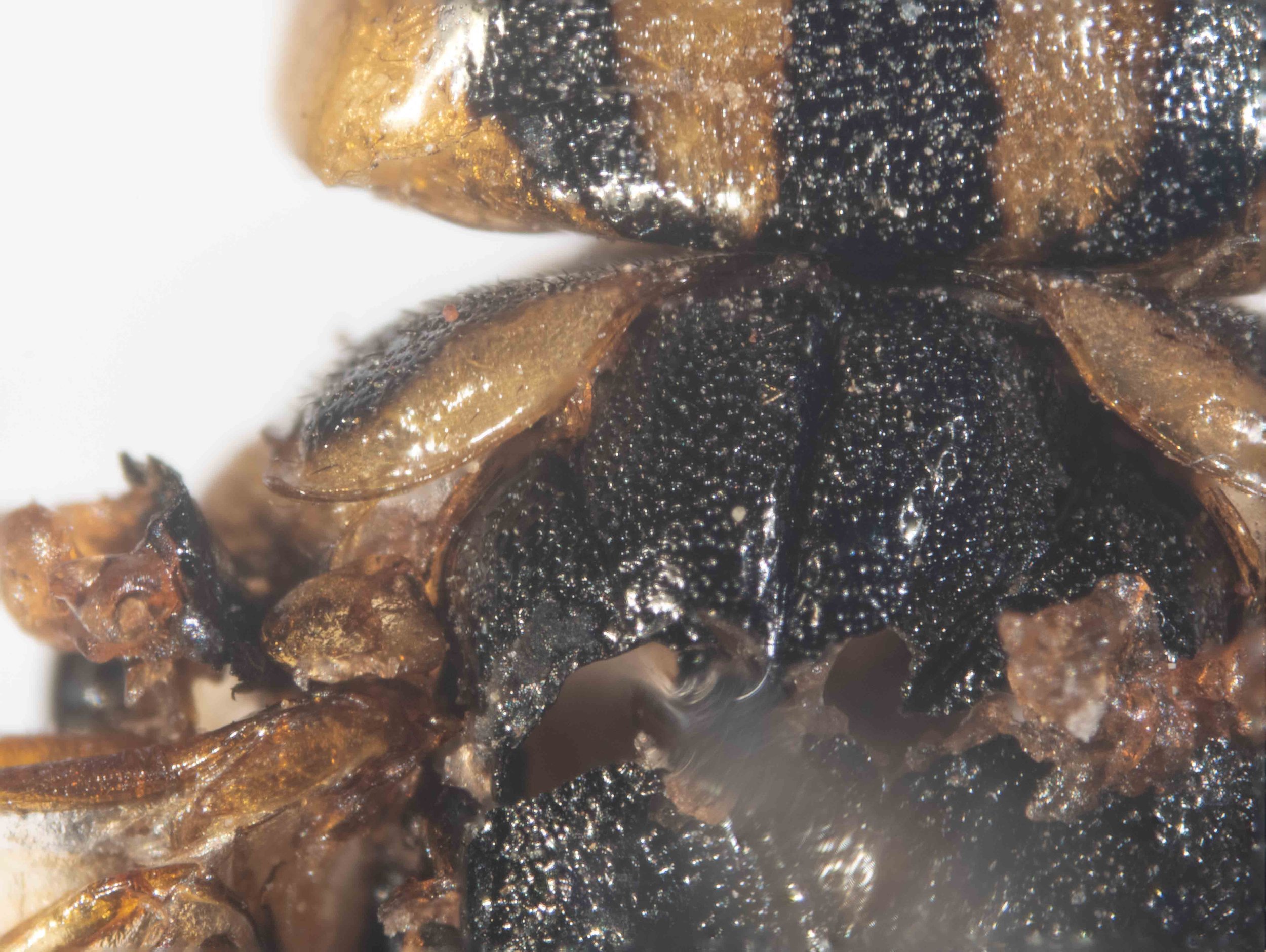
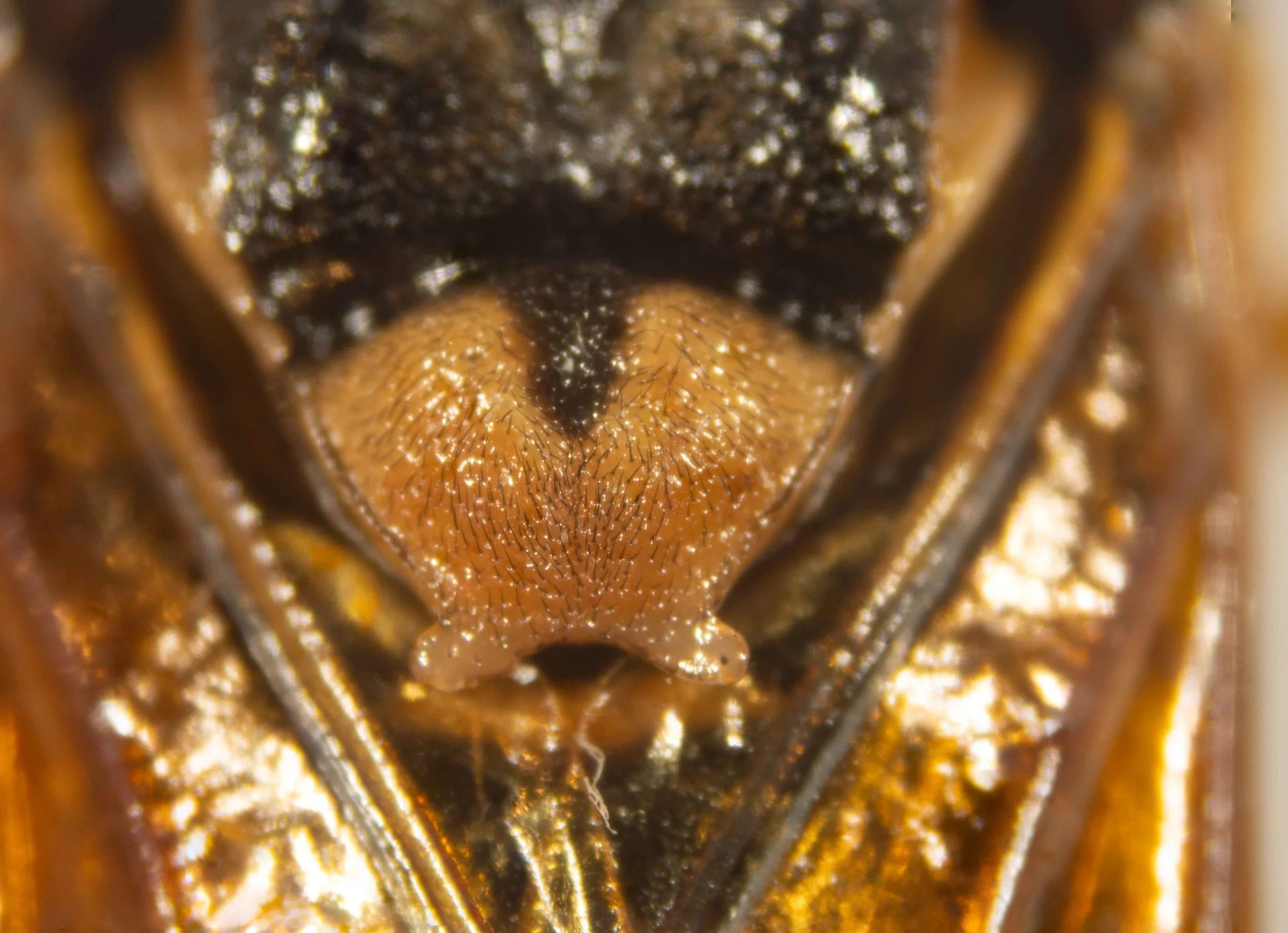
Scutellum of X. halidaii is yellow and thickly punctured. In X. amenaida the midline furrow is deep and yellow in colour, sometimes with a brownish-black (in male) or reddish-yellow (in female) area in the apical half.
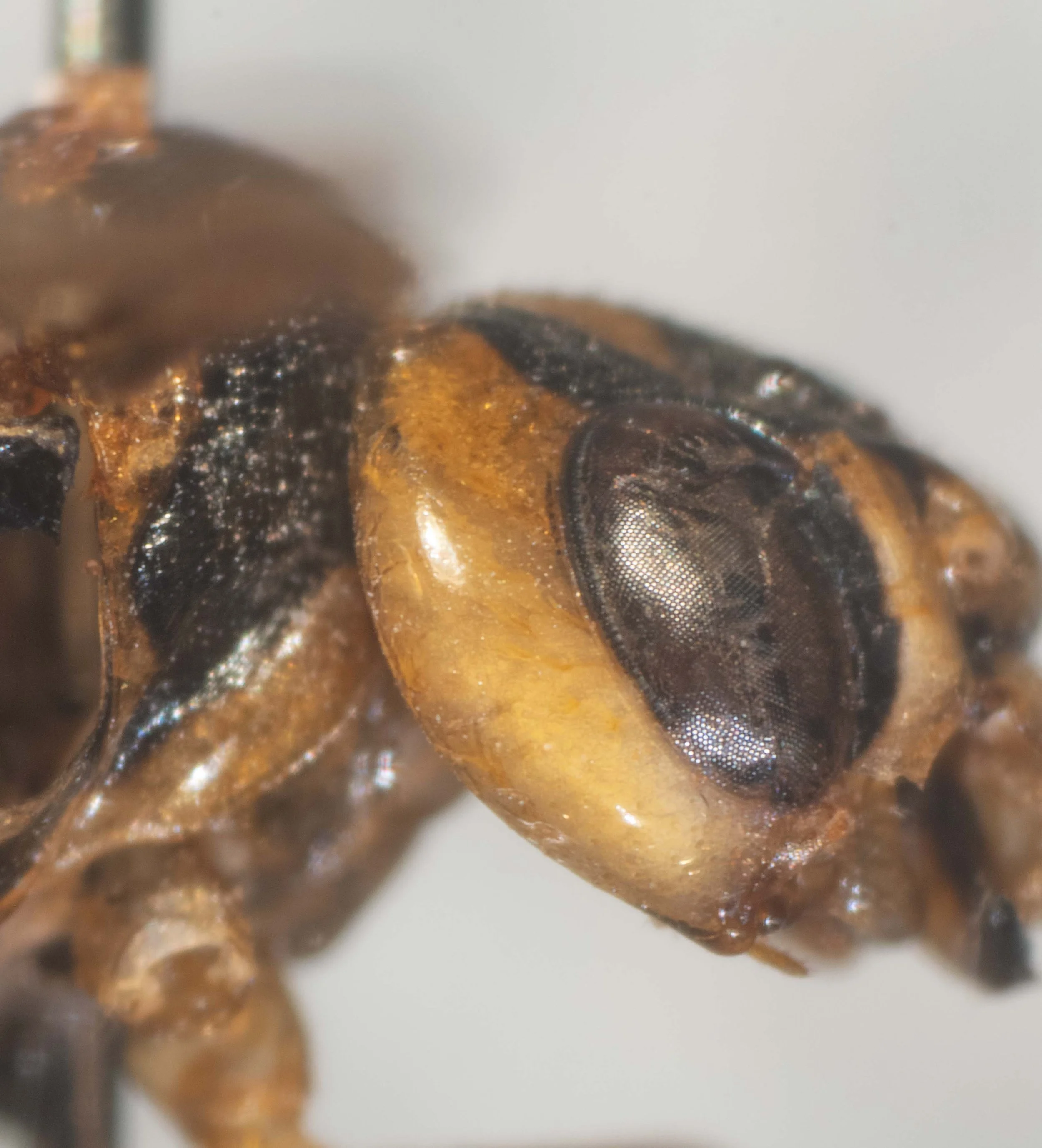
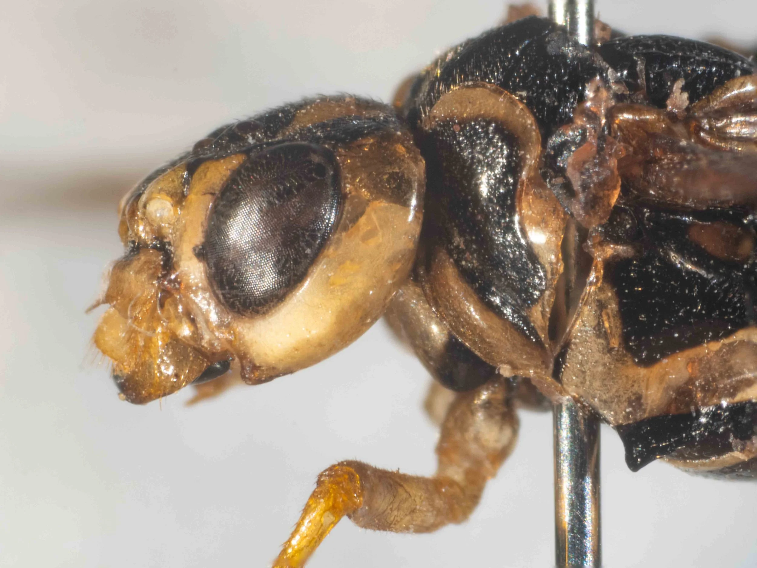
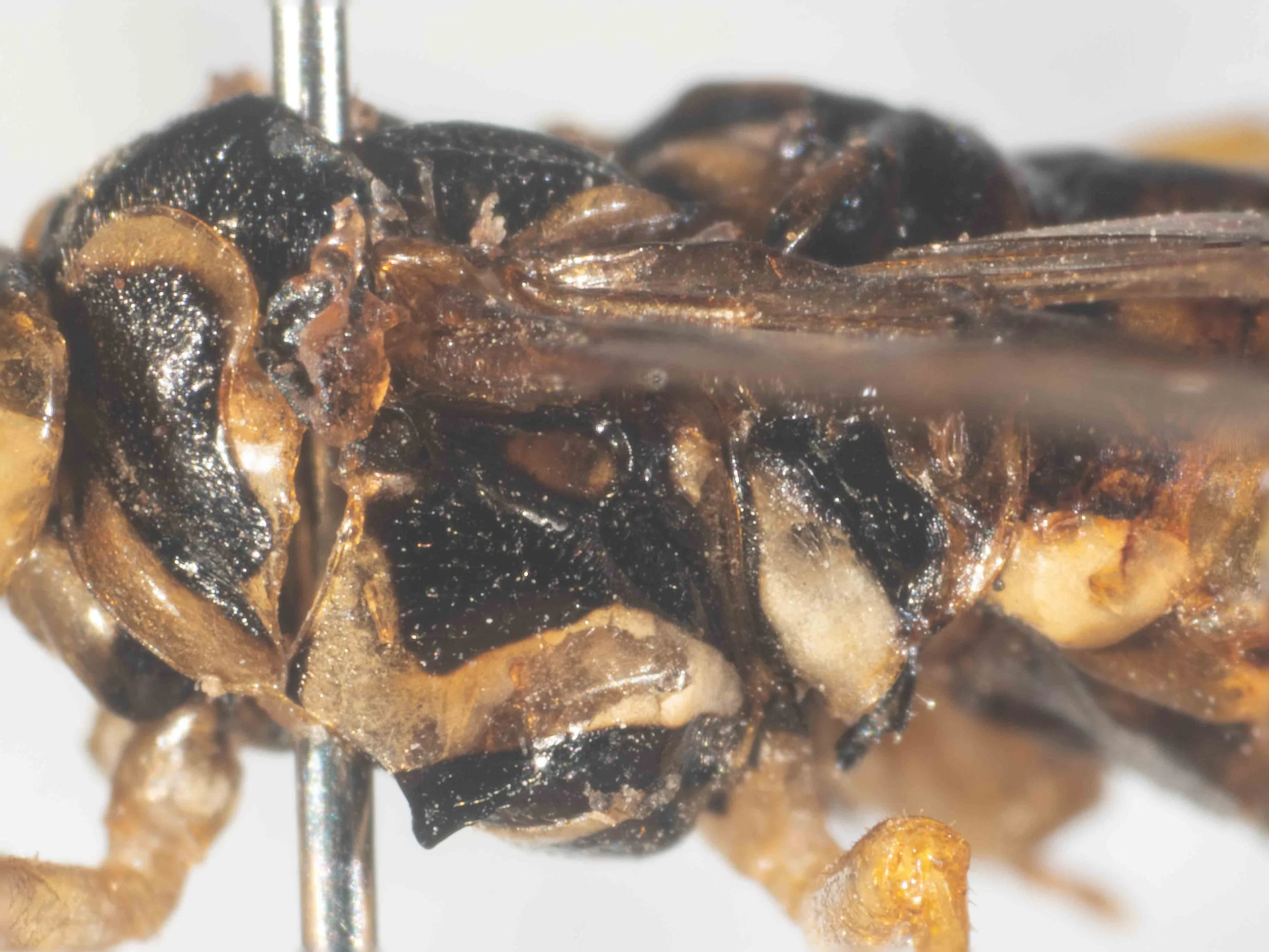
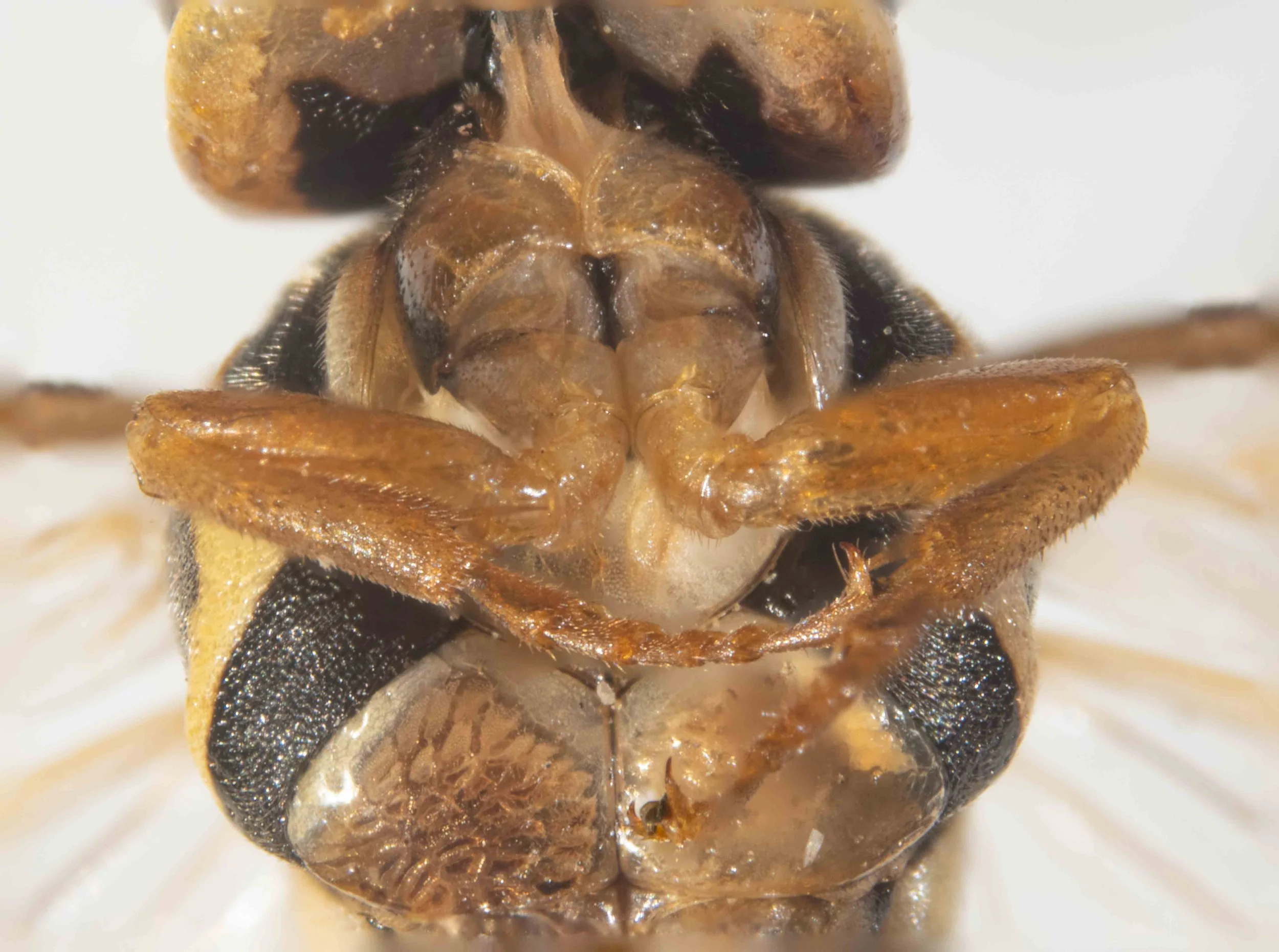
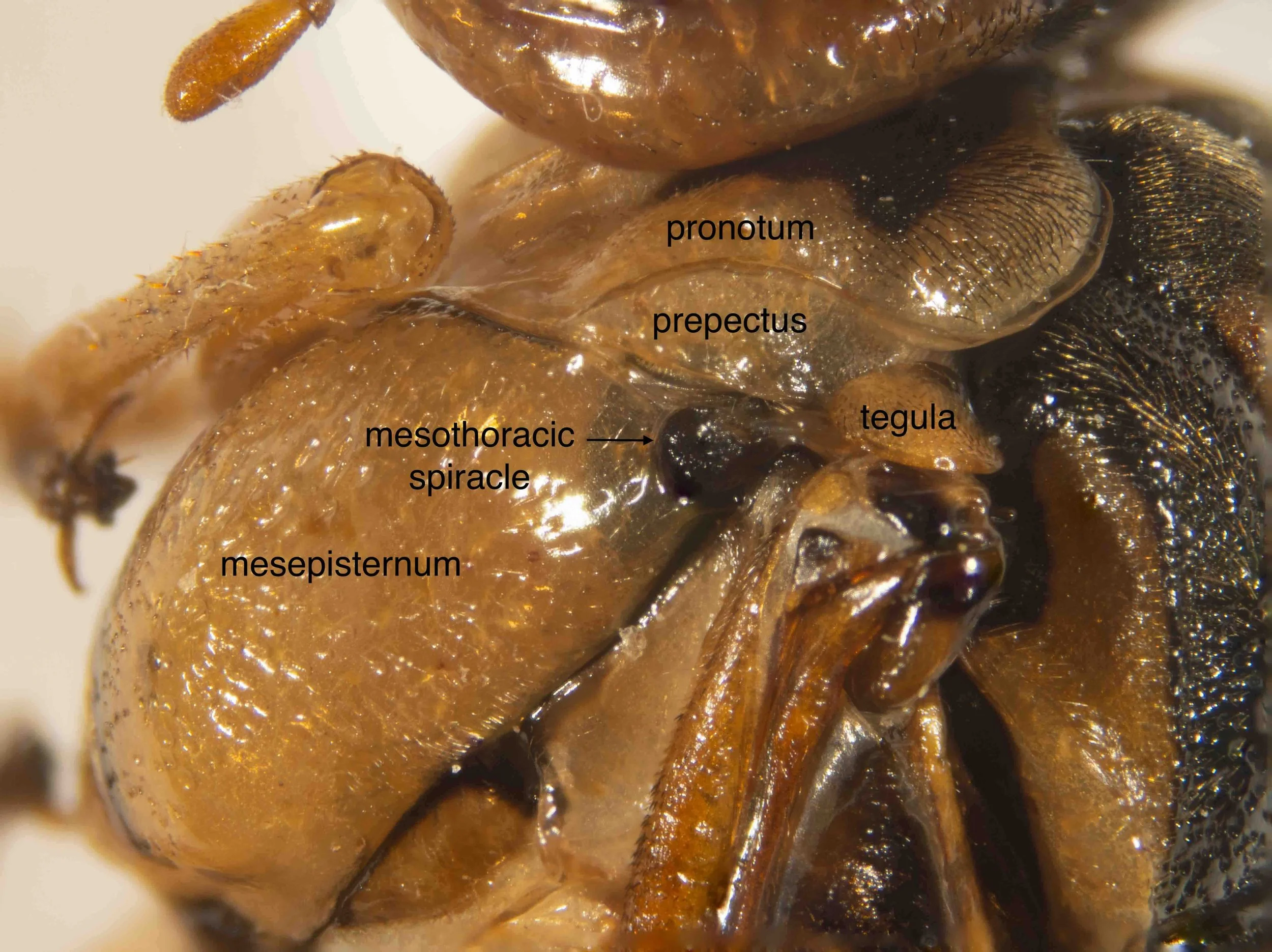
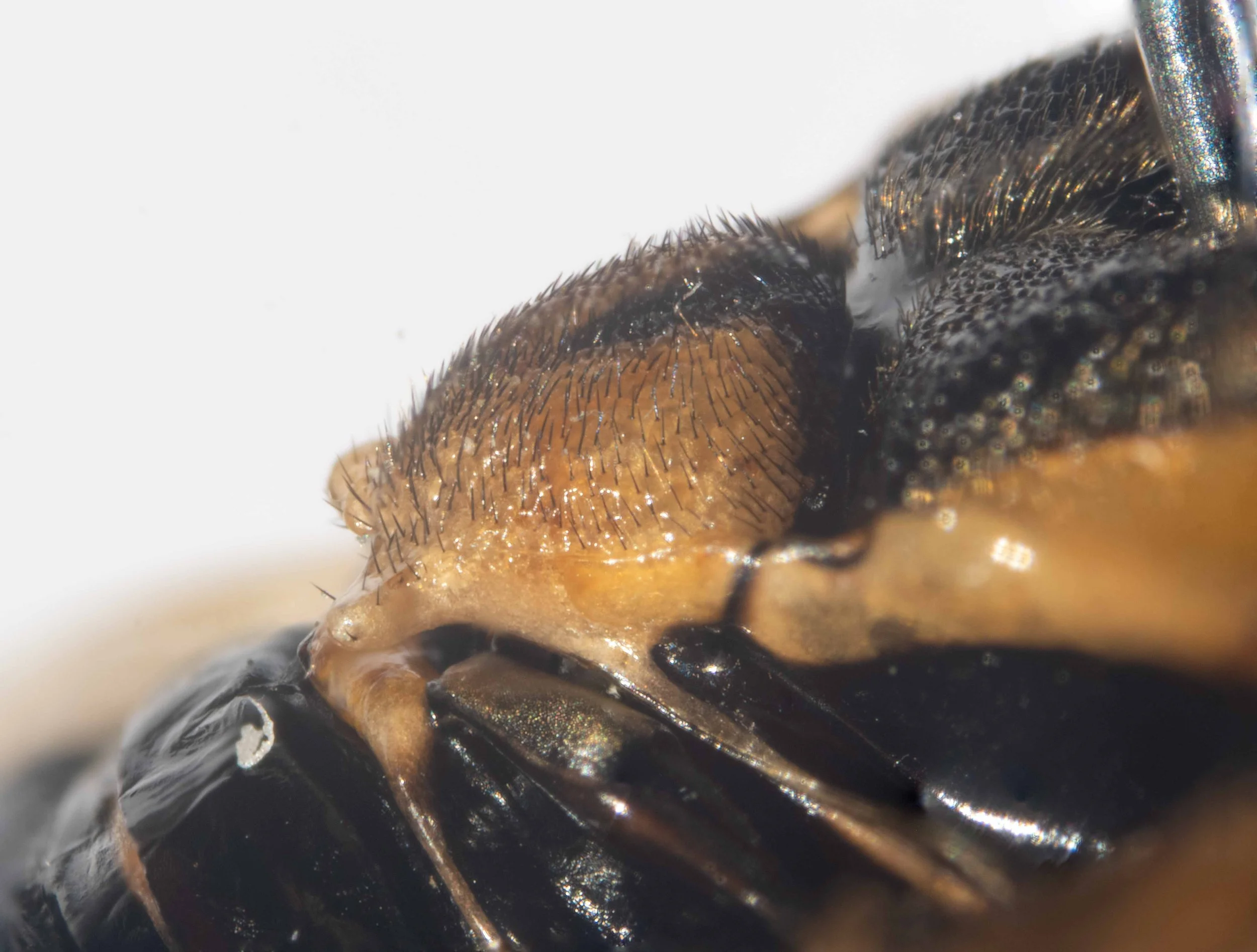
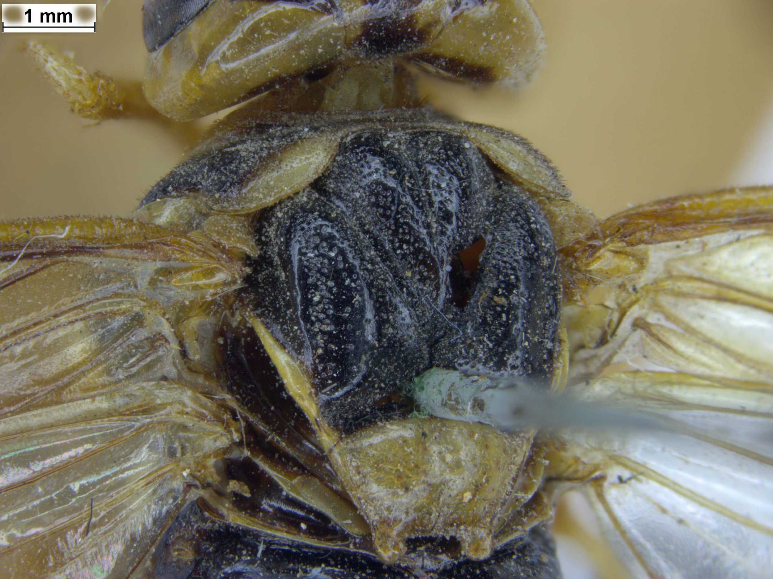
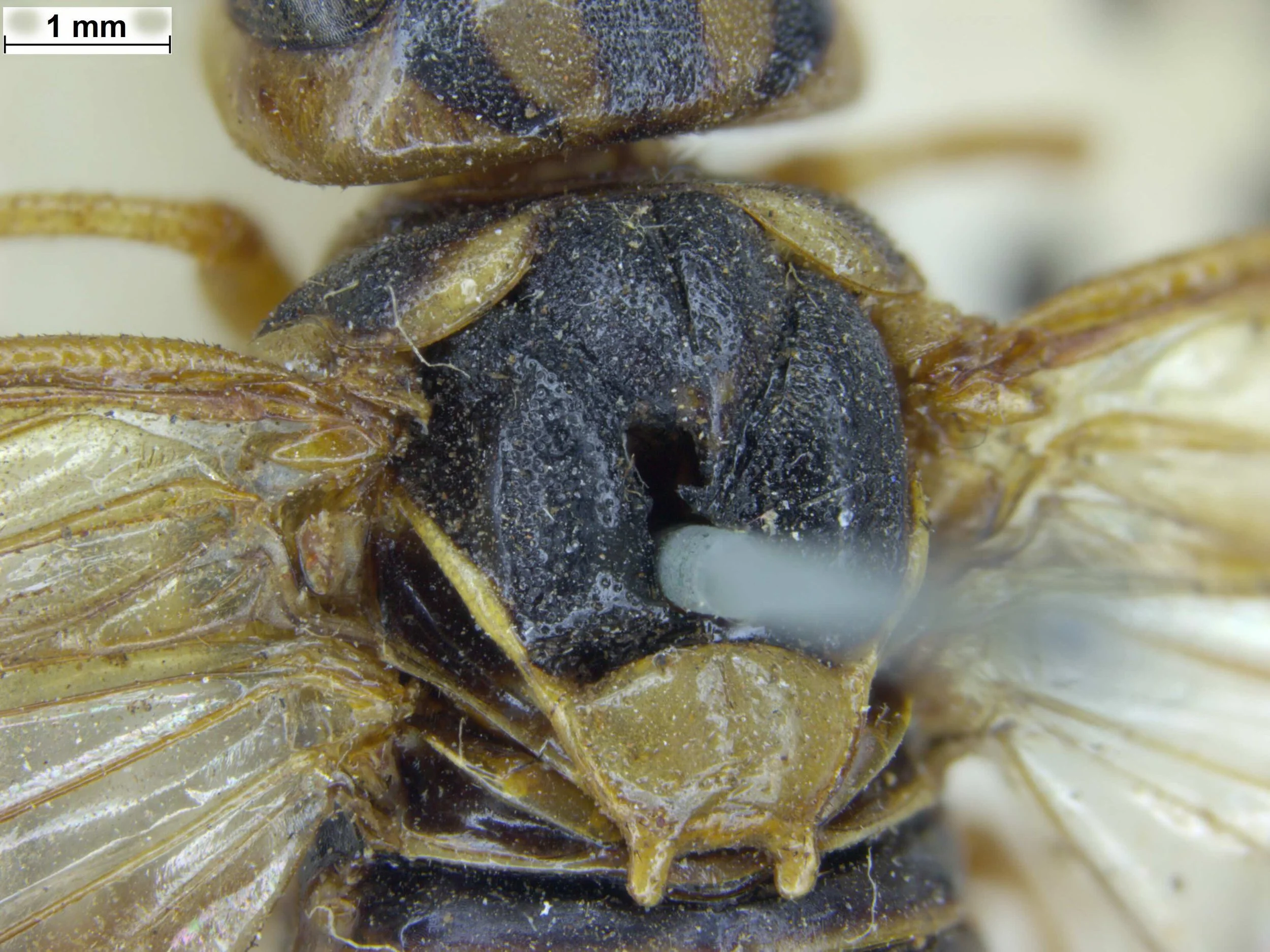
Mesepisternum of X. amenaida is yellow (in male) or red (in female), whereas in X. halidaii it is shiny black with a yellow transverse band
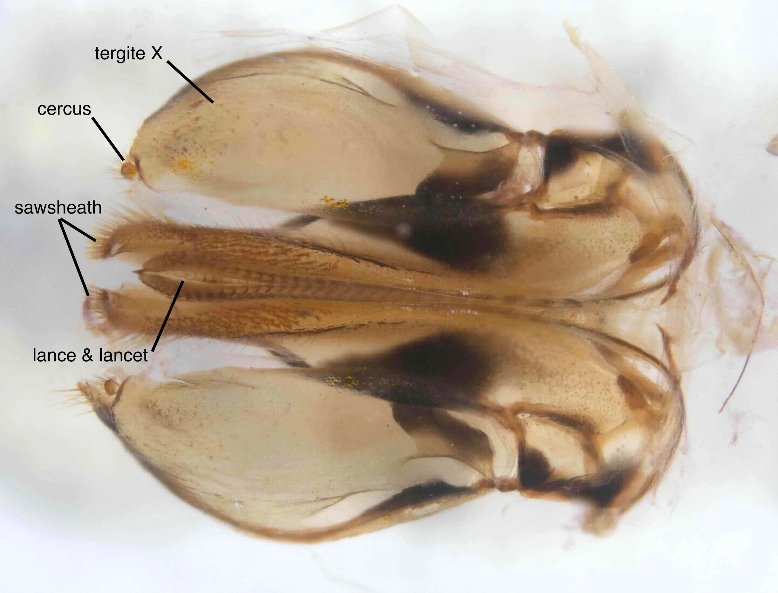
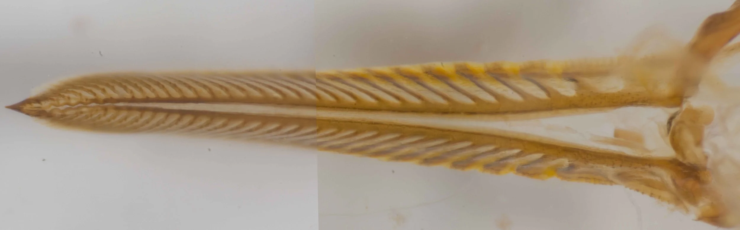
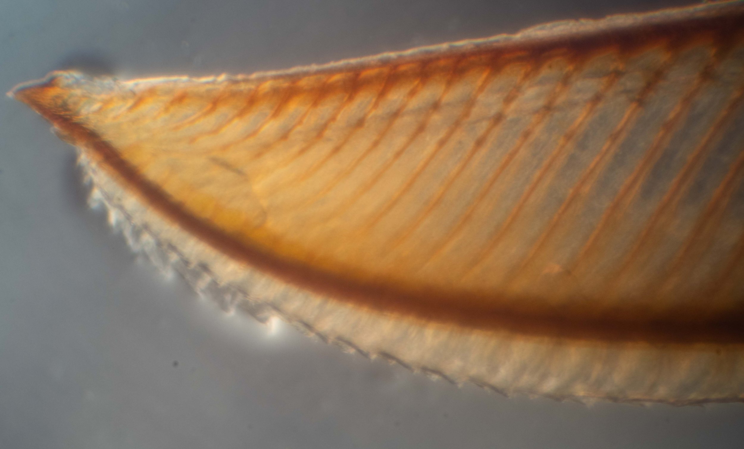
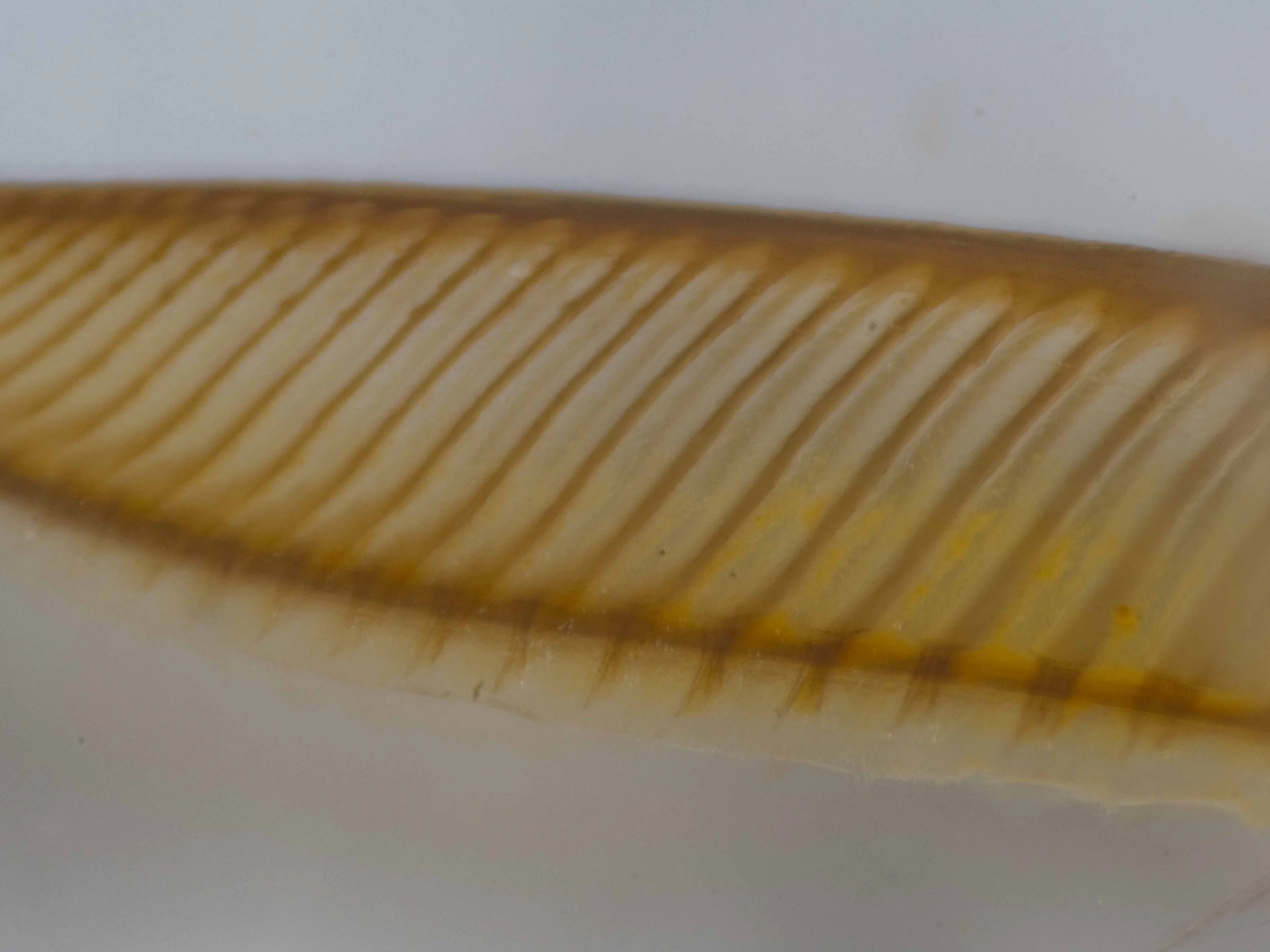
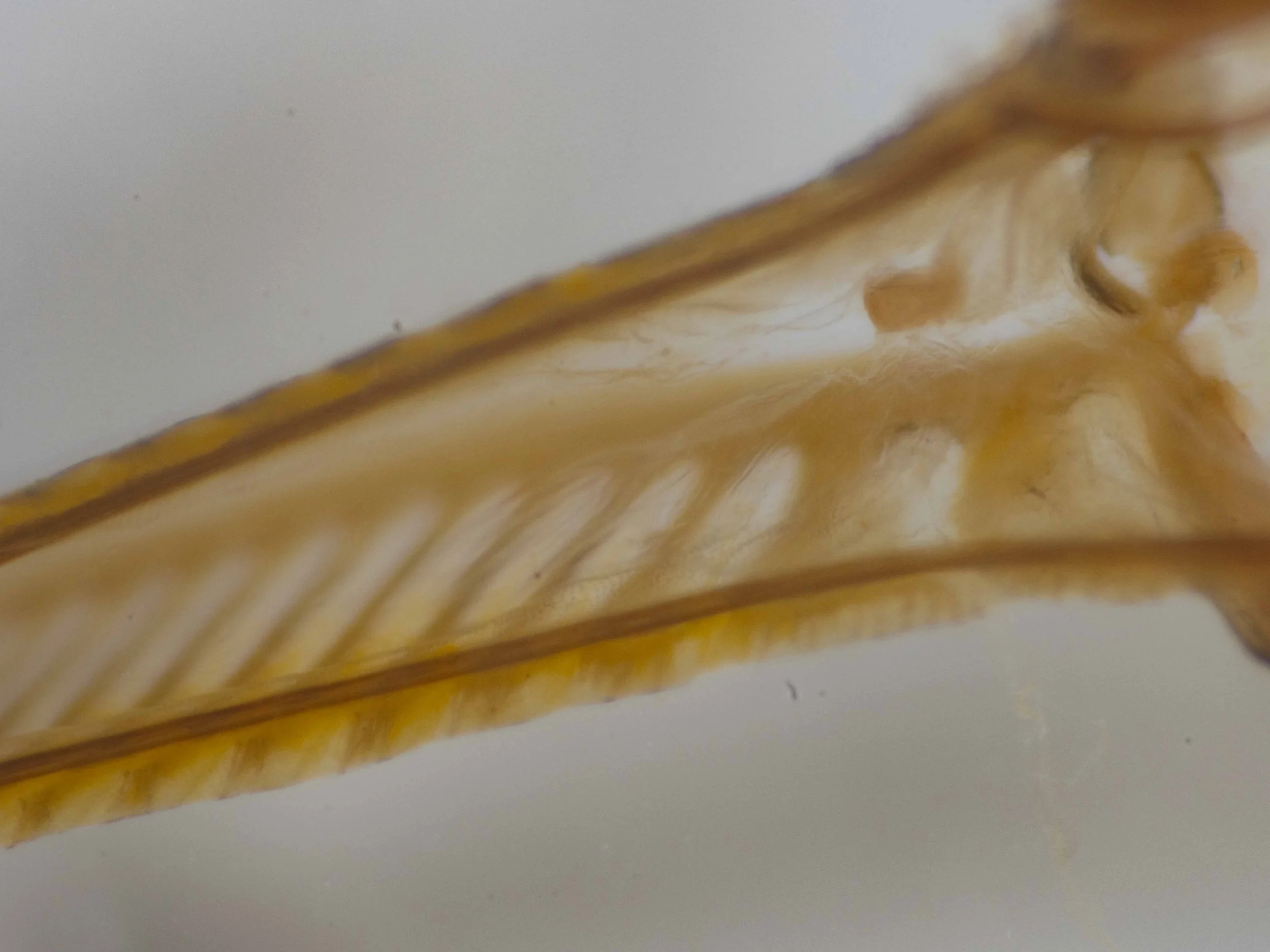
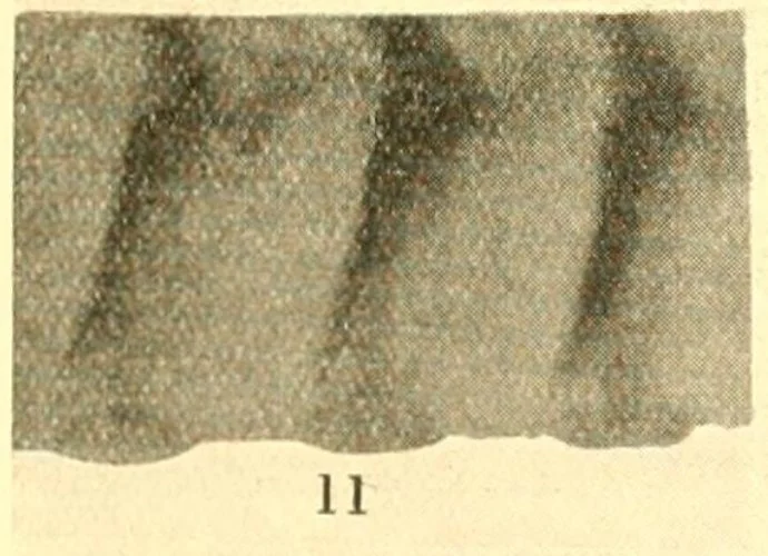
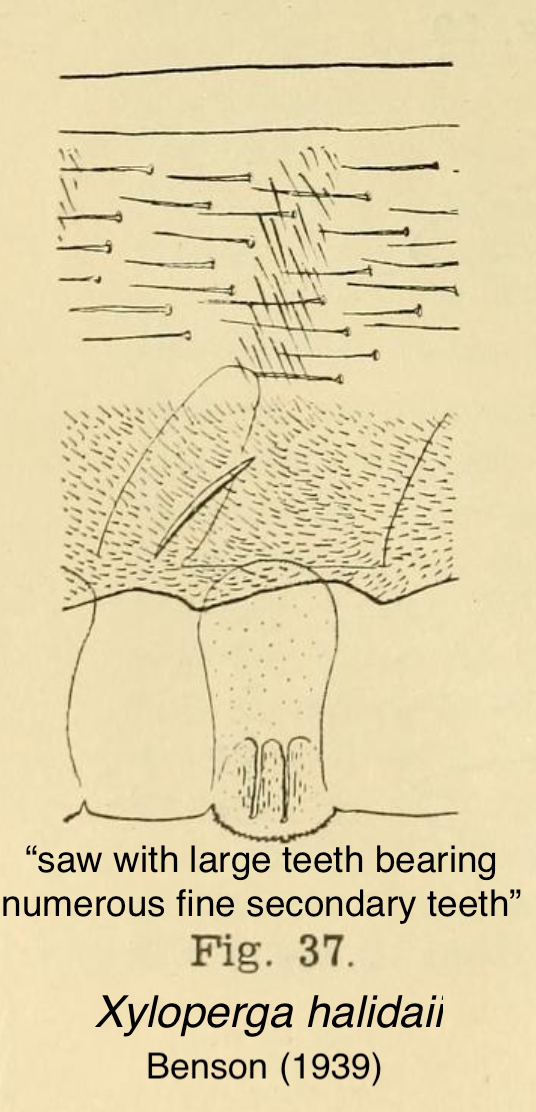
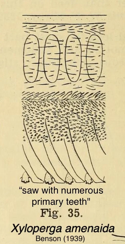
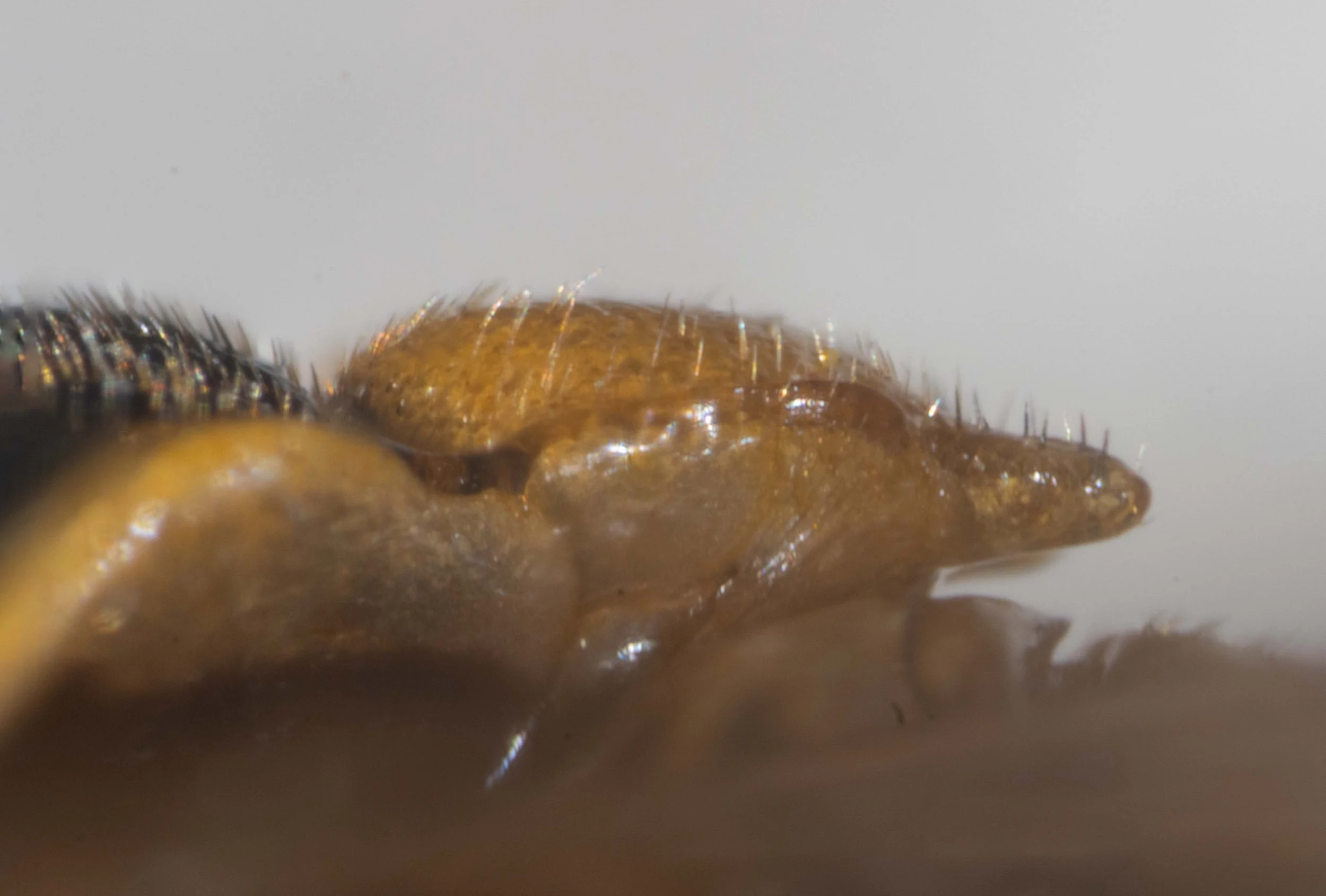
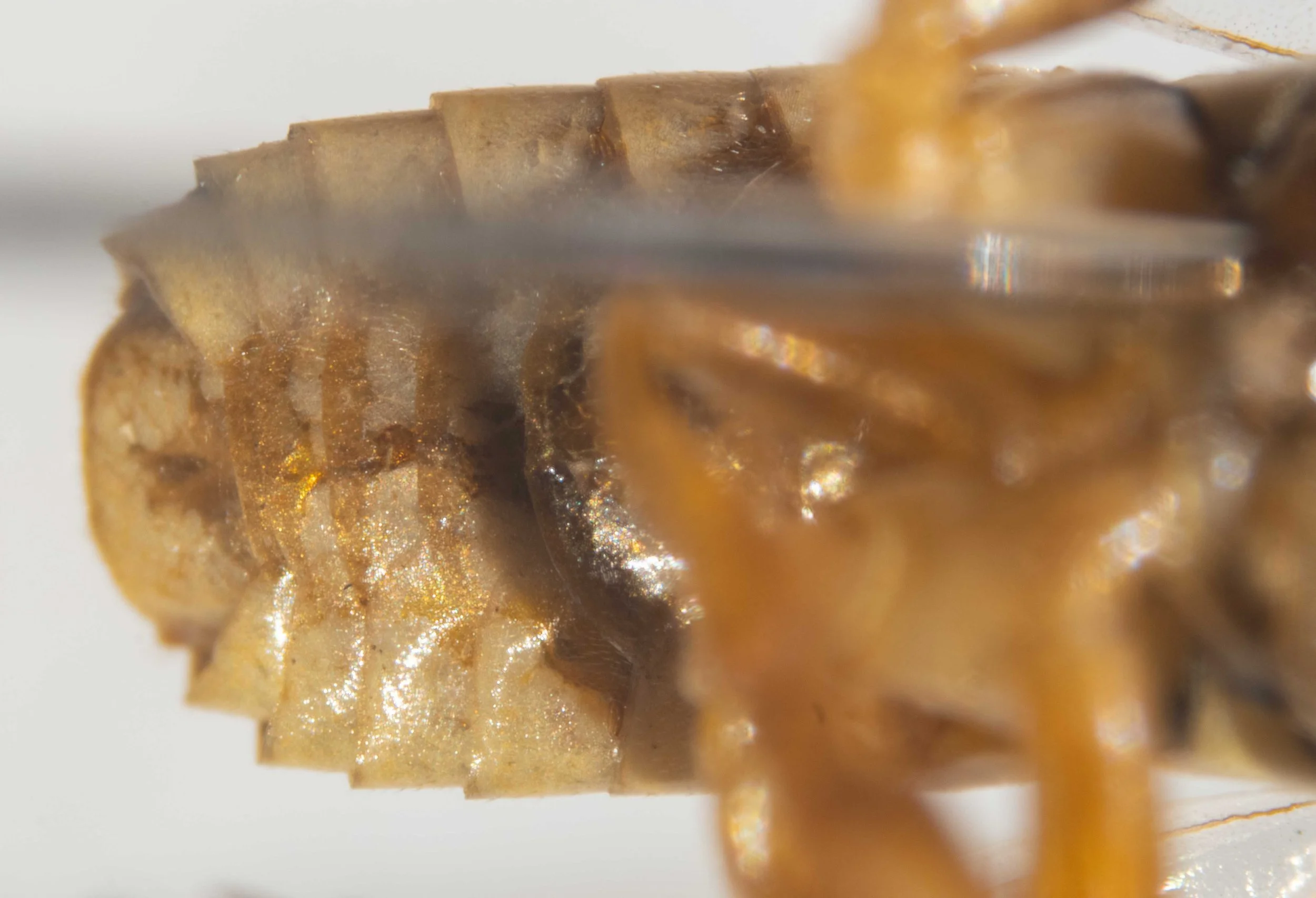
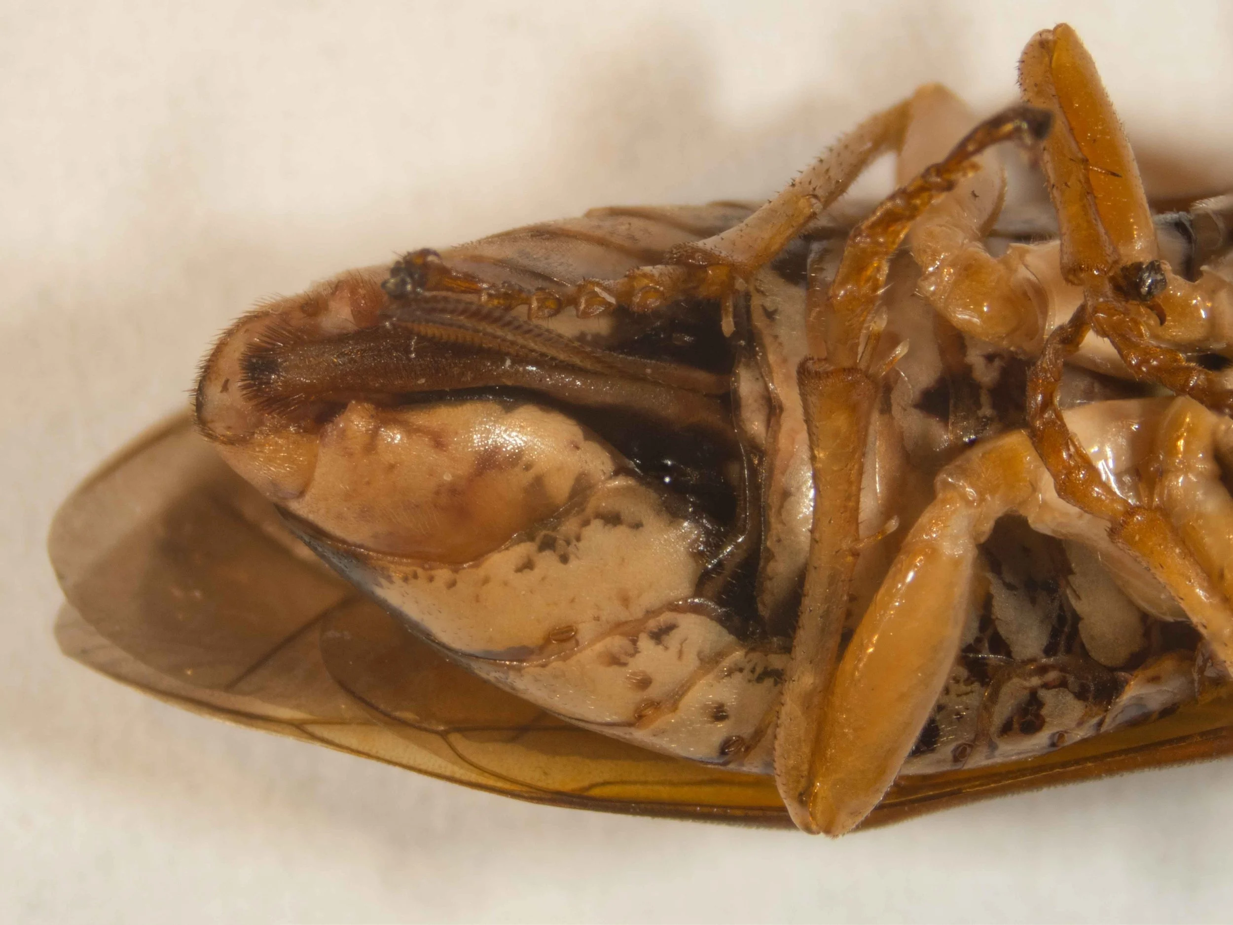
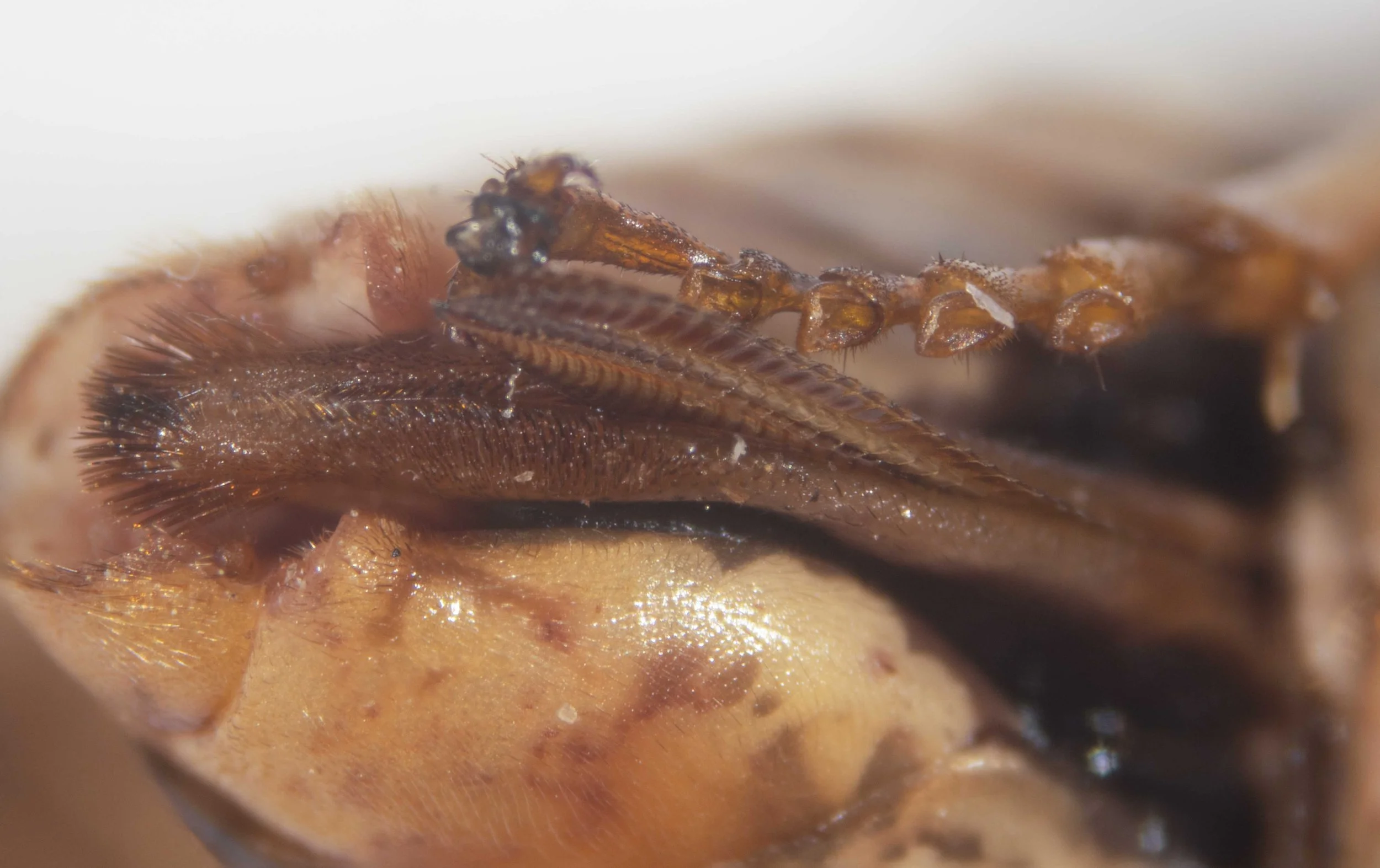
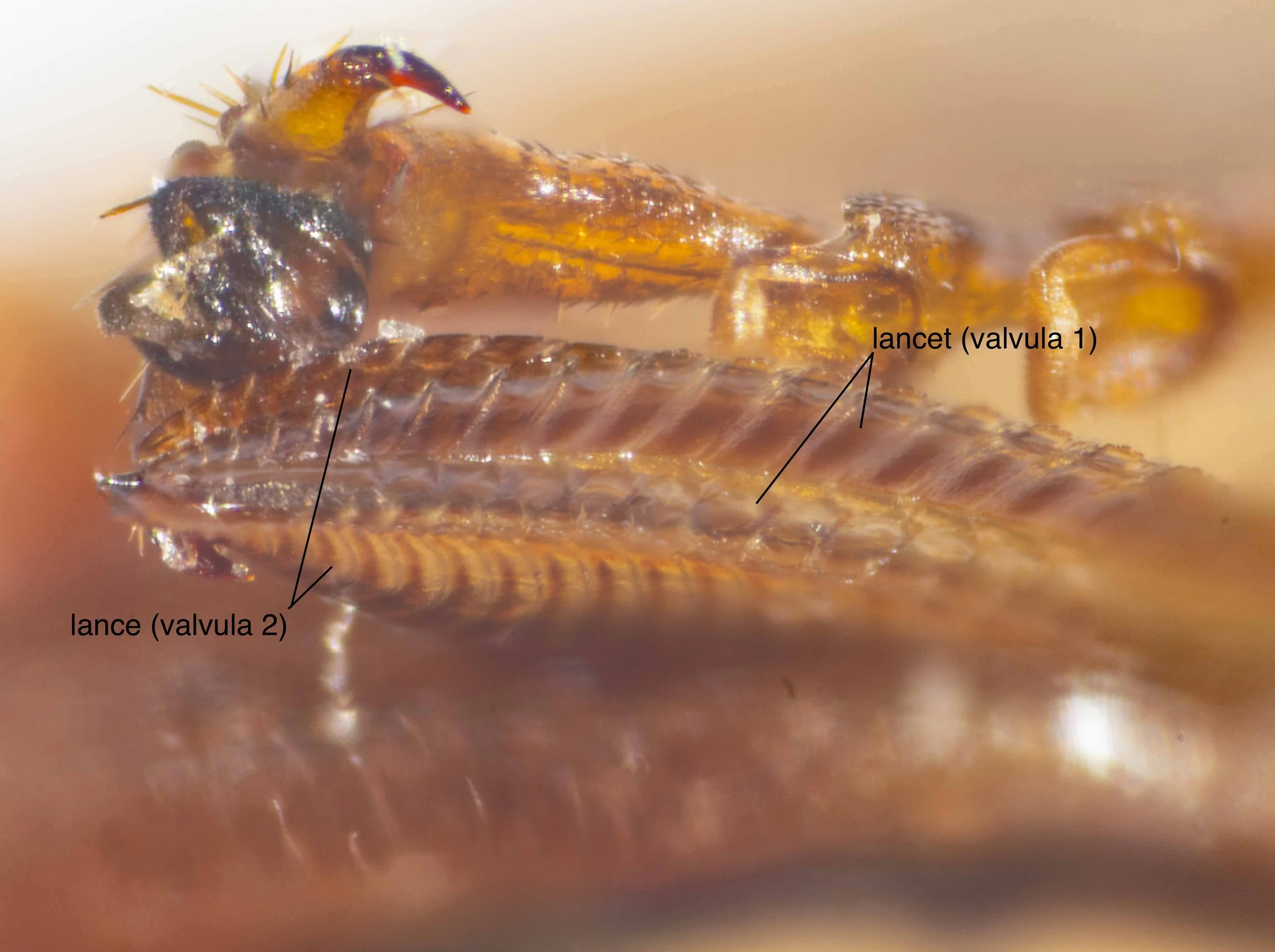
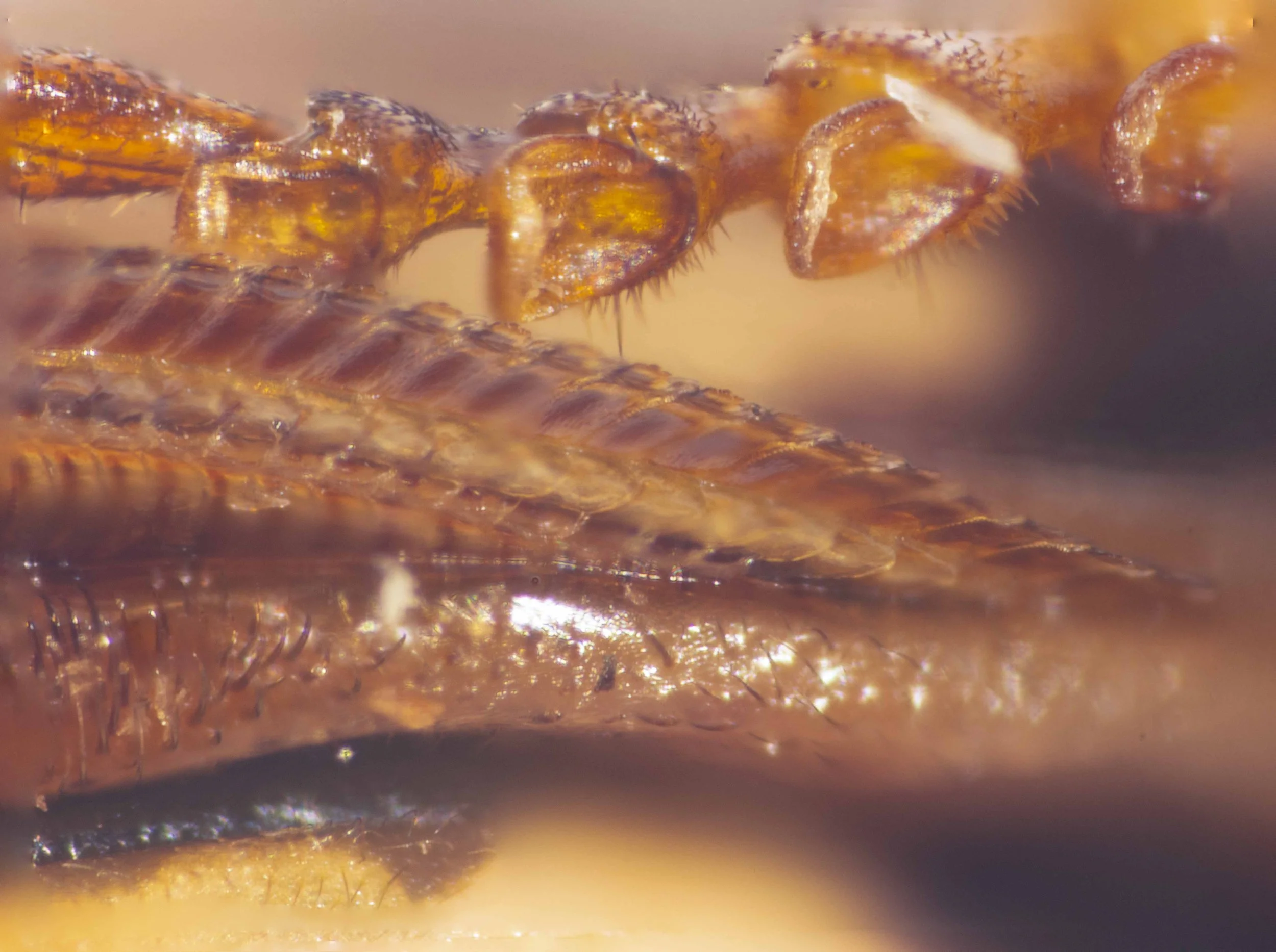
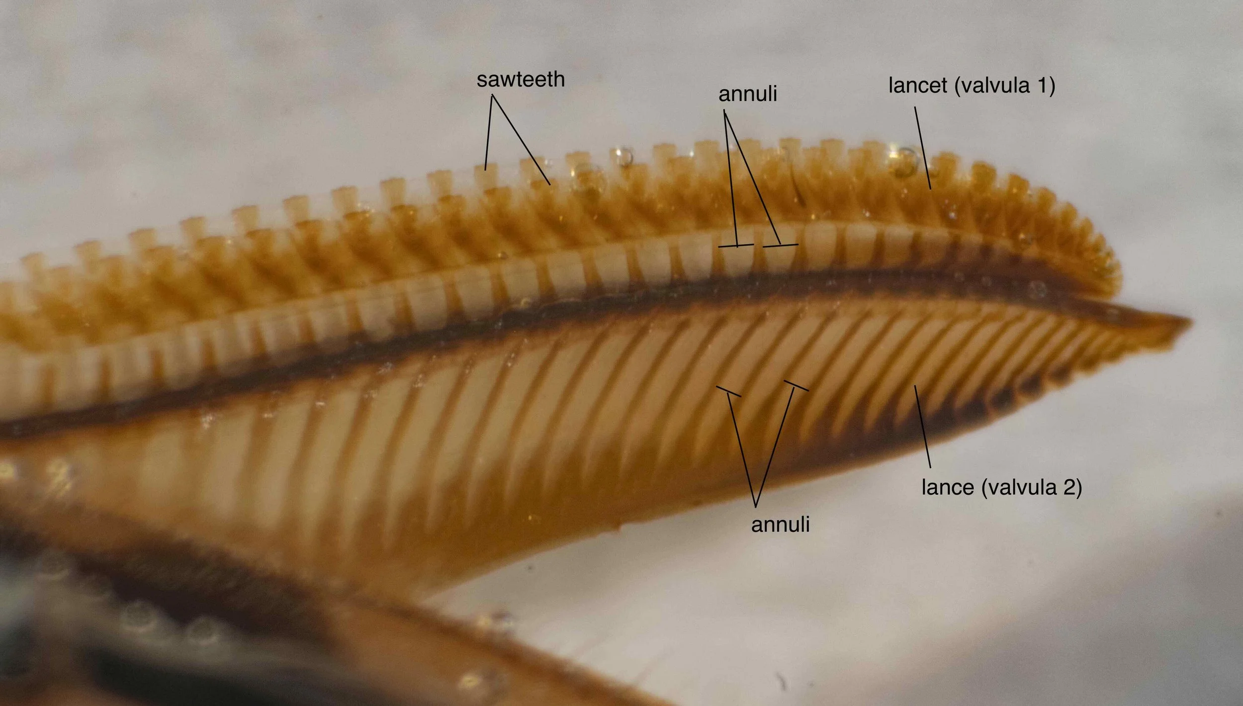
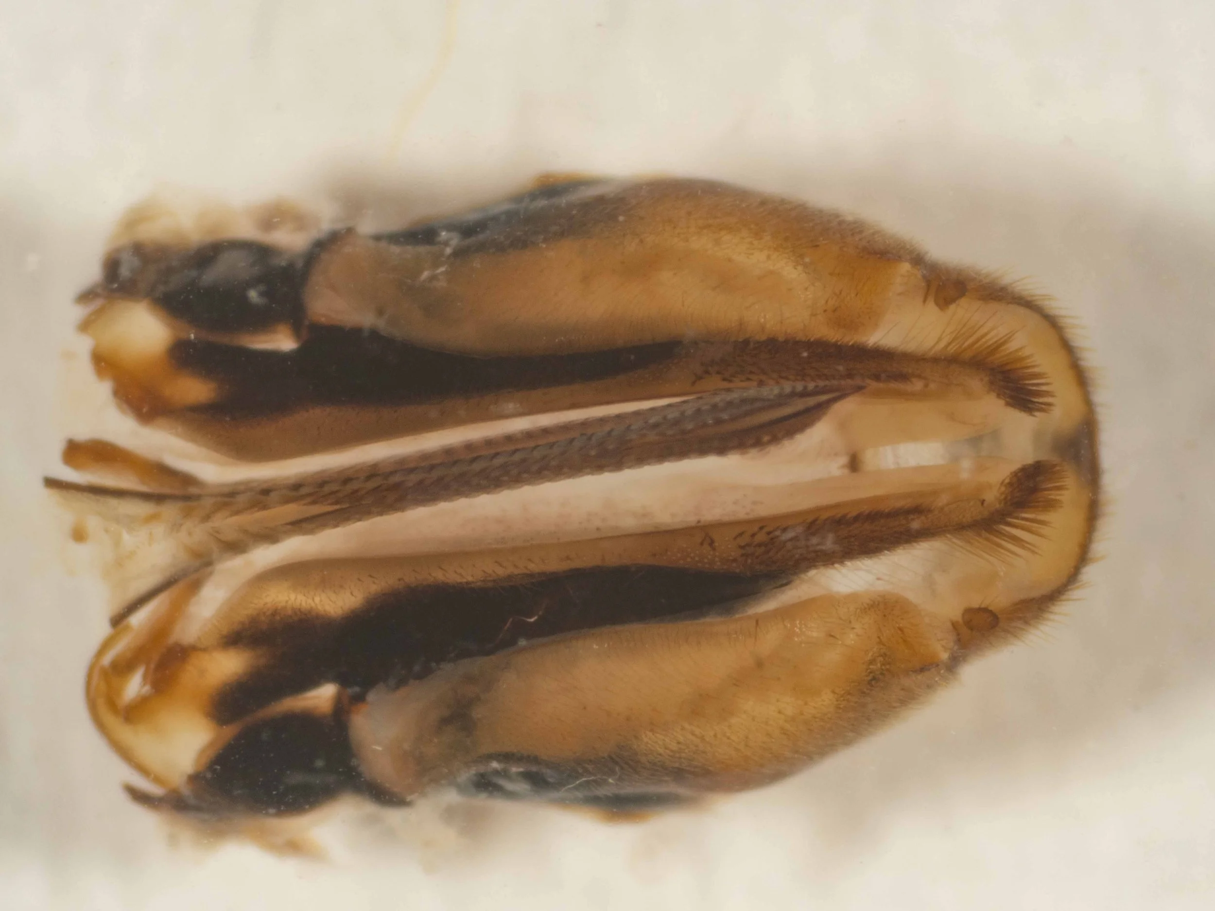
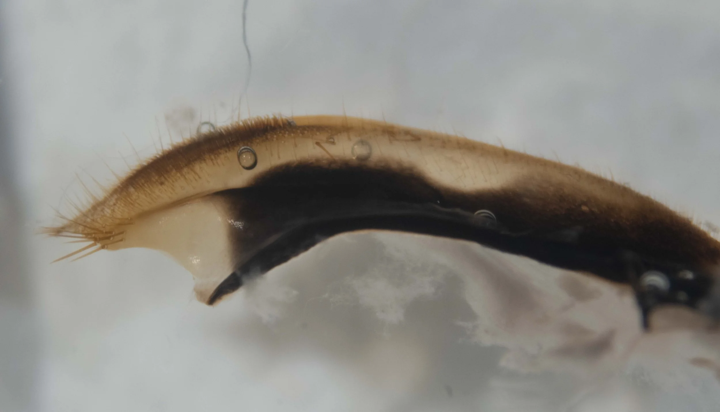
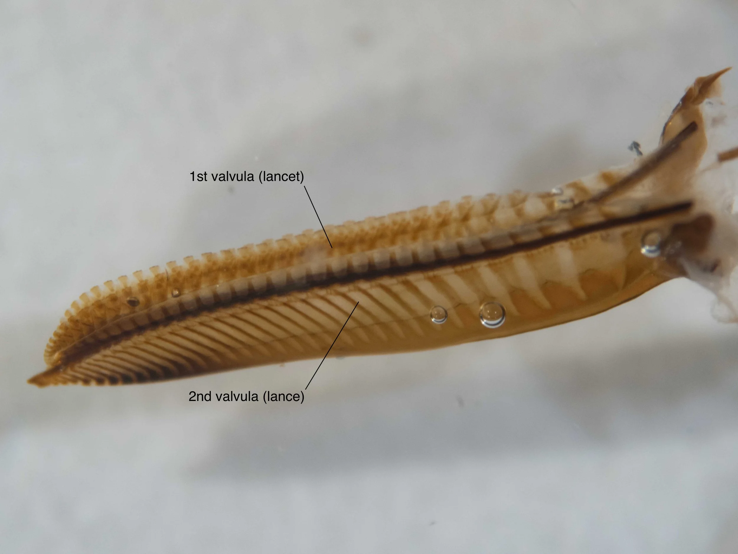
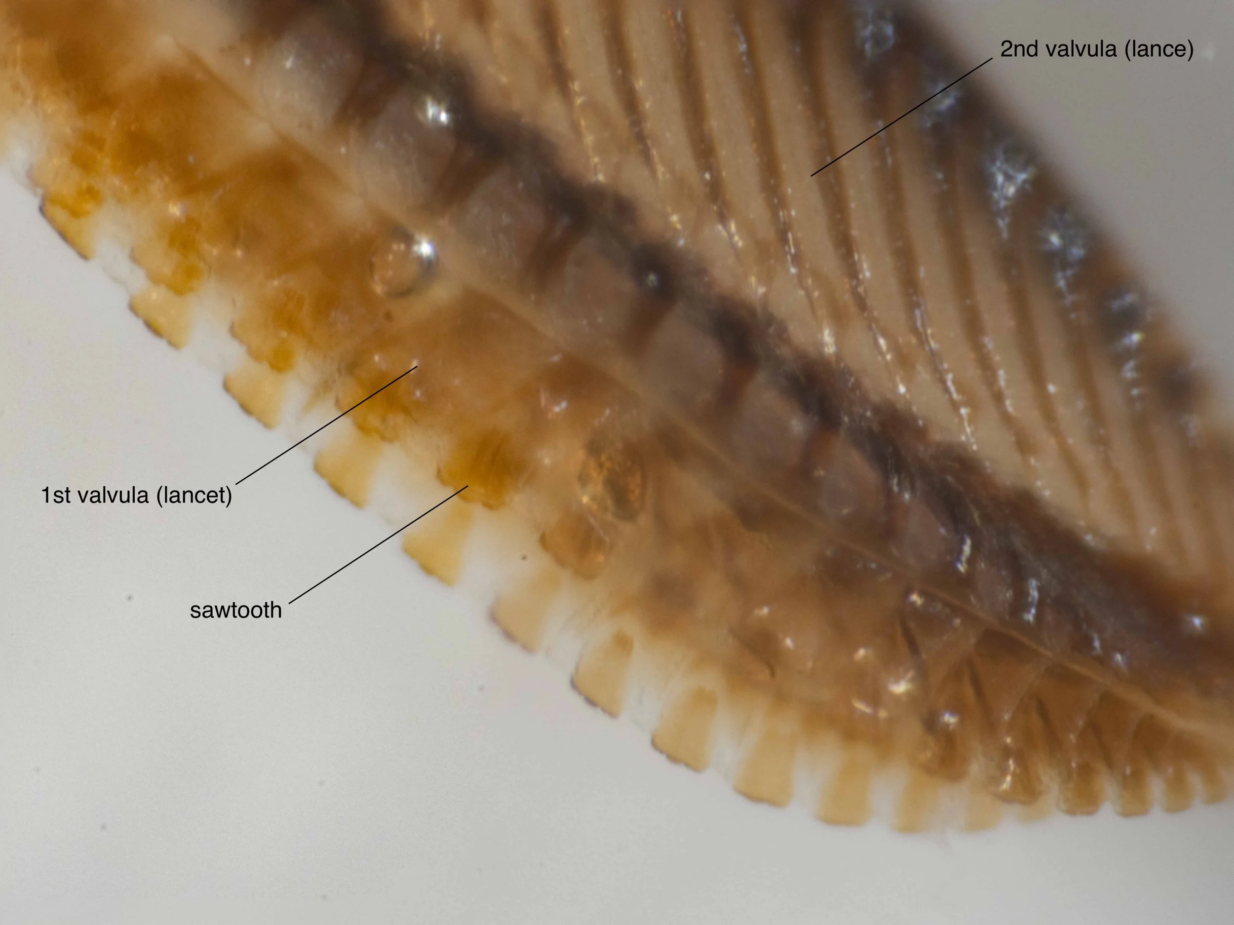
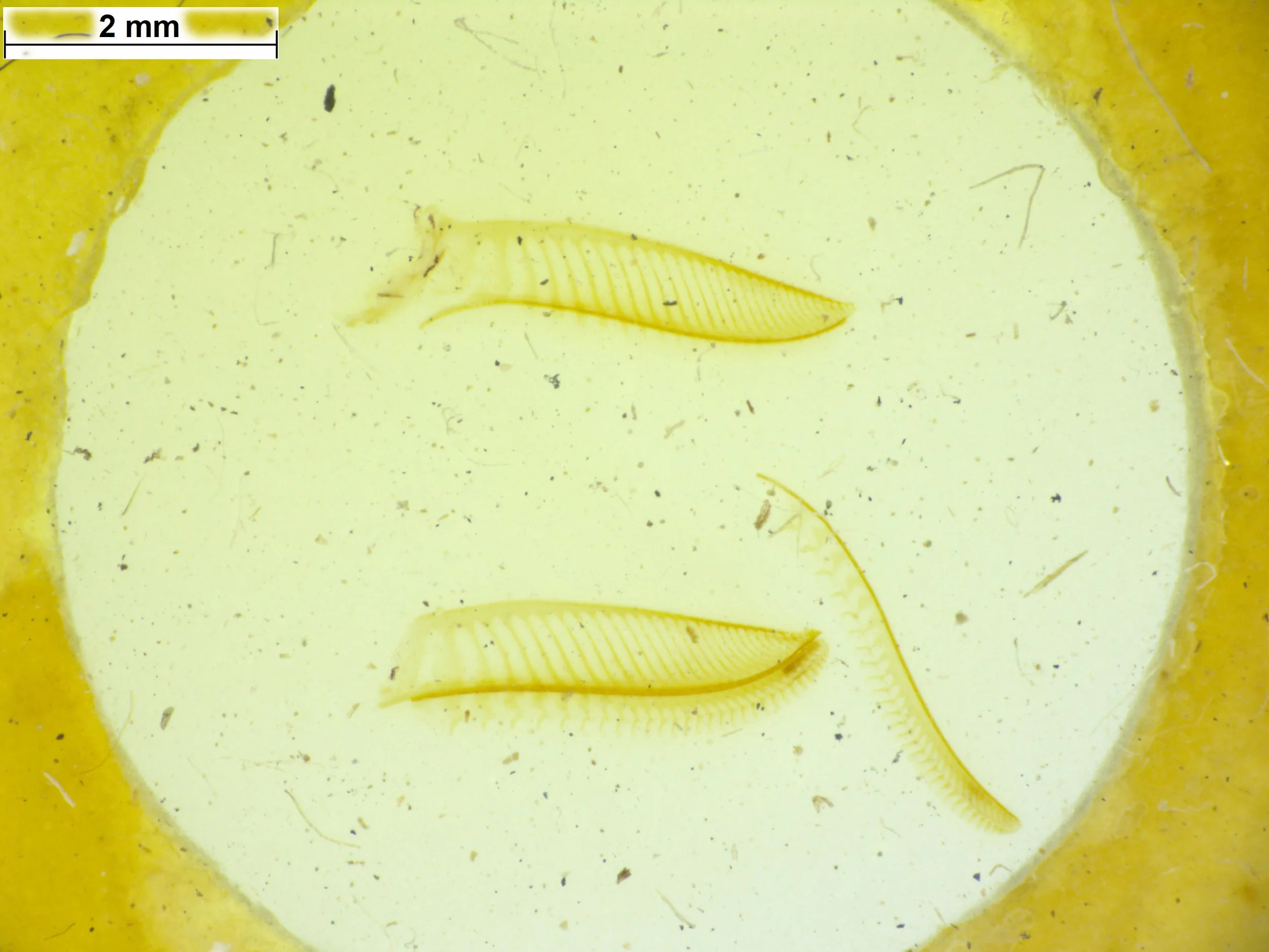
Saw of X. halidaii has large teeth with numerous fine secondary teeth, whereas in X. amenaida it has numerous primary teeth
Hind femur of male of X. amenaida is dilated. There is no mention of this in X. halidaii.
Thickened (incrassate) hairs on apex of front tibia in X. amenaida, whereas these are absent in X. halidaii.
Hind basitarsus in X. amenaida is only as long as breadth of apex of tibia and scarcely 1.5x its own apical breadth, whereas in X. halidaii it is longer than the apex of hind tibia and more than 2x as long as its own apical breadth.
Comparing specimens A, B, C to published descriptions
I have chosen features 1-5 from the list above. Features 6 and 7 could not be assessed with confidence in the specimens.
Click on the image to download a pdf of the table (29KB)
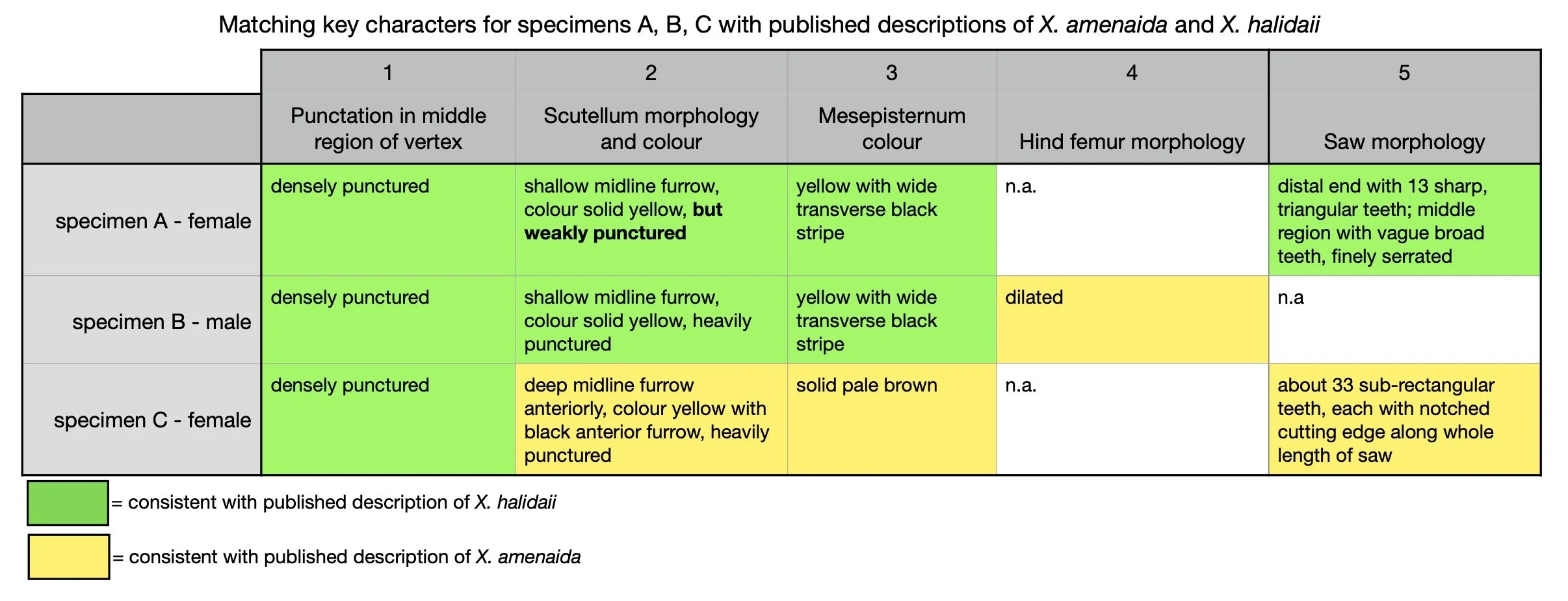
Conclusions:
There is a match between features in specimen A and diagnostic characters for X. halidaii. Notably, this includes saw morphology.
Features in specimen B match the diagnostic characters for X. halidaii except for the hind femur morphology. X. amenaida males are reported to have a dilated hind femur, but there is no mention of this for X. halidaii. However, iNaturalist observations of males indicate this may be a variable feature within a species and therefore not a suitable diagnostic character.
There is a match between features in specimen C and nearly all of the diagnostic characters for X. amenaida, including the saw morphology. The one character that does not match is the punctation of the middle region of the vertex, which is reported to be unpunctured in X. amenaida but which appears to be densely punctured in specimen C.
Examination of the holotype specimen of X. amenaida may help to clarify this discrepancy.
Additional information for species delimitation
BOLD specimen of male Xyloperga halidaii - SICOD1430-19 Specimen determination: Jeremy deWaard
Specimen B matches this BOLD specimen of X. halidaii closely in all visible features.
2. Images of type specimens of X. halidaii and X. amenaida from SAMA,
determined by Forsius (1927). Images kindly supplied by Ben Parslow, SAMA.
Xyloperga amenaida type specimen
Male specimen shares following features with BOLD specimen of male X. halidaii above:
- genae narrower than eye width
- mesepisternum with a wide yellow transverse stripe (however background colour is brown rather than black)
- hind femur dilated
This may suggest that the species determination of either the SAMA specimen or the BOLD specimen is incorrect. Further information - in particular, images of the holotype of both X. halidaii and X. amenaida - is required to decide between these alternatives.
Xyloperga halidaii type specimen
The preservation of this specimen is too poor to decide whether the species determination by Forsius of X. halidaii is correct or incorrect.
Images of holotypes of X. amenaida and X. halidaii
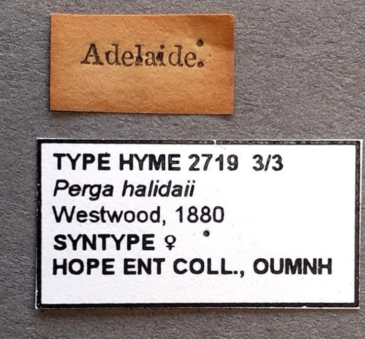
The following images show two syntypes- 2/3 and 3/3 - of X. halidaii female ©️ Oxford University Museum of Natural History CC by 4.0. Photos kindly provided by Dr. James Hogan.
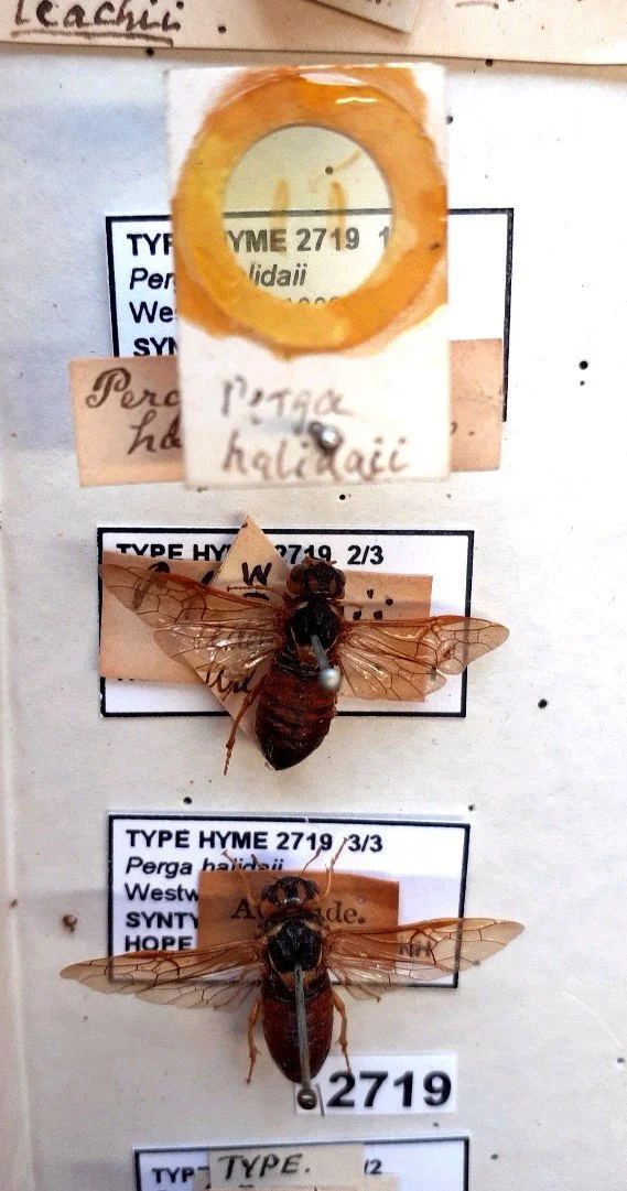

The syntypes of female X. halidaii show the following features which separate them from published descriptions of X. amenaida (see “Differences between X. amenaida and X. halidaii in published descriptions” section above):
Scutellum is all yellow, with no trace of black at midline, and with shallow midline furrow
Mesepisternum is yellow with a wide transverse black stripe - visible in syntype 3/3.
In addition, there is a striking similarity between the syntypes and the female specimen A. These similarities include:
Head - vertex bright yellow with 3 wide, longitudinal black stripes which extend anteriorly to cover ocellar region; genae yellow, narrower than eye width; antennal scape and pedicel yellow.
Pronotum - black with wide yellow posterior border, covered in short, brown erect hairs.
Mesoscutum - black. Note that the syntypes lack the yellow patch towards the rear of the middle lobe of the mesoscutum which is present in specimen A - although syntype 2/3 does show a small faint patch in this position (last photo in panel above).
Note that the 5 iNaturalist observations of female X. halidaii specimens in which the dorsal side of the mesoscutum is visible - #334295790 (specimen A), #257318398, #66817614, #36182382 and #35402759 - all show a yellow patch of variable but consistently small size.Scutellum semi-oval, convex, solid yellow with obtuse long yellow hind lobes and shallow midline furrow and black anterior border; long yellow extensions from anterior corners towards tegulae.
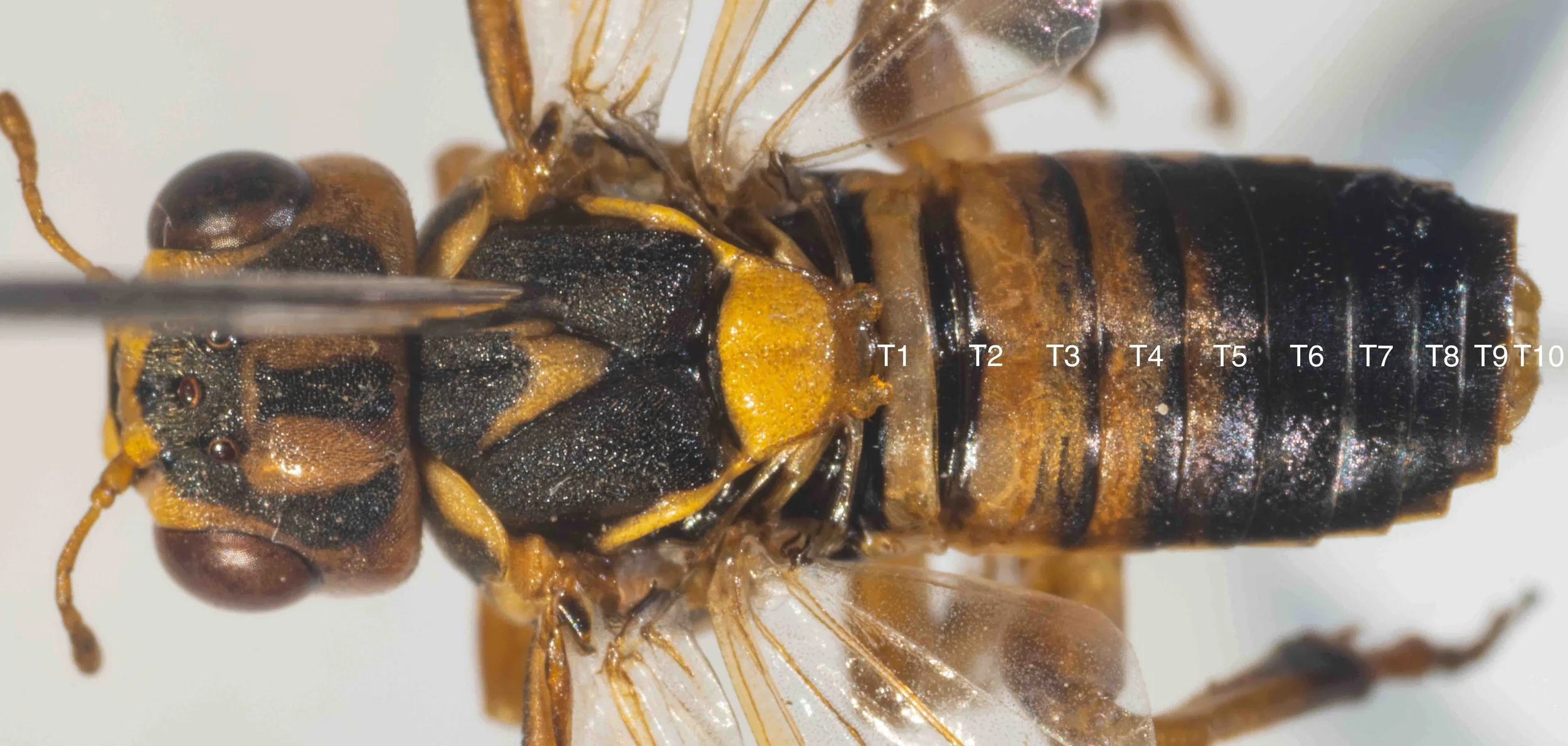
Abdomen orange, speckled with black with T1 solid black except for thin yellow posterior border and T2 thin solid black anterior border.
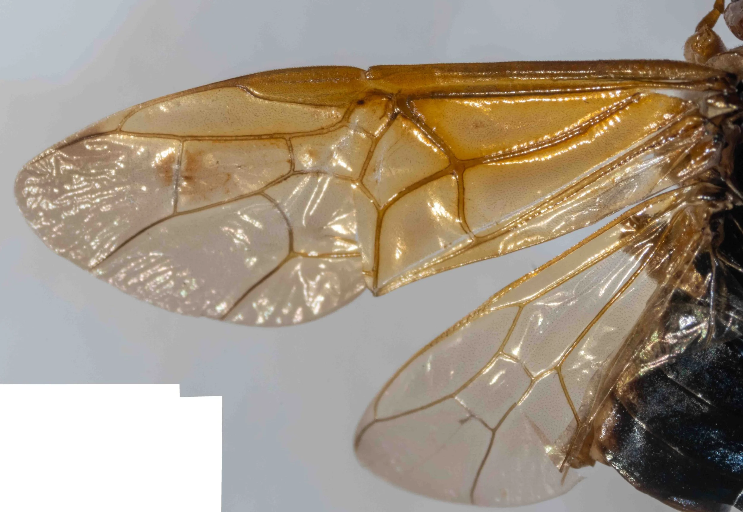
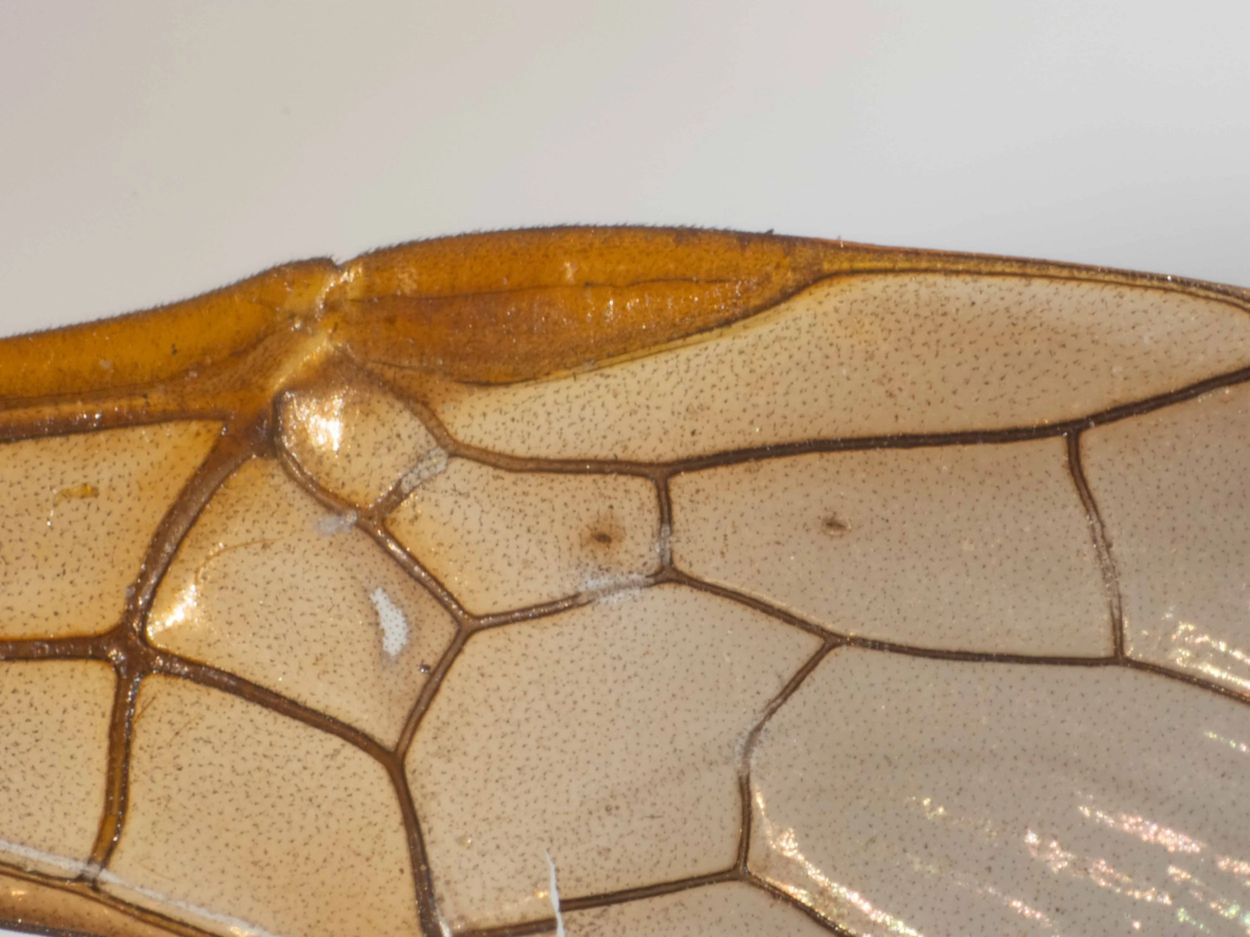
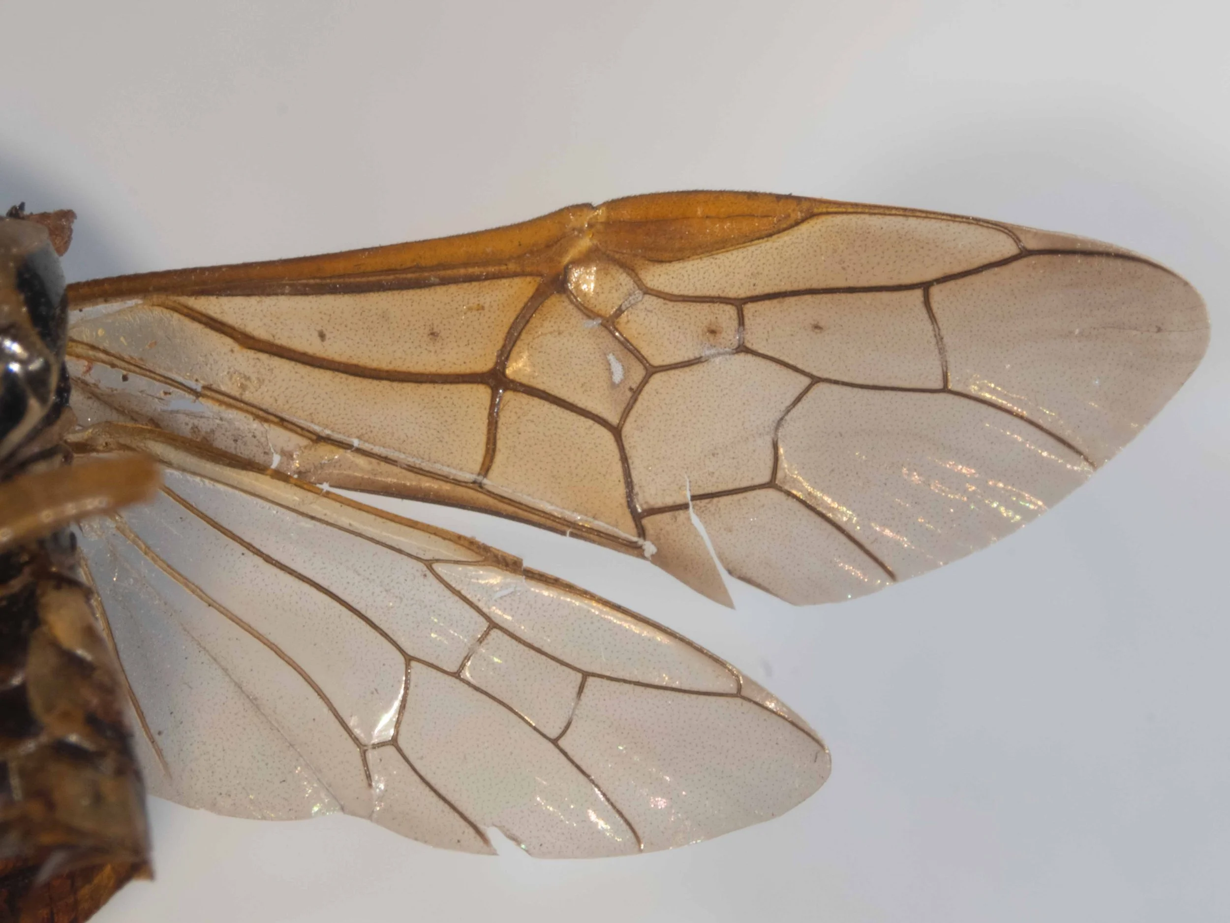
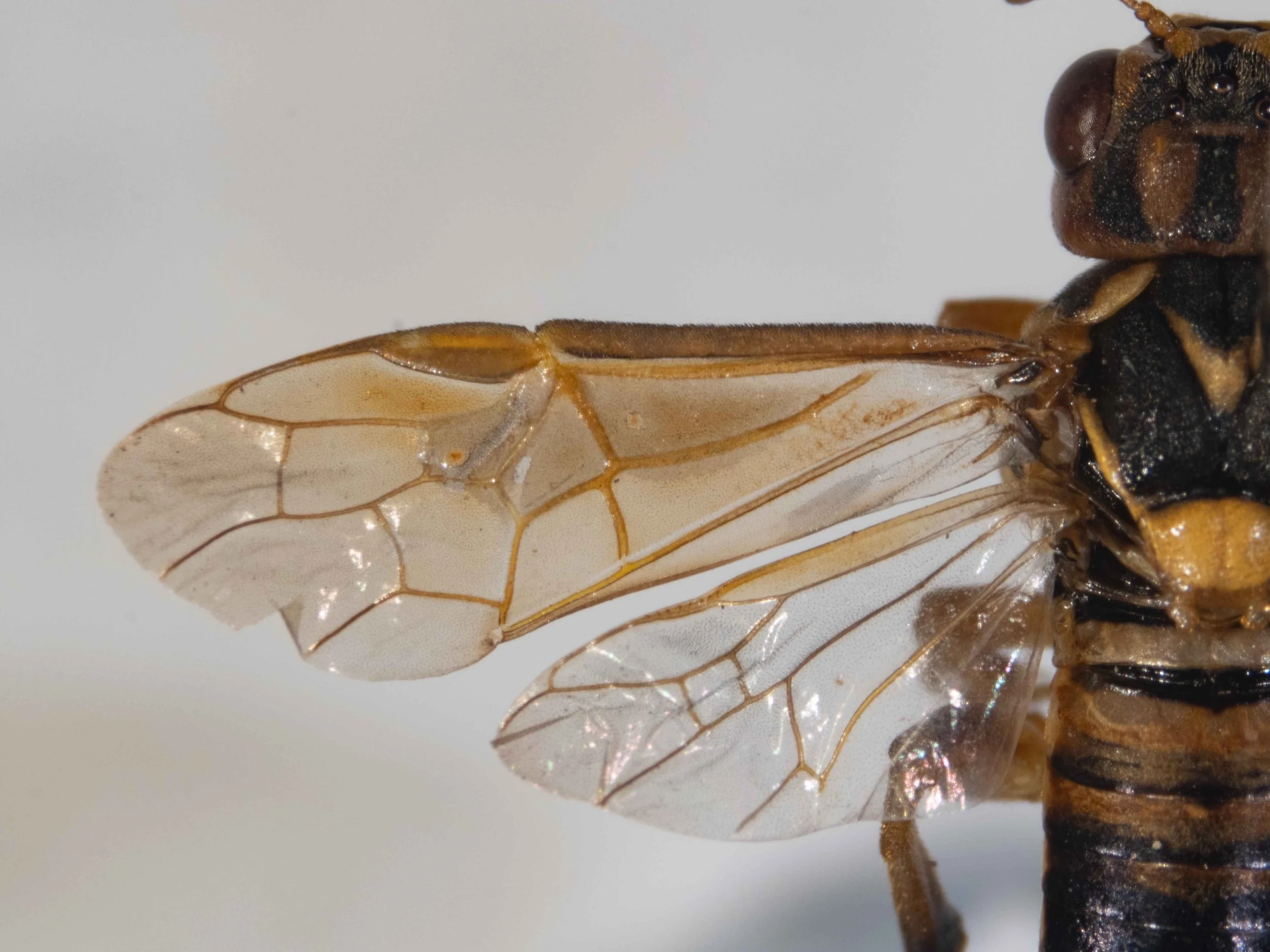
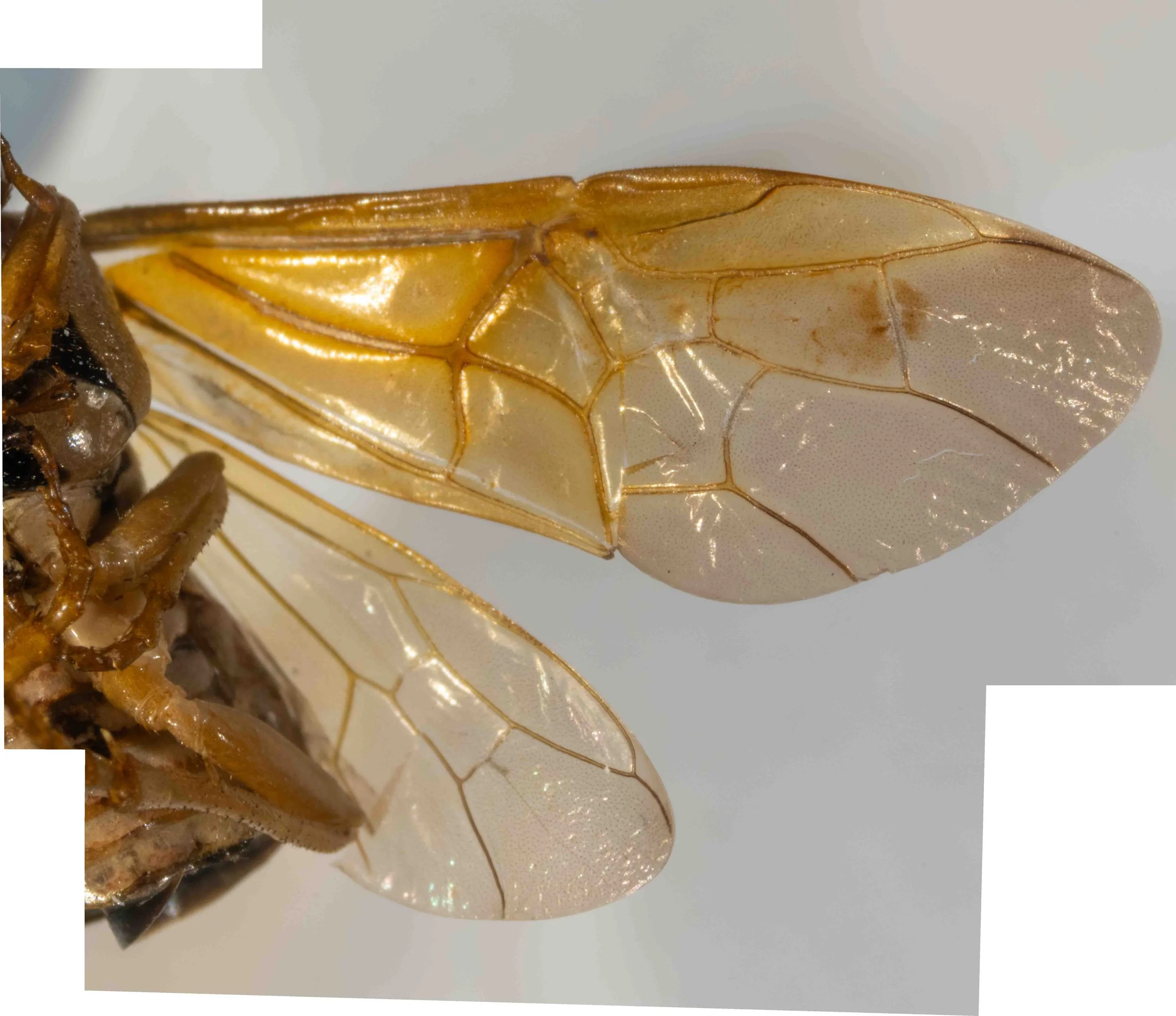
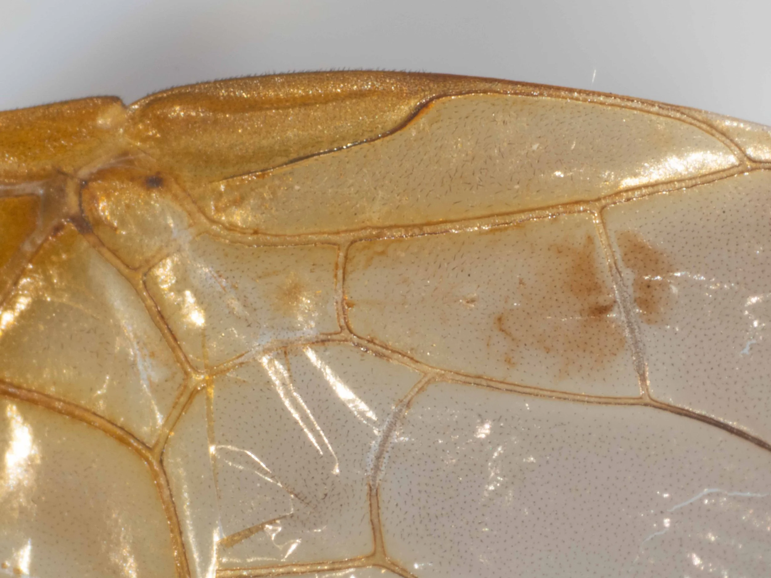
Fore and hind wings with identical venation; fore wings pale yellow hyaline, costa and stigma orange, venation brown; dark spots in corresponding positions in cubital 2, cubital 3 and radial cells.
The following images show two syntypes- 1/2 and 2/2 - of X. halidaii (Perga jurinei Westwood) male ©️ Oxford University Museum of Natural History CC by 4.0. Photos kindly provided by Dr. James Hogan.


The syntypes of male X. halidaii (Perga jurinei Westwood) show the following features which separate them from published descriptions of X. amenaida (see “Differences between X. amenaida and X. halidaii in published descriptions” section above):
Scutellum is all yellow, with no trace of black at midline, and with shallow midline furrow.
Mesepisternum is yellow with a wide transverse black stripe, although that stripe is pale in syntype 2/2.
In addition, there are strong similarities between the two syntypes and the specimen B male. These similarities include:
Head: vertex bright yellow with 3 longitudinal black stripes which extend anteriorly to cover ocellar region - these stripes are bold and wide in syntype 1/2 but faint and narrow in syntype 2/2; genae yellow, narrower than eye width; antennal scape and pedicel yellow.
Pronotum: black with wide yellow posterior border, covered in short, brown erect hairs.
Mesoscutum - black. Specimen B has a yellow V towards the rear of middle lobe which is continuous across the midline. Syntype 2/2 has a similar wide yellow stripe which crosses the midline on each side of the middle lobe. Westwood’s 1880 drawing of Perga jurinei (Plate 37, Fig. 6, last image in panel above), which is apparently a drawing of syntype 2/2, shows these yellow stripes. There is no sign of this stripe in syntype 1/2.
In 17 of the 19 iNaturalist observations of male X. halidaii, a yellow V at the rear of the middle lobe, continuous across the midline is apparent.
Scutellum semi-oval, convex, solid yellow with obtuse long yellow hind lobes and shallow midline furrow; long yellow extensions from anterior corners towards tegulae.
Dorsum of abdomen solid black, except for yellow posterior half of T2. Side of tergites yellow, venter solid yellow.
Hind femora dilated.
Fore wings have identical venation and infuscation in syntypes and specimen B; fore wings pale yellow hyaline, costa and stigma orange, venation brown.
The following images show holotype of Perga rufomaculata (Xyloperga amenaida) female ©️ Trustees of the Natural History Museum, London CC by 4.0. Photos kindly provided by Suzanne Ryder.

Comparison between X. amenaida (Perga rufomaculata) holotype and original description.
Comparison between holotype and specimen C.
The following images show holotype of Perga (Xyloperga) amenaida Kirby male ©️ Trustees of the Natural History Museum, London CC by 4.0. Photos kindly provided by Suzanne Ryder.

Comparison between X. amenaida holotype and original descriptions.
Conclusions
Specimen A matches the syntype of X. halidaii (a female).
Characters for X. halidaii which are apparent in most iNaturalist observations include: mesepisternum yellow with a wide transverse black stripe; scutellum solid yellow with shallow midline furrow; vertex bright yellow with 3 wide, longitudinal black stripes which extend anteriorly to cover ocellar region; mesoscutum black with an uninterrupted yellow V or spot at the rear of the middle lobe.
I have recently received photos of the types of the male and female X. amenaida from the BMNH (see panel above). I am currently assessing the differences between these and the X. halidaii type photos to establish diagnostic characters for species separation which are usable in iNaturalist photos.
References:
Benson, R.B. 1939. A revision of the Australian sawflies of the genus Perga Leach, sens. lat. (Hymenoptera, Symphyta). The Australian Zoologist 9: 324-357
Kirby, W.F. 1882. List of Hymenoptera, with descriptions and figures of the typical specimens in the British Museum. Vol. I. Tenthredinidae and Siricidae. London: British Museum, xxviii
Morice, F.D. 1919. Notes on Australian sawflies, especially the “Authors' Types” and other specimens in the British Museum of Natural History and the Hope Collections of the Oxford University Museum; with diagnostic synopses of the genera and species, and photographs illustrating their structural characters. Transactions of the Entomological Society of London 66: 247-333, pls XI-XV.
Westwood, J.O. 1880. A monograph of the sawflies composing the Australian genus Perga of Leach. Proceedings of the Zoological Society of London 1880: 359-379
This is a workbook page … a part of our website where we record the observations and references used in making species identifications. The notes will not necessarily be complete. They are a record for our own use, but we are happy to share this information with others.