
Apiomorpha ovicola (Eriococcidae: Apiomorphinae)
Workbook
Apiomorpha ovicola - gall Apiomorpha #5
A single female gall was found near the Apiomorpha pharetrata #4 gall, on the same Eucalyptus globoidea sapling.
It was on a stem, not at the base of a leaf, had a different shape to the pharetrata female gall and is larger (15mm).
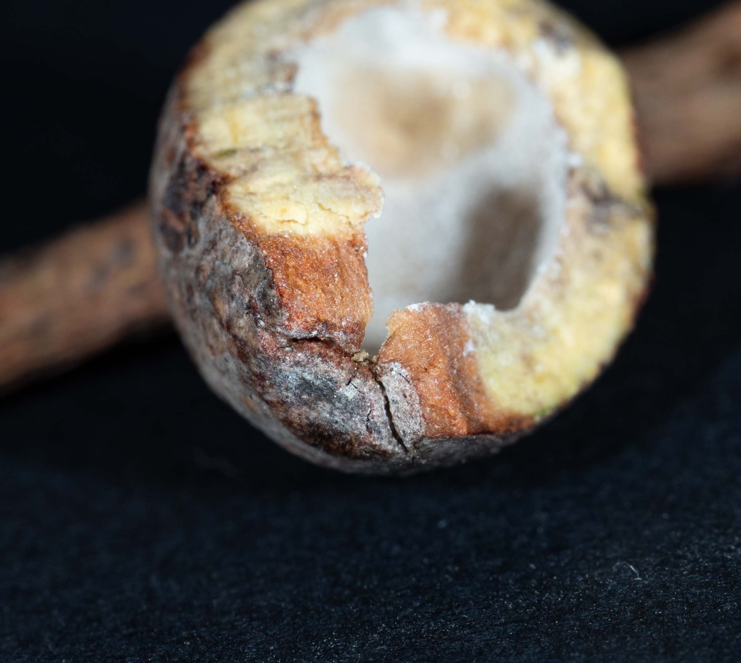
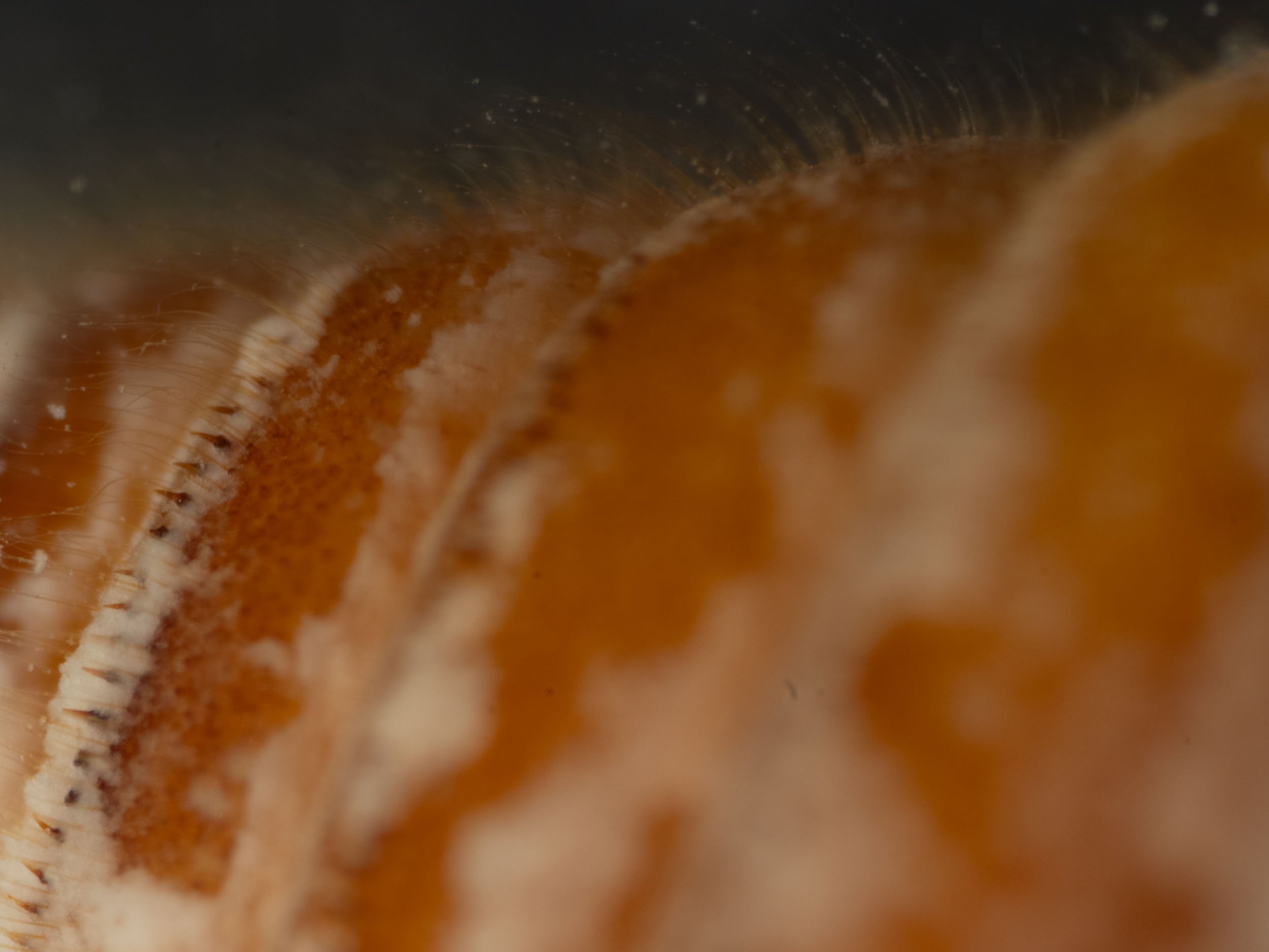
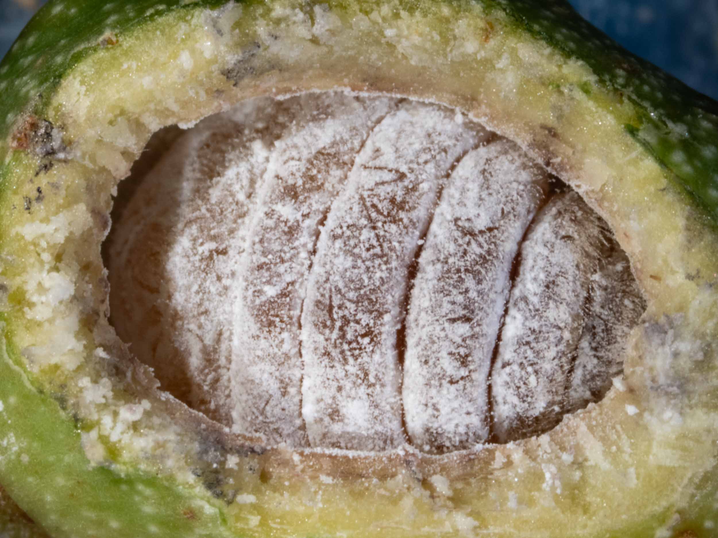
I collected it on 29/2/24 and cut slices off the top to reveal contents.

Eucalyptus globoidea sapling on which A. ovicola gall was found


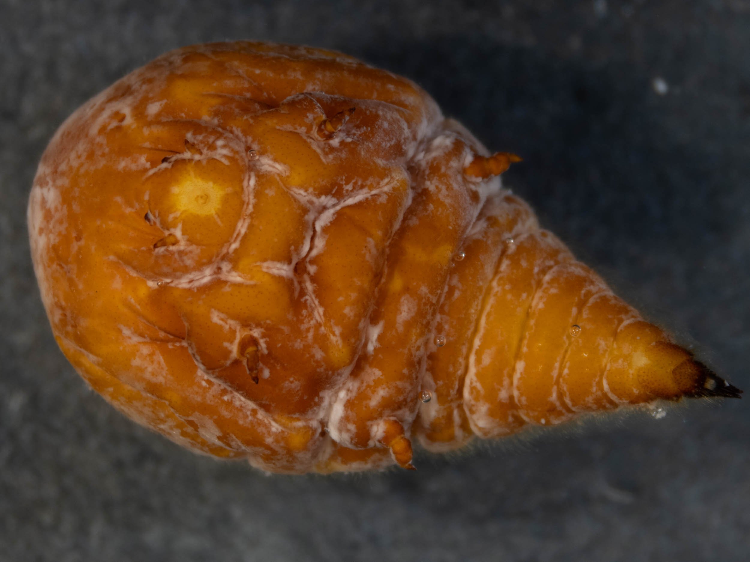
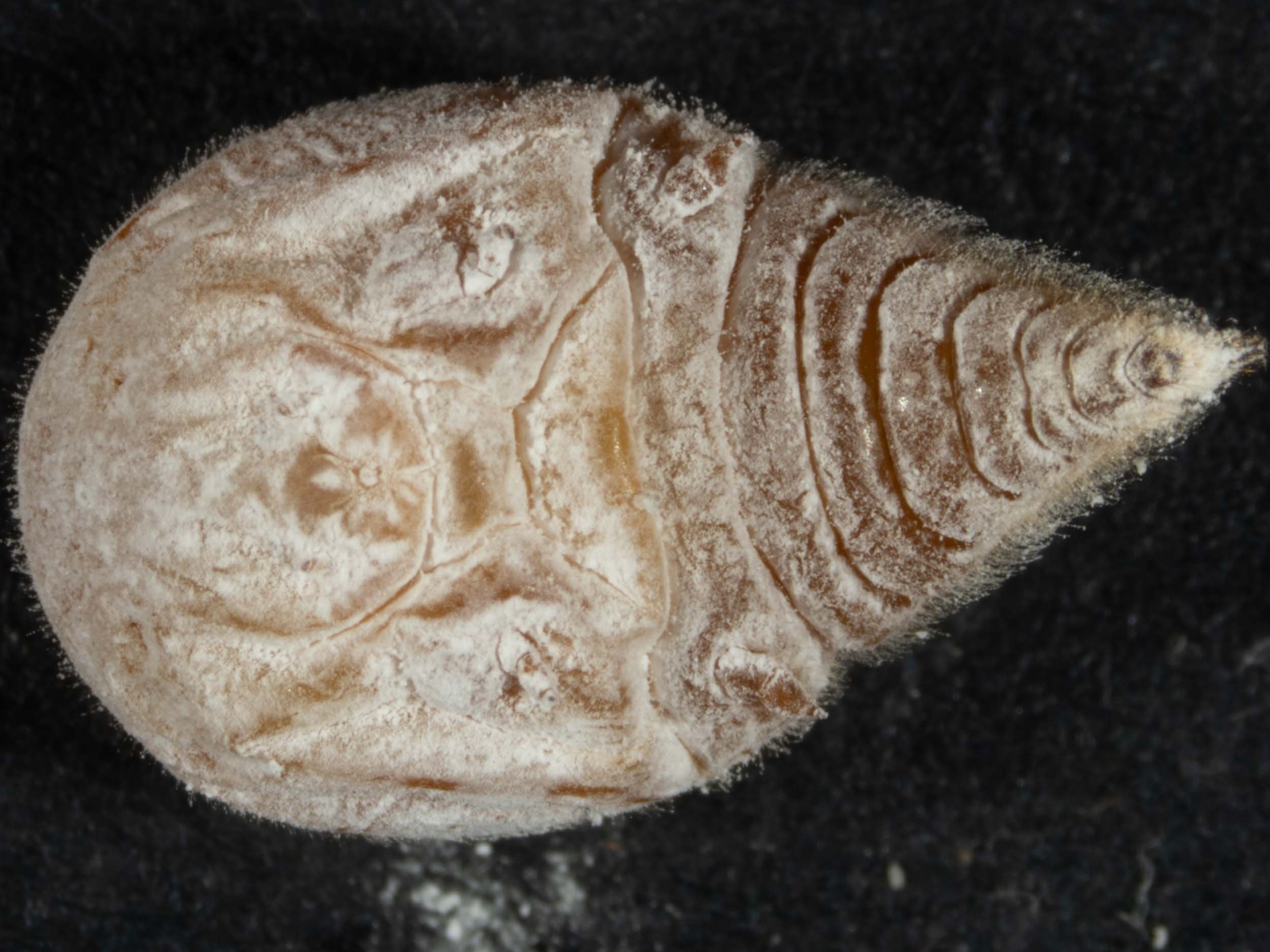
A living female was present inside. I photographed her live, then killed her (in a killing jar) for additional photographs. I removed the body wax by soaking in a detergent solution for an hour before fixing in ethanol.


A comparison of the shape of the female coccoid to drawings in Gullan 1984 suggested Apiomorpha minor and Apiomorpha variabilis as likely species. But Penelope Mills has pointed out that Apiomorpha minor is a junior synonym of Apiomorpha ovicola. (See Mills et al. 2017: Nomenclatural changes in the Australasian gall-inducing genus Apiomorpha Rübsaamen (Hemiptera: Coccomorpha: Eriococcidae); DOI: 10.11646/zootaxa.4250.5.6)
Species identification from adult female morphology
How well does my female match Gullan’s description of Apiomorpha ovicola ?
Images of the female are shown below.


Gullan’s 1984 description of the adult female coccoid is shown below with my observations of the female in bold.
Body. 7.9-26.0 (l8.4±4.l) mm long, 2.9-14.0 (9.2±2.5) mm wide.
Integument. Mostly membranous with 2 shallowly invaginated, sclerotized dorsal patches intersegmentally between thoracic segments I and I1, II and III, and thoracic segment III and abdominal segment II, with posterior abdominal segments lightly sclerotized, especially dorsally from abdominal segment IV posteriorly on mature specimens.
Antennae. Apparently 5-segmented, 165-300 (233±29) µm long, with last division indistinct. Apex with 4-6 (mostly 5) stout, long (31-36 µm) fleshy setae and 1 or 2 (mostly 2) slender, shorter (15-22 µm) fleshy setae. 5-10 (mostly 8) hair-like setae, 18-100 µm long, distributed on segments as follows: 3-4 on I, 1-3 (mostly 2) on II, 0 on III, 1-4 on IV, 0 or 1 on V.
Labium. 150-200 (172±15) µm long, 150-240 (204±21) µm wide
Legs. Often with many hair-like setae on coxa and femur of middle and hind legs, especially on coxa. Forelegs 400-880 (670±110) µm long; tarsal claw distinct. Middle legs 850-1350 (1160±120) µm long; tarsal claw sometimes indistinct. Hind legs 950-1600 (1290±150)µm long; coxa and femur entirely covered with pustules; tarsal claw indistinct..
Spiracles. Of typical trilabiate type, sometimes with posterior plate subfimbriate. Mesothoracic spiracles 270-510 (403±59)µm long, 180-370 (278±50)µm wide. Metathoracic spiracles 310-570 (458±65) µm long, 200-410 (316±5I) µm wide.
Abdominal segment IX. Lightly to moderately sclerotized, longer ventrally than dorsally, 670-1 170 (900±110) µm wide, without spine-like setae.
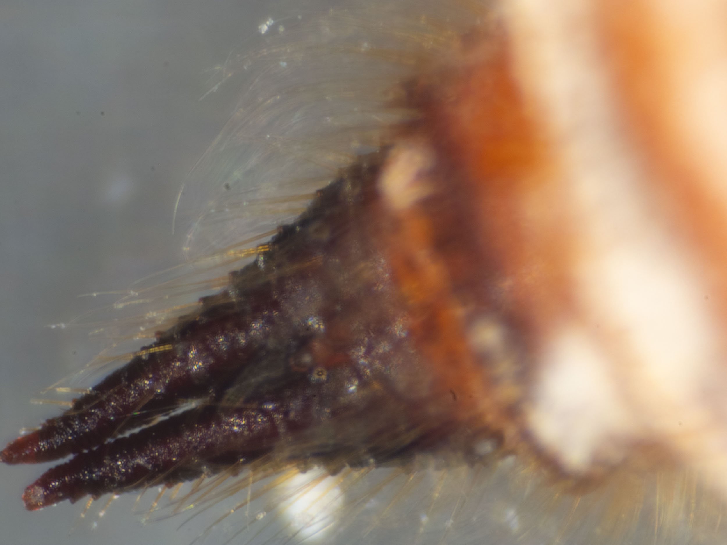
Anal lobes. Moderately sclerotized, 740-1280 (950±120)pm long, attenuate until near apex, slightly divergent apically. Each apex distinctly bifurcate, terminating in 2 subequal, spine-like processes, 40-60 µm long. Lobes papillose owing to spine-like projections each bearing 1 hair-like seta, 125-230 µm long, from near its base; with 2 or 3 long (150-230 µm), more robust setae originating from ventral or exterolateral projections near each apex; projections absent from inner margins of lobes; a few hair-like setae without projections present ventrally near base of lobes.
Venter. Without spine-like setae. Hair-like setae on all body segments, 50-800 µm long, longest on posterior abdominal segments excluding IX. Multilocular disc pores sparsely scattered on head, sparsely to densely scattered on thorax and anterior 2/3 of abdominal segments III-VIII, absent from IX; pores of 5-13 locules present, 9- and 11-locular pores predominant.
Dorsum. With some spine-like setae, 30-180 µpm long, with larger setae (with region of sclerotization surrounding setal base) forming a prominent, mediolongitudinal band with smaller setae scattered laterally; setae of medial band on head, thorax and abdominal segment II somewhat larger than those of medial band on remainder of abdominal segments; some specimens with bifid setae; setae distributed on head and thoracic segments as follows: 93-173 (30-180 µm long) on head and I; 67-255 (30-180 µm long) on II; 51-246 (50-160 µm long) on Ill; and on abdominal segments: 39-155 (50-150µm long) on 11; 34-112 (40-130 µm long) on III; 27-55 (50-110µm long) on IV; 22-39 (60-120 µm long) on V; 12-31 (60-130µm long) on VI; 8-25 (60-130 µm long) on VII; 6-25 (70-130 µm long) on VIII; 0 on IX. Abdominal segments IV-VIII possessing a well defined, posteromarginal row of spine-like setae with setae anterior to this row numbering: 16-39 on IV; 11-27 on V; 4-18 on VI; 1-15 on VII; 0-13 on VIII. Hair-like setae on all body segments, 50-800 µm long, longest near spine-like setae on posterior abdominal segments excluding IX, which has only short hair-like setae. Multilocular disc pores sparsely to densely scattered on head, thorax and abdominal segment II but rare or absent medially amongst spine-like setae, densely scattered on anterior 2/3 of abdominal segments III-VII, a few laterally on abdominal segment VIII, absent from IX; pores of 5-1 1 locules present, 9- and 11-locular pores predominant.

from Gullan 1984
Conclusion:
My adult female coccoid is a good match to the description of Apiomorpha ovicola in Gullan 1984.
Species identification from female gall morphology
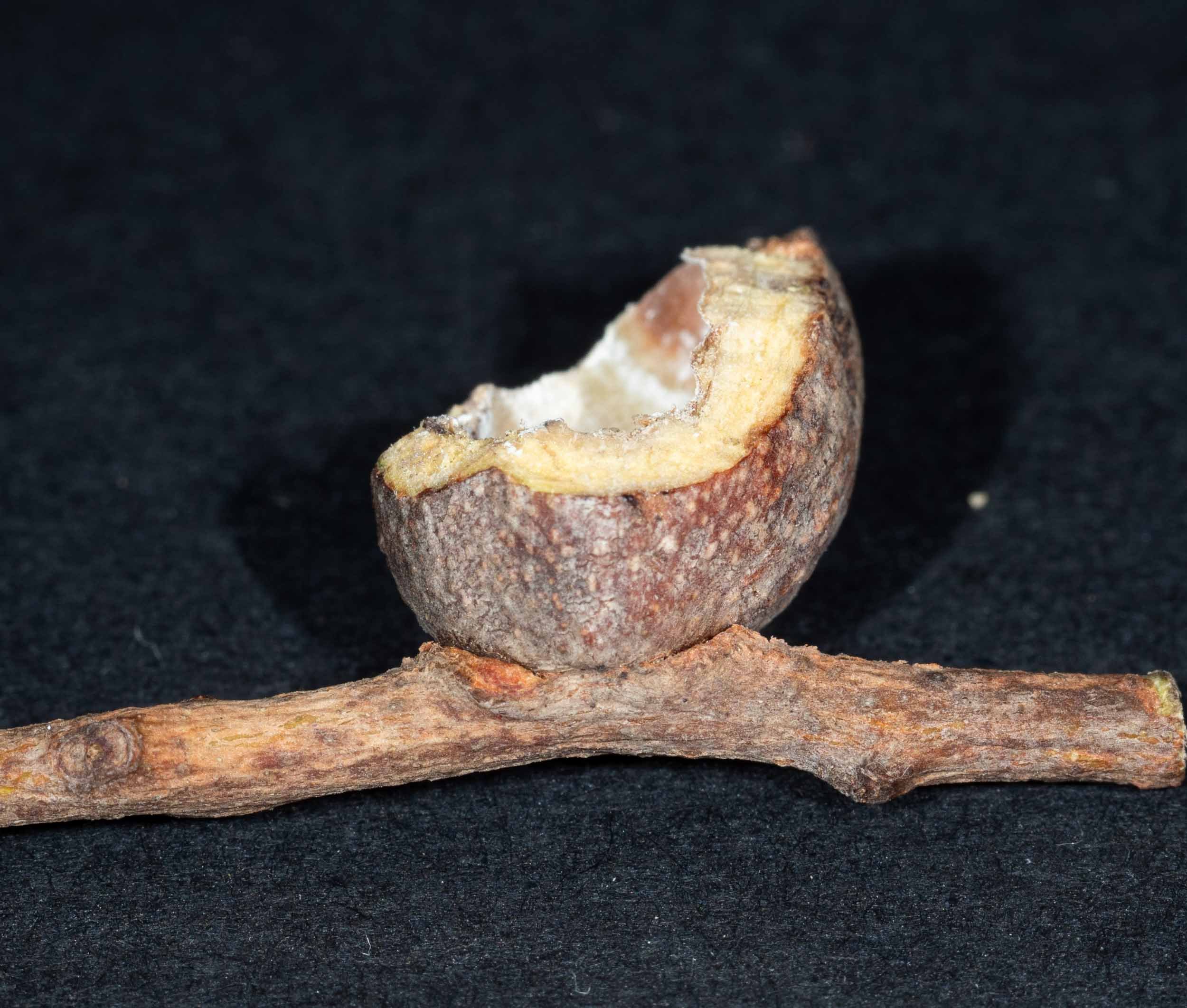
This is Gullan’s description of the Apiomorpha ovicola female gall, which is a close match to my gall (bold text my observations). Compare gall images in the panel above to the description and image from Gullan’s paper..
“Mature gall ovoid to fusiform, with curvature of upper surface greatest especially near base and sometimes distinctly tapered towards apex, 18.0-38.0 mm long, 9.0-19.0 mm wide, sessile, usually dependent, with basal attachment 2.0-5.0 mm in diameter. Apex rounded to distinctly truncate, if truncate then 2.0-5.0 mm in diameter; apical orifice circular, 0.3-0.6 mm in diameter, usually situated in slight depression. Gall cavity ovate, approximating contour of outer gall surface, but apical end acuminate; wall 1.5-2.5 mm thick. Living gall bright green to pale brownish green with surface relatively smooth; dry gall finely wrinkled longitudinally.
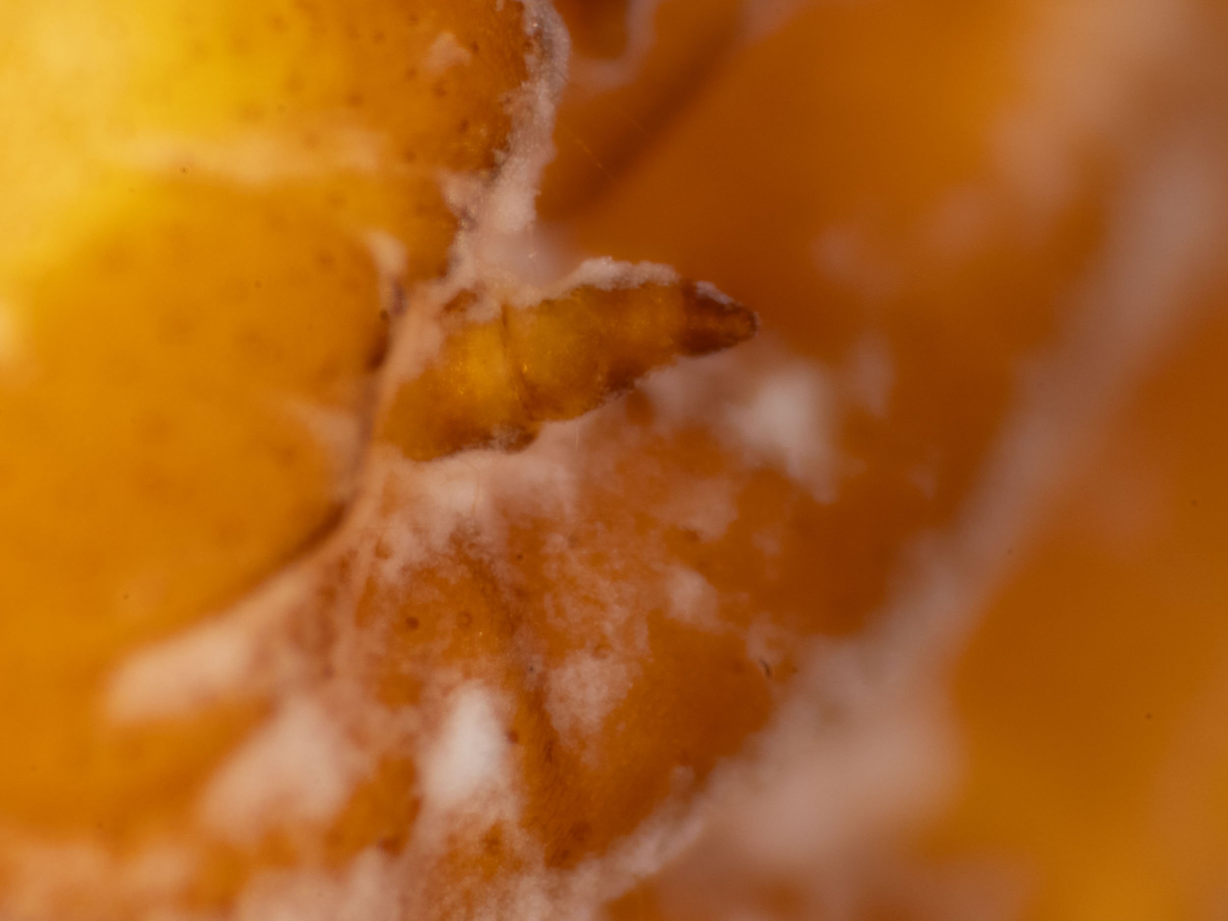
Immature gall almost cylindrical; apex truncate with apical orifice plugged by a small, cylindrical or mammilliform projection.”
Gullan states that galls of females of Apiomorpha ovicola are mostly produced on stems but occasionally on the calyx tube of a bud or fruit; they occur either singly or in aggregations, often with galls of males. Galls of males are mainly found on leaves, on either abaxial or adaxial surfaces, especially on or near the midrib, but sometimes they occur on stems. A. ovicola has been reported (Froggatt 1921, 1931; Brimblecombe 1959b; specimen labels) from a large number of host plants of the eucalypt subgenus Symphyomyrtus (see below).

Gullan 1984

Gullan 1984
Distribution
A. ovicola and specimens that have been tentatively identified as belonging to this species have been recorded (Froggatt 1931; Brimblecombe 1959b; specimen labels) from many localities in south-east Queensland with one record from Winton in central Queensland, from central and non-coastal, eastern New South Wales, from a few localities in the western half of Victoria, from south-east and north-east South Australia and from central and south central Northern Territory.
Overall conclusion:
Based on female coccoid morphology and female gall morphology, the current specimen is most likely to be Apiomorpha ovicola
Apiomorpha ovicola (Apiomorpha #10)
A single female gall was found on the stem of a young Eucalyptus globoidea sapling on Link trail by Kerri on 8/3/24. This was photographed in situ with iPhone and camera on 9/3/24 and collected. There were many ants around the opening of the gall, collecting honeydew.
Photographs were taken in the lab after removing the branch from the tree. see iNaturalist observation.
I cut slices off the top to reveal the female inside. She was very active. There was no sign of crawlers or parasite inside the gall.
Antennae. 5-segmented, 150-250 (~200) µm long. (Apex with 4 or 5 (mostly 5) stout, long (28-56 µm) fleshy setae and 1 or 2 (mostly 2) slender, shorter (15-23 µm) fleshy setae. 6-9 (mostly 7) hair-like setae, 15-64 µm long, distributed on segments as follows: 3 on I, 1 or 2 (mostly 2) on 11, 0 on 111, 2 on IV, 0-2 on V)?.
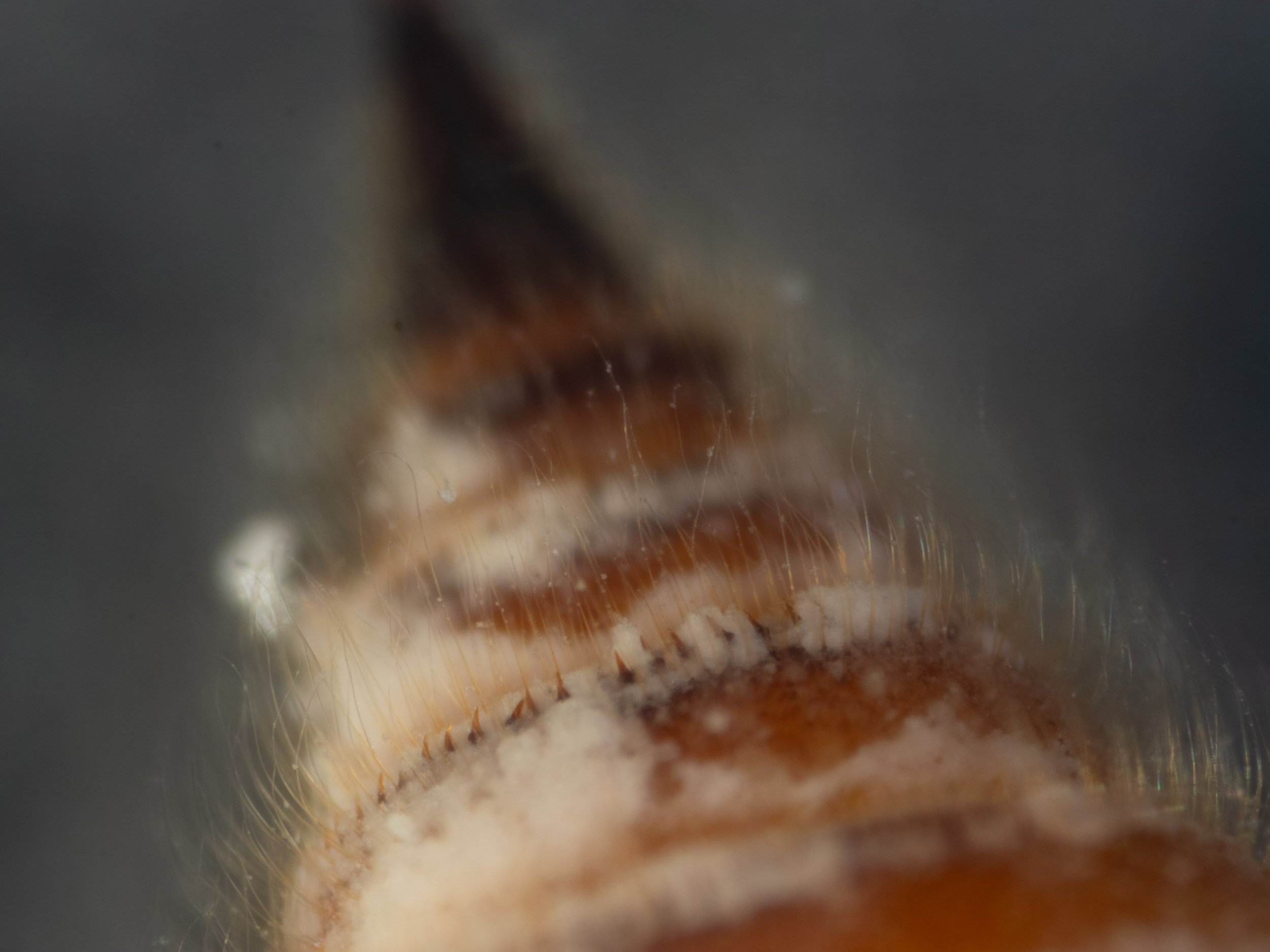
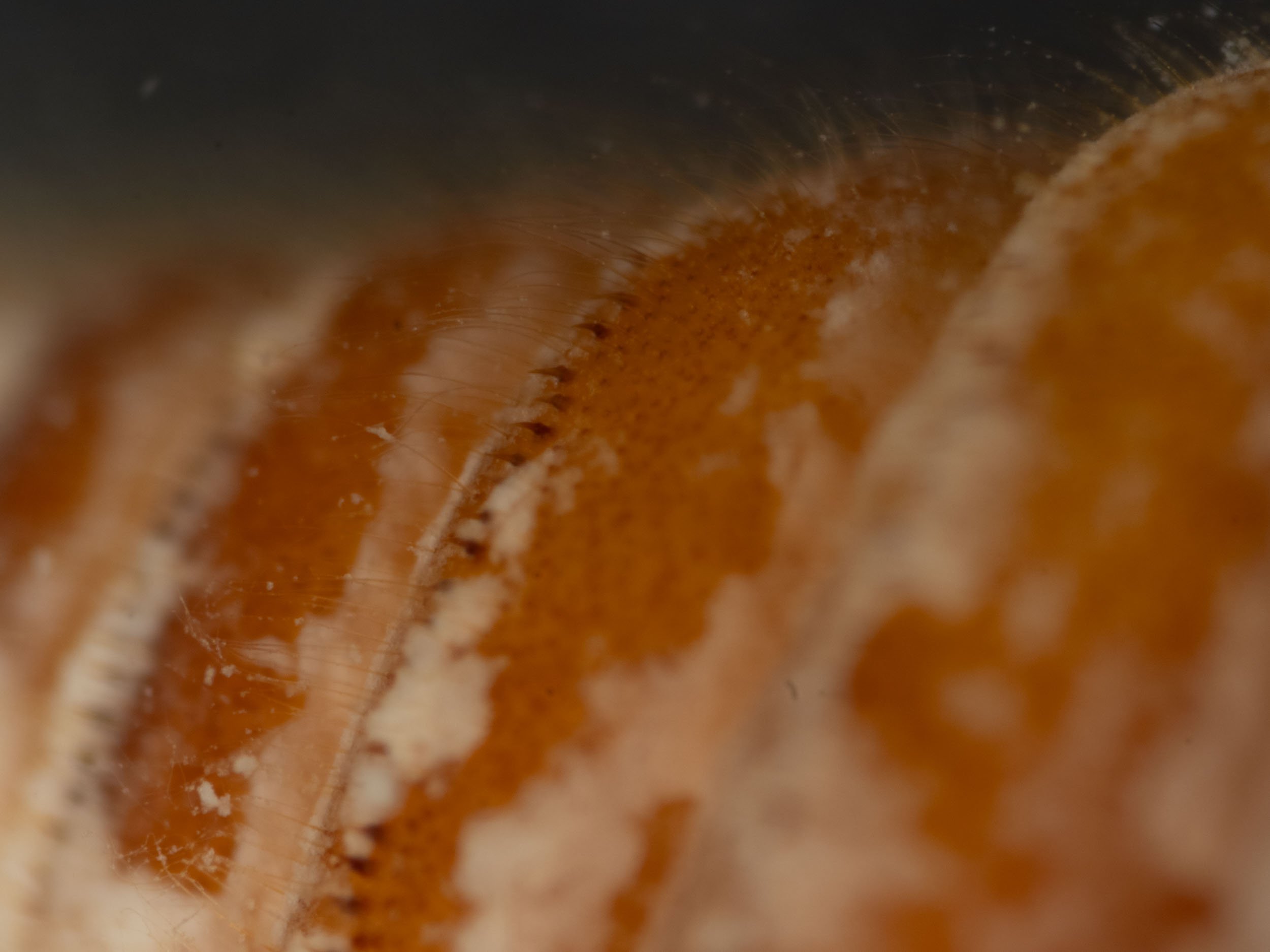
Venter. With some spine-like setae, 40-150 µm long, singly or in row extending from posterolateral margin often on each side of abdominal segments V-VIII, but sometimes absent from 1 or more of these segments; distributed as follows: 0-6 (mostly 0) (50-70 µm long) on V (none present); 0-8 (mostly 0) (60-120 µm long) on VI (2 on each side); 0-10 (40-150 µm long) on V11 (4 on each side); 0-9 (50-135 µm long) on VIII (none present); 0 on IX. Hair-like setae on all body segments, 30-400 µm long, longest laterally on posterior abdominal segments excluding IX (yes). (Multilocular disc pores rare on head, most abundant on thorax, confined to lateral and anterior margins of abdominal segments III-VII, absent from abdominal segments VIII and IX; pores of 5-9 locules present, 7-locular pores predominant). not visible with observation methods used.
Dorsum. With spine-like setae, 25-140 µm long, on at least thoracic segment III and abdominal segments II-VIII, usually absent from head and thoracic segment I and sometimes absent from thoracic segment II; distributed on head and thoracic segments as follows: 0-8 (singly on 2 specimens, 8 on another specimen) (30-40 µm long) on head and I (12 on head and 14 on I); 0-13 (25-60 µm long) singly or in irregular row on II (24 in irregular row); 4-47 (30-120 µm long) in irregular row or narrow, irregular band on III (28 in irregular row); and on abdominal segments: 4-59 (30-130 µm long) in irregular row or narrow, irregular band on II (28 in irregular row); 9-59 (30-140 µm long) in irregular row on III (29 in irregular row); 14-58 (35-135 µm long) in irregular row on IV (24 in regular row); 13-45 (35-130 µm long) in regular to irregular row on V (~24 in regular row); 17-45 (35-120 µm long) in regular row on VI (19? in regular row); 18-50 (35-120 µm long) in regular row on VII (19 in regular row); 12-37 (35-120 µm long) in regular row on VIII (16 in regular row); 0 on IX (0). Hair-like setae on all body segments, 50-550µm long (up to 500µm), longest near spine-like setae on posterior abdominal segments (yes). (Multilocular disc pores sparsely distributed and mainly confined to margins of head, thoracic segments and abdominal segment II, densely distributed on anterior half of abdominal segments III to VI, mainly confined to anterolateral part of abdominal segment VII, absent from VlII and IX; pores of 5-9 locules present, 7-locular pores predominant) not visible with observation methods used.
Anal lobes. Lightly to moderately sclerotized (moderately), 350-620 (480±80) (520) µm long, tapering until near apex, slightly divergent apically (yes). Each apex distinctly bifurcate, terminating in 2 subequal, spine-like processes 20-50 µm long (yes). Hair-like setae, 60-400 µm long, (yes) mostly confined to ventral and outer surfaces of lobes (yes), with 1 longer (400-470 µm), more robust seta originating ventrally near each apex.
Apiomorpha ovicola - gall Apiomorpha #10
9/3/24 This single female gall, positioned on a stem, was collected from small sapling of E. globoidea on Link Trail. Many ants were collecting honeydew from the opening to the gall.


Slices of the gall were removed to exposed a large, live female was inside. She was very responsive to touch. I took images of her live, then chilled here and put her in the killing jar. After overnight fixation in alcohol, I washed here with detergent solution.


This is a workbook page … a part of our website where we record the observations and references used in making species identifications. The notes will not necessarily be complete. They are a record for our own use, but we are happy to share this information with others.