Apiomorpha pharetrata (Eriococcidae: Apiomorphinae)
Workbook
Notes about the gall-forming coccoid genus Apiomorpha pharetrata
Several of these galls were found on eucalypt saplings on our property.
We have collected several of these galls from different trees.
Species ID of Apiomorpha pharetrata based on following descriptions in Gullan (1984).
Description of Gall of Female (based on 26 mature galls and several immature galls; Fig. 102)
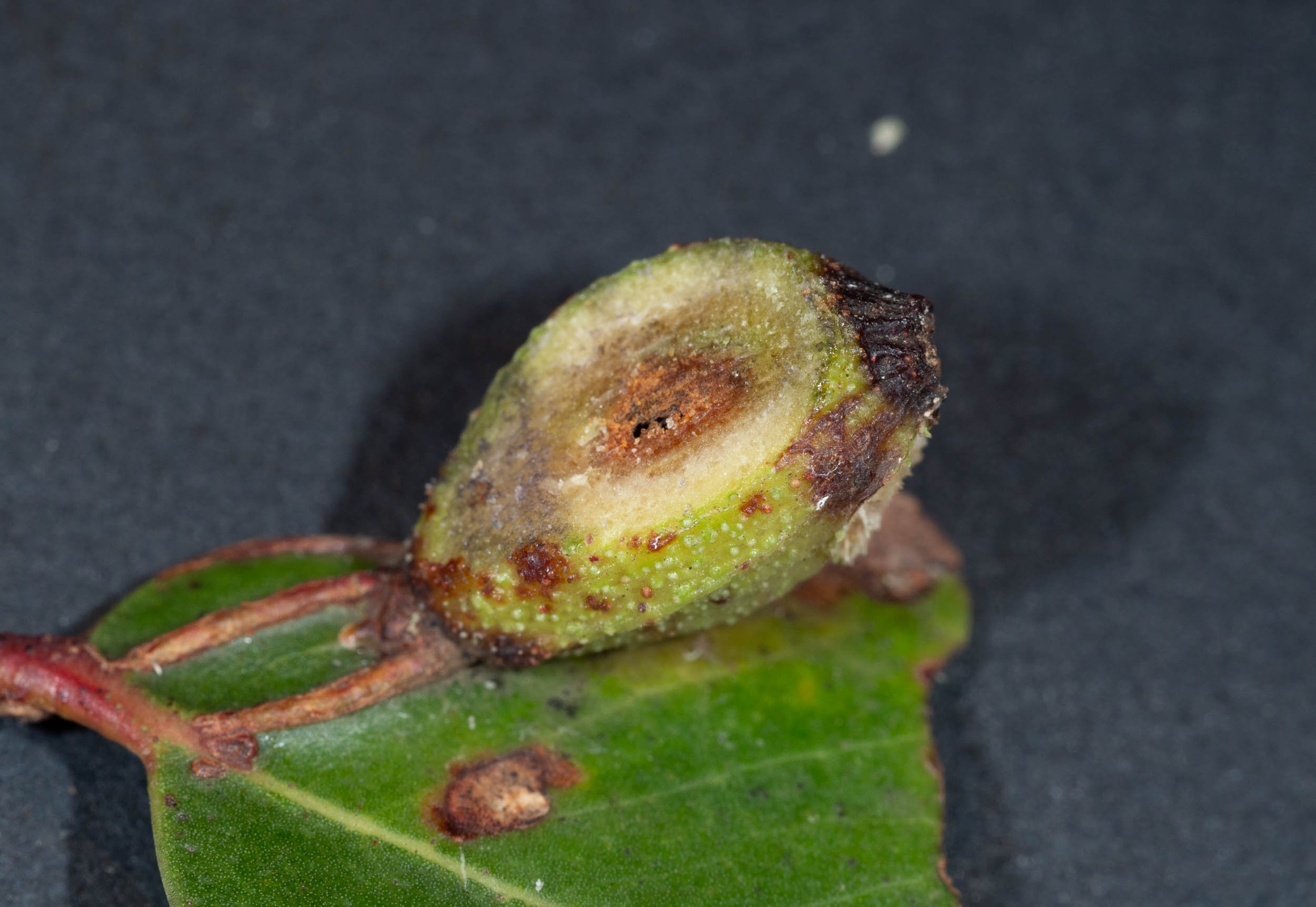
Mature gall fusiform to ellipsoidal, rarely cylindrical, sometimes ribbed, 5.5-24.0 mm long, 4.0-9.5 mm wide, usually widest medially, sessile with basal attachment 2.0-5.5 mm in diameter. Apex rounded to truncate, if truncate 1.5 - 2.5 mm in diameter but without a raised, outer rim; apical orifice mostly circular, 0.25-0.50 mm in diameter, sometimes in slight depression, with orifice sometimes surrounded by slightly serrate or crenate rim. Gall cavity fusiform with apical end acuminate, basal end rounded; wall 0.5-2.0 mm thick. Living gall green to crimson or burgundy red; dry gall green, brown or black; surface either granulate or with prominent narrow, longitudinal ribs from base to apex.
Immature gall cylindrical or ellipsoidal, apical orifice closed if very immature.
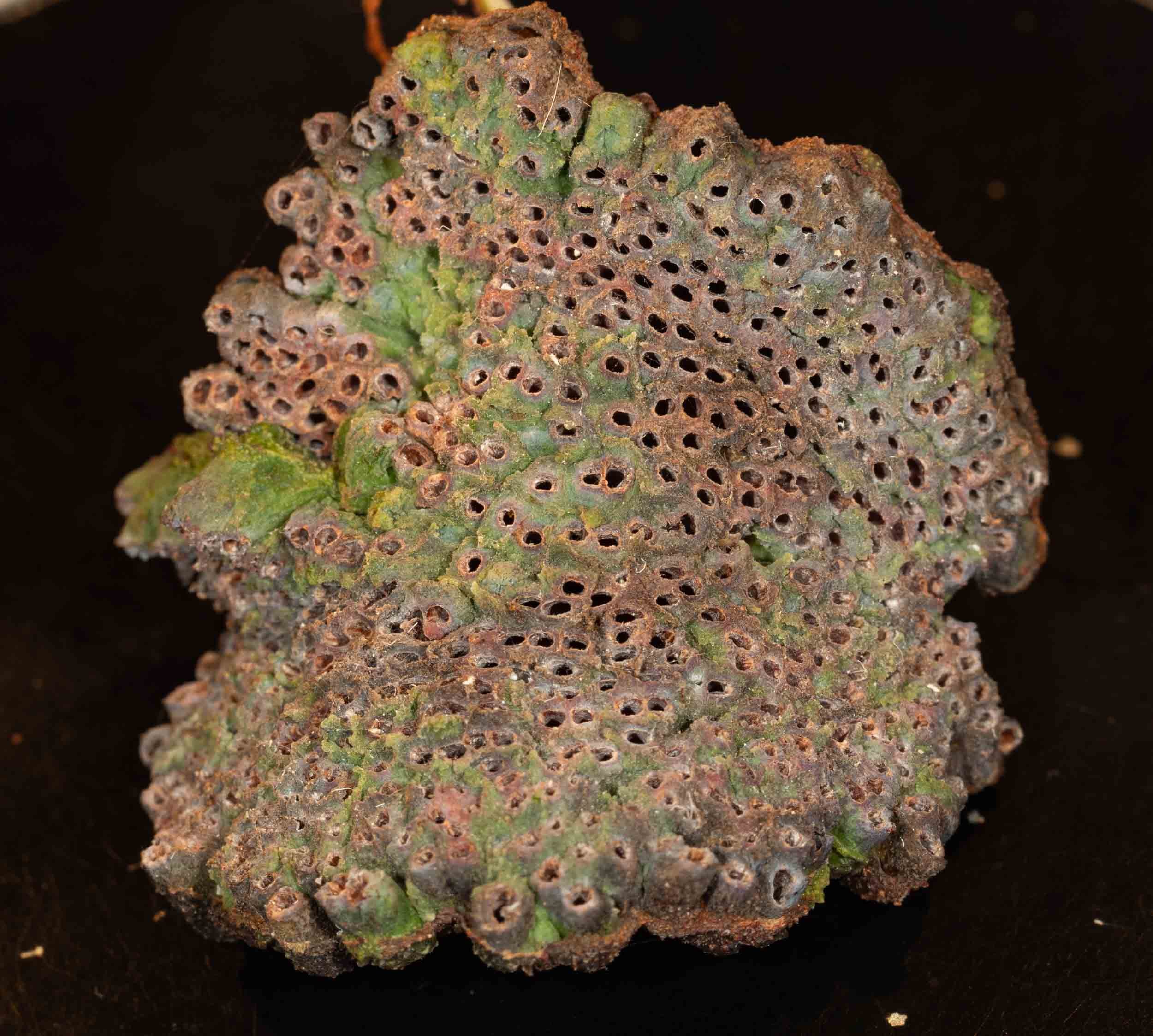
Description of Compound Galls of Males (based on 14 mature compound galls and several immature compound galls; Figs 100, 101)
Mature compound gall produced by male 1st-instar nymphs consists of an irregular, plate- like structure bearing many partially coalesced, tubular galls; whole structure attached to side of maternal gall near apex by junction 3.5-7.0 mm across widest part, with apex of maternal gall barely projecting beyond attachment area. Individual gall of each male tubular, 3-0-6.0 mm long, 0.8-1.7 mm in diameter at apex; apical orifice approximately circular, 0.4-1.1 mm in diameter, with outer surface of rim irregular or jagged; outer gall wall when visible often rough, either partially or almost completely coalesced with walls of surrounding galls. Galls clustered together on plate-like structure of irregularly circular to elliptical outline, often divided into several partially attached pieces, 12.0-35.0 mm across widest part, with lower surface usually convex, upper surface either eoncave or convex depending on maturity of specimen; up to 300 or 350 individual galls in each complex. Living com- pound gall with lower surface green to reddish green, with tubes of upper surface yellowish brown to pinkish brown, usually without white wax around orifices of individual galls.
Immature compound gall with 2 sides inturned to enclose developing mass of small, tubular galls.
Apiomorpha pharetrata #1
collected from this tree on the left side of the path to the Biolytix tank - Eucalyptus globoidea?
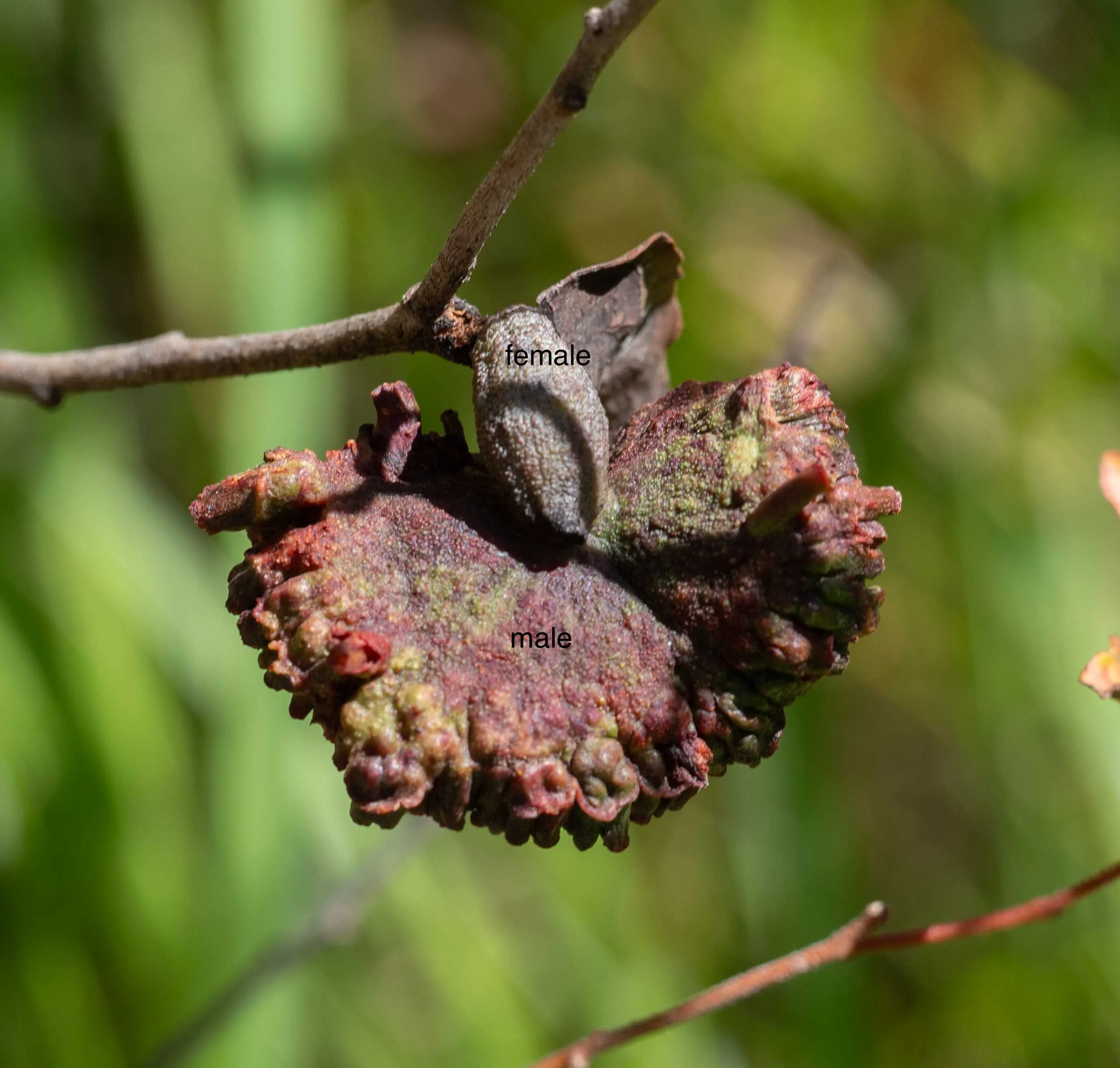
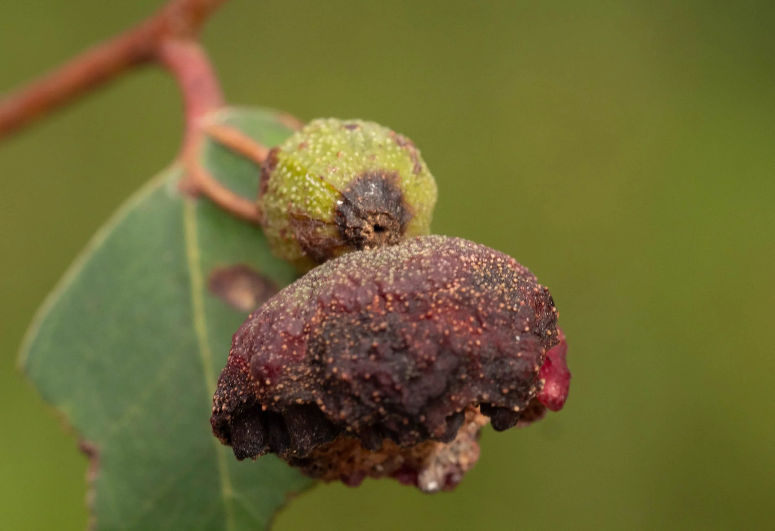
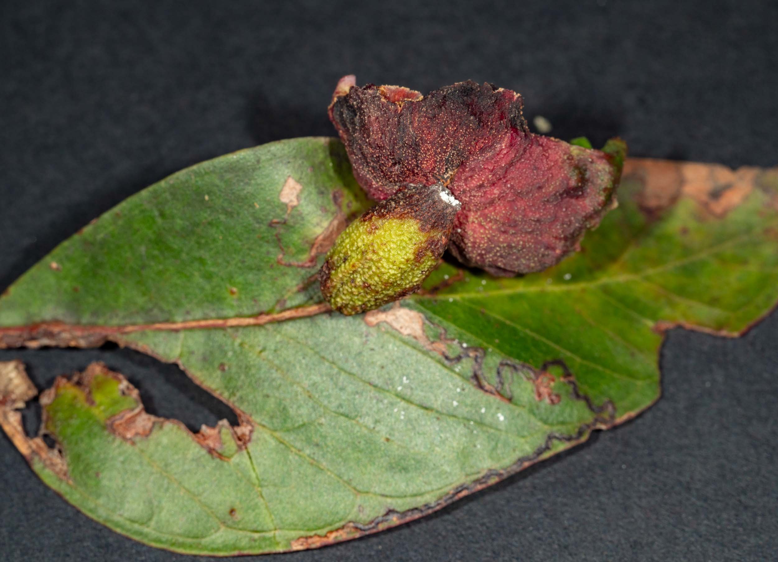
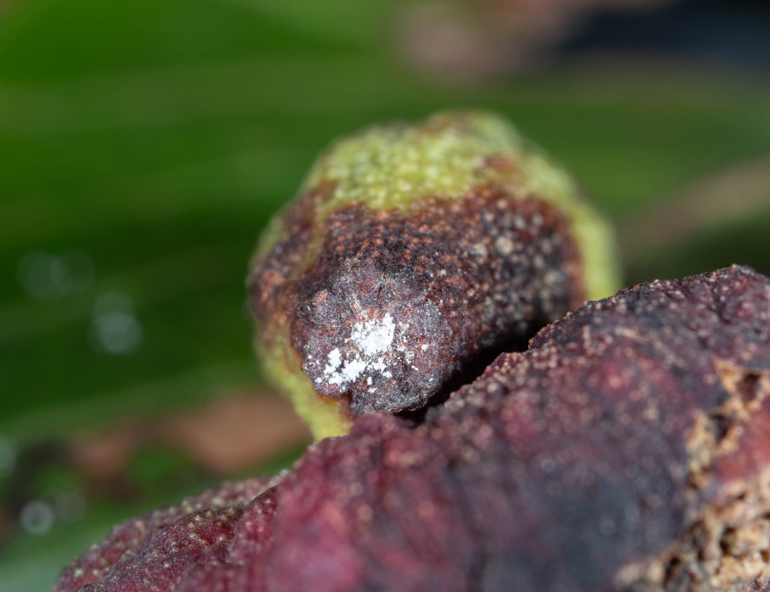
the gall in situ - female gall is at base of petiole of a leaf, which has died. The compound male gall is attached to the female gall near the apical pore.

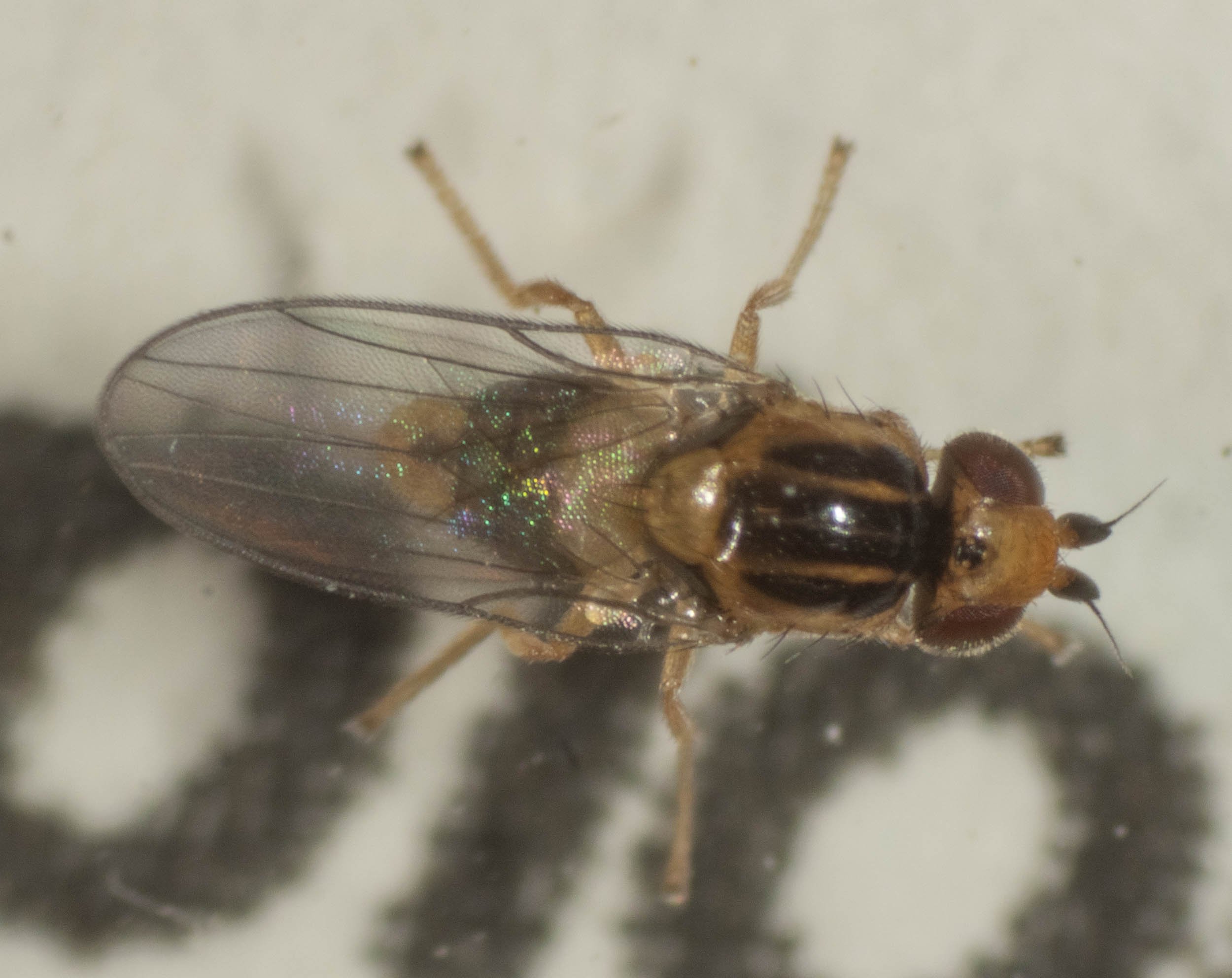
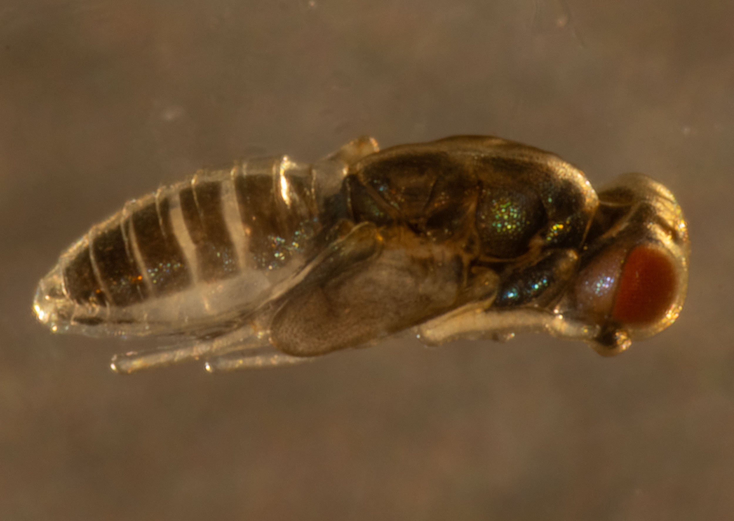
I cut open the gall of the female. She had been parasitised and was now an empty shell. Two dipteran larvae were crawling around inside the gall and there was an empty dipteran puparium there as well. On 5/3/24 a fly had eclosed from one of the larvae/pupae in the female gall. This was identified as a member of the subfamily Oscinellinae (frit flies) by Tony Daley on iNaturalist.


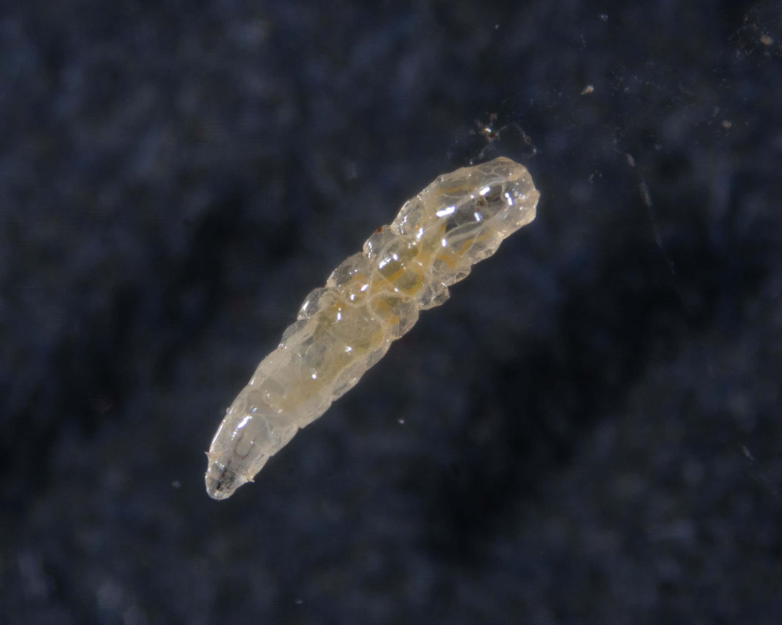


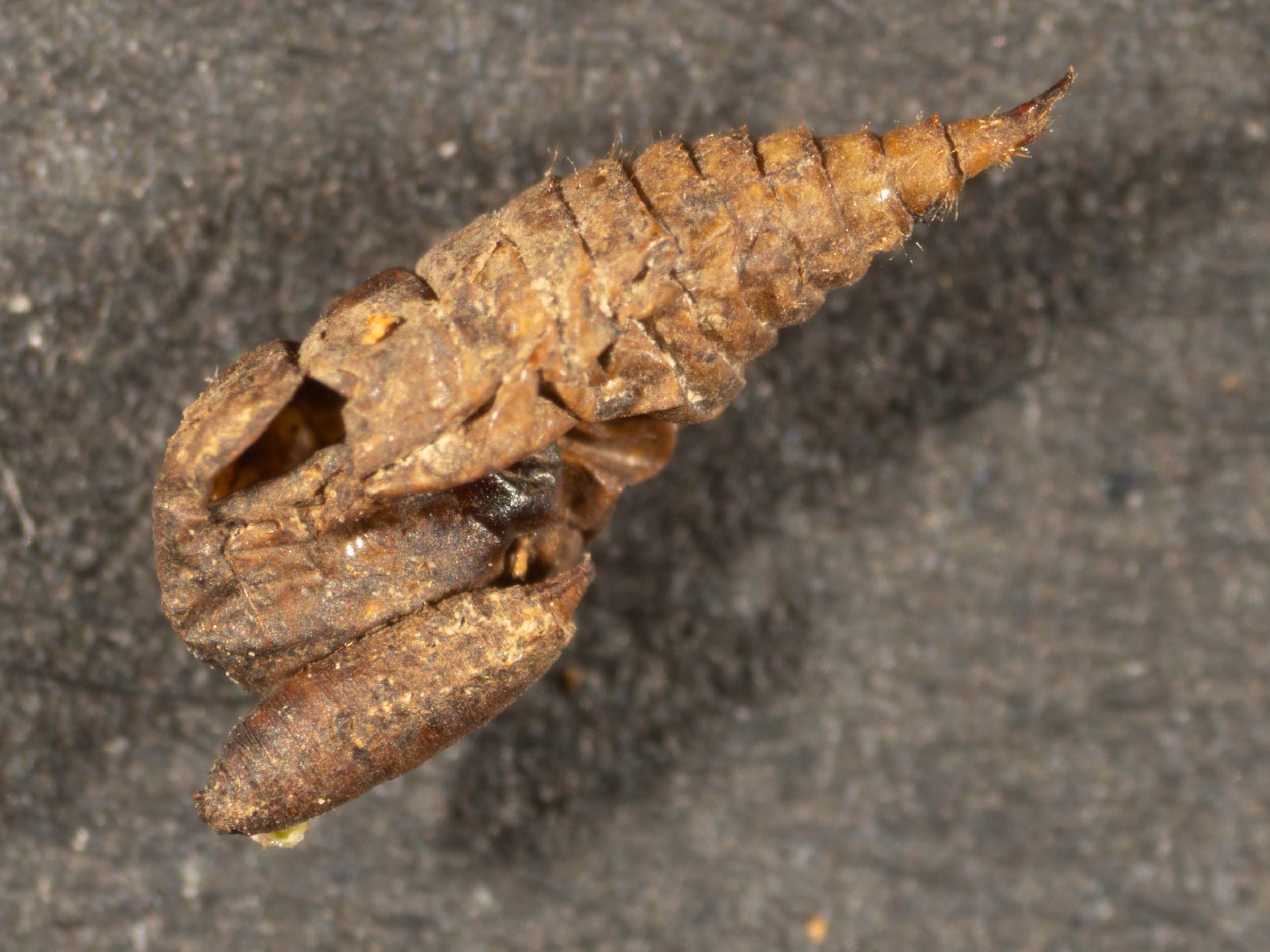
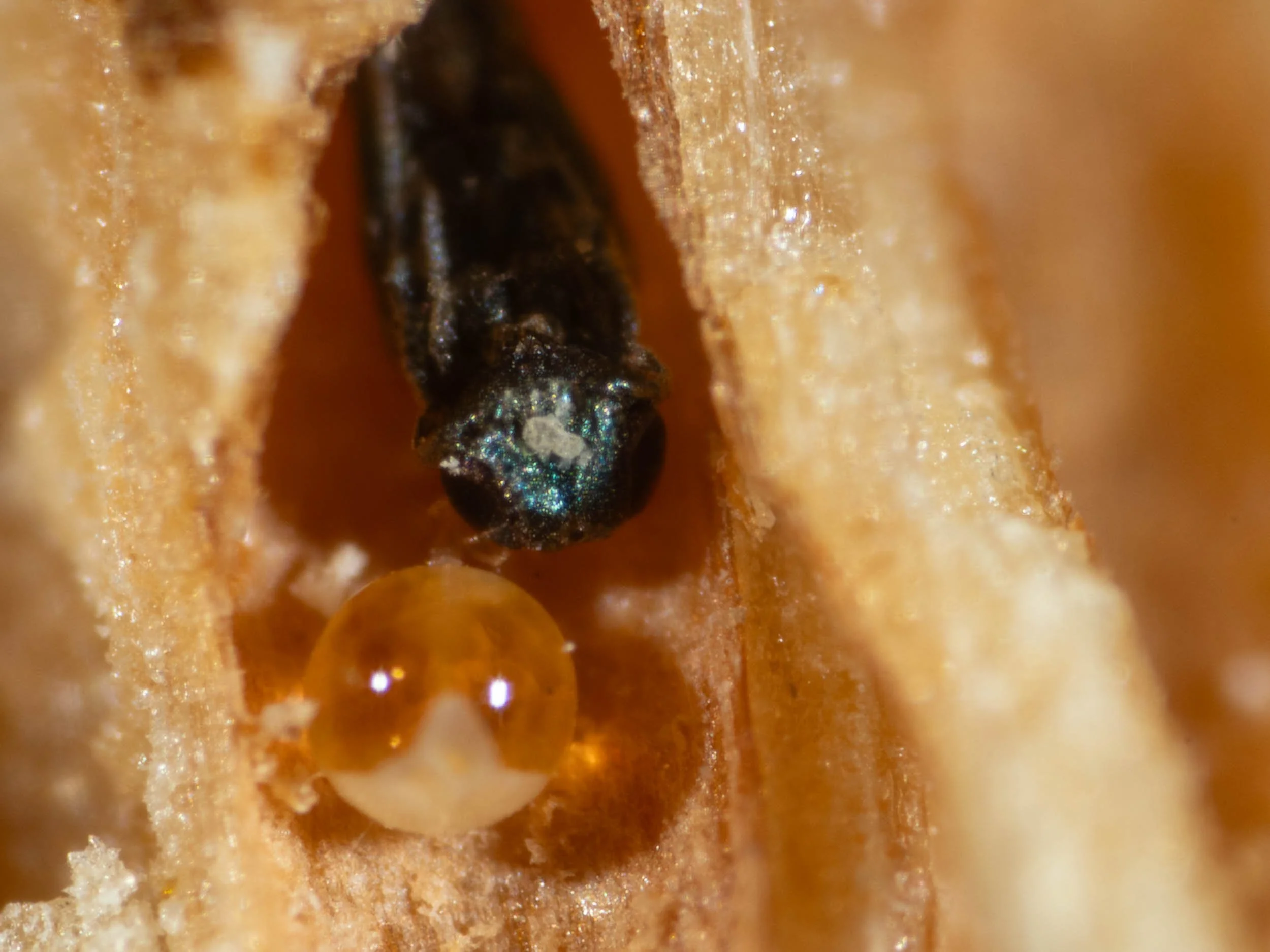
After removing the gall of the males from the female gall, I made vertical slices to expose the contents of individual cells. In two cells, a parasitic wasp crawled out. One of these eclosed immediately after the cell was opened.


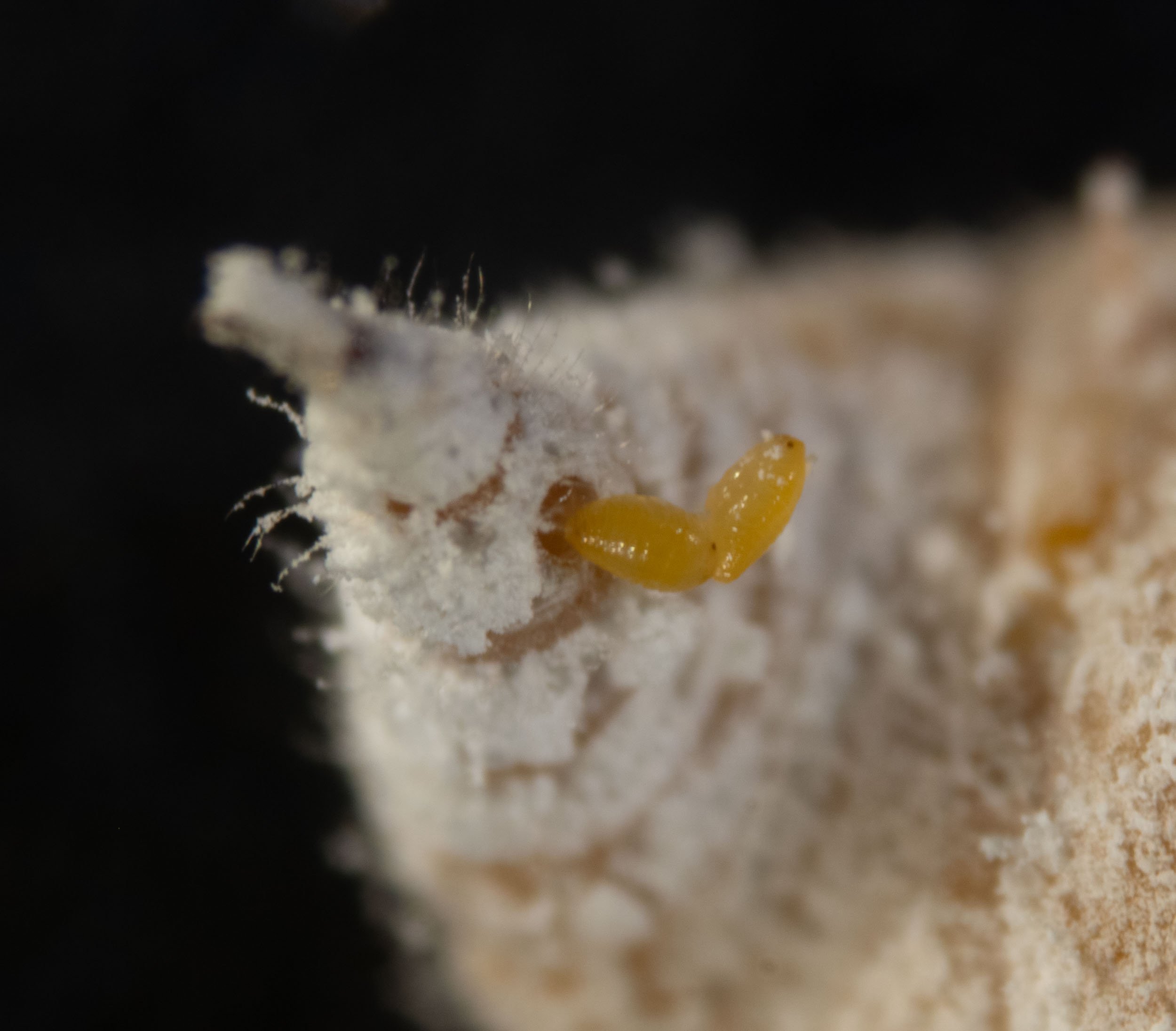
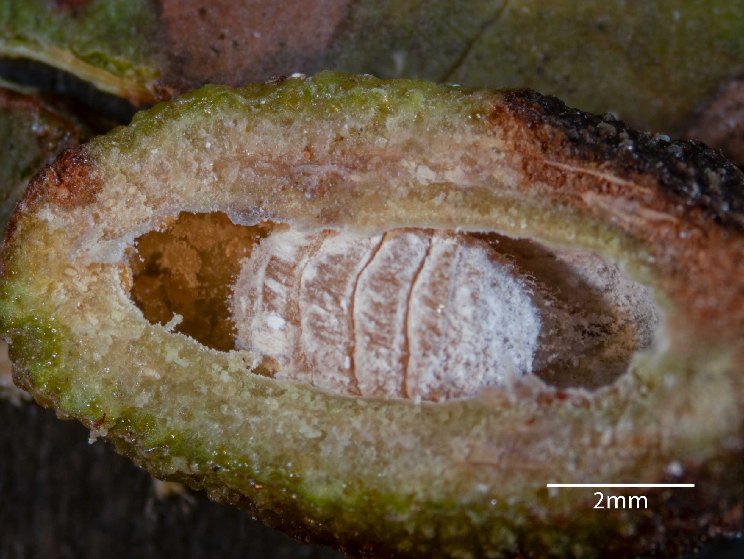
A range of developmental stages of male nymphs was found in other cells of the male gall. This panel shows two of the youngest nymphs.


a 2nd instar nymph is shown in the next panels


3rd instar? nymph in shown in the next panels


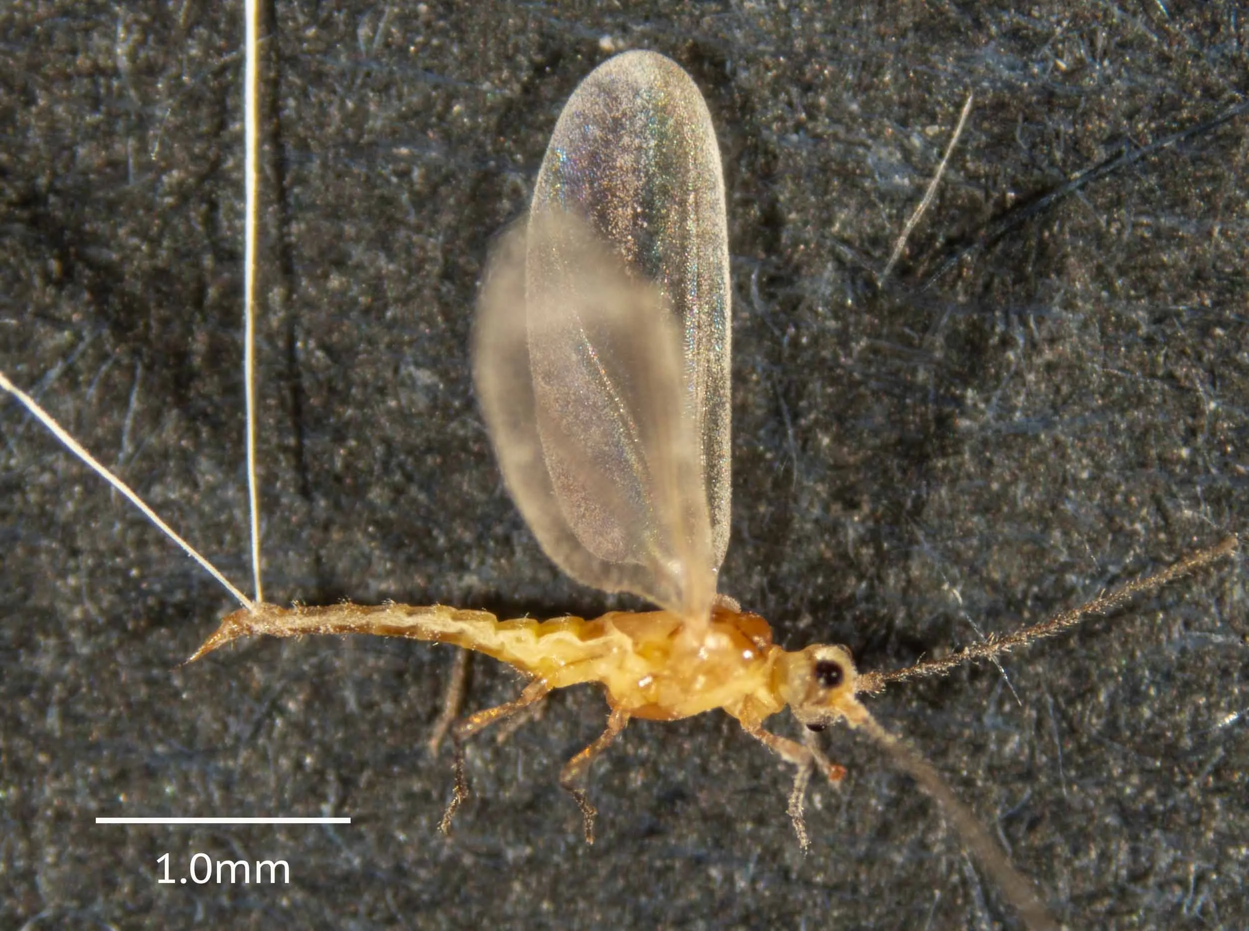
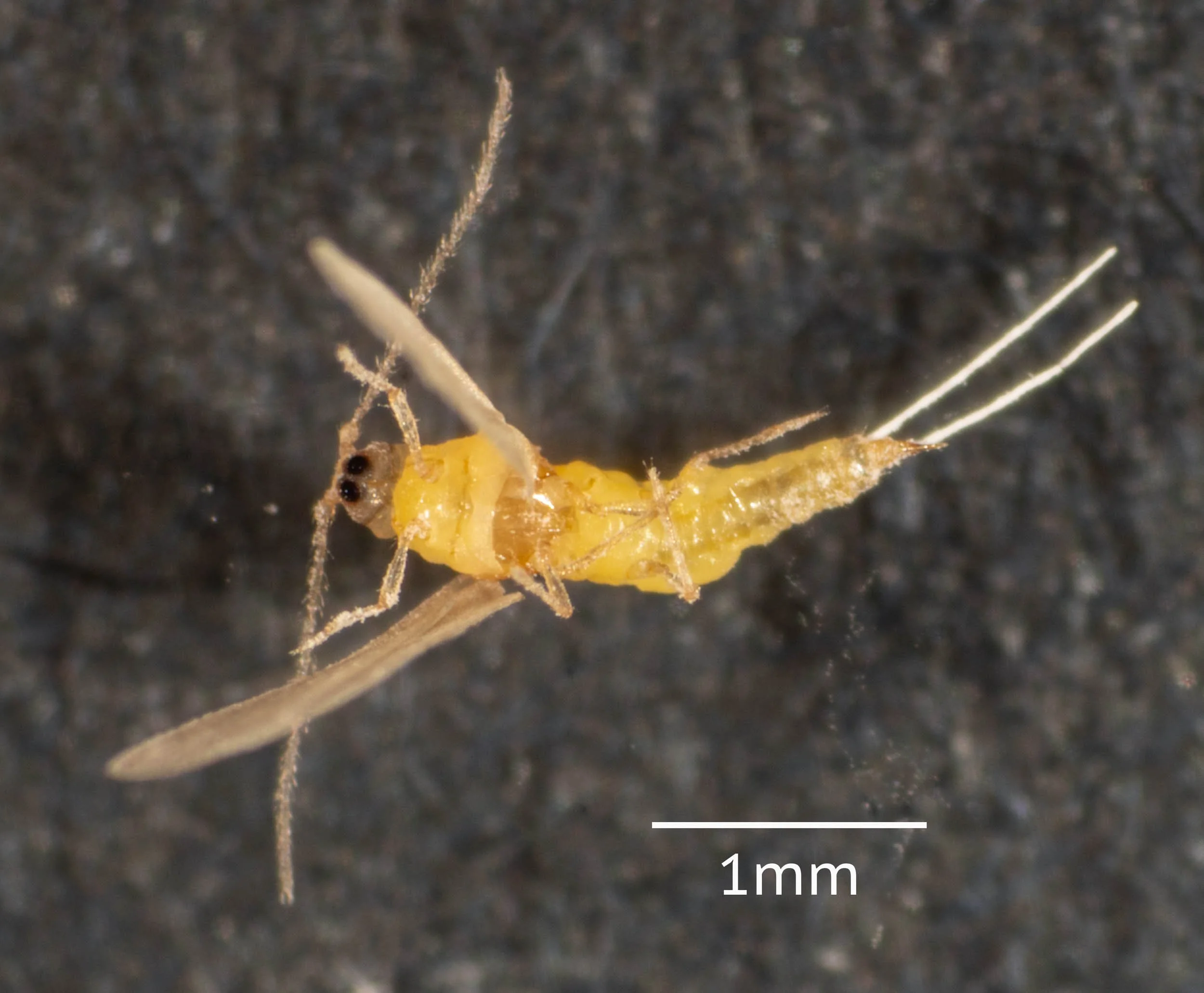
adult males are shown in the next panel


Apiomorpha pharetrata #2
collected from this smaller tree, closer to the Biolytix tank - Eucalyptus globoidea?
update 30/9/24 this very small tree is still barely living with just a few green leaves at the top
arrow shows gall
the gall in situ - female gall is at base of petiole of a leaf, which has died. The compound male gall is attached to the female gall near the apical pore.


after removal from tree


I removed a slice from the female’s gall, revealing several dipteran pupae inside as well as a couple of late stage larvae. The shell of the parasitised female Apiomorpha lay inside. I kept the female gall in a covered petri dish. On 5/3/24 an adult fly was seen in the dish. I imaged this live under the Tessovar, then placed it in the killing jar. Identified as subfamily Oscellinae. iNaturalist observation here.


late stage wasp larvae were found in two of the cells of the male gall


a mature nymph was present in one cell and an adult eclosed from another after its cell was opened


Apiomorpha pharetrata #N
collected from small eucalypt (Angophora floribunda?) on northern side of house.
Kerri took these photos of the gall on 15th January, 2024. Note that the male galls were closed at the top and were green in colour.
iNaturalist observation identified as Apiomorpha pharetrata by penelopeuq (Secretary of Entomological Society of Queensland).
30/9/24 update: we haven’t yet been able to locate this tree to check species ID. It is among a stand of several eucalypts to the north of the house


Apiomorpha pharetrata #3
I took these photos on 25 Feb 2024. Update 30/9/24 based on the time of photograph, branch thickness and background on photos, this is likely to be a medium sized eucalypt sapling to the west of the tree #1, just to the east of the house alongside the track. This candidate tree is alive, large and healthy.


The gall was collected on 29 Feb 2024.


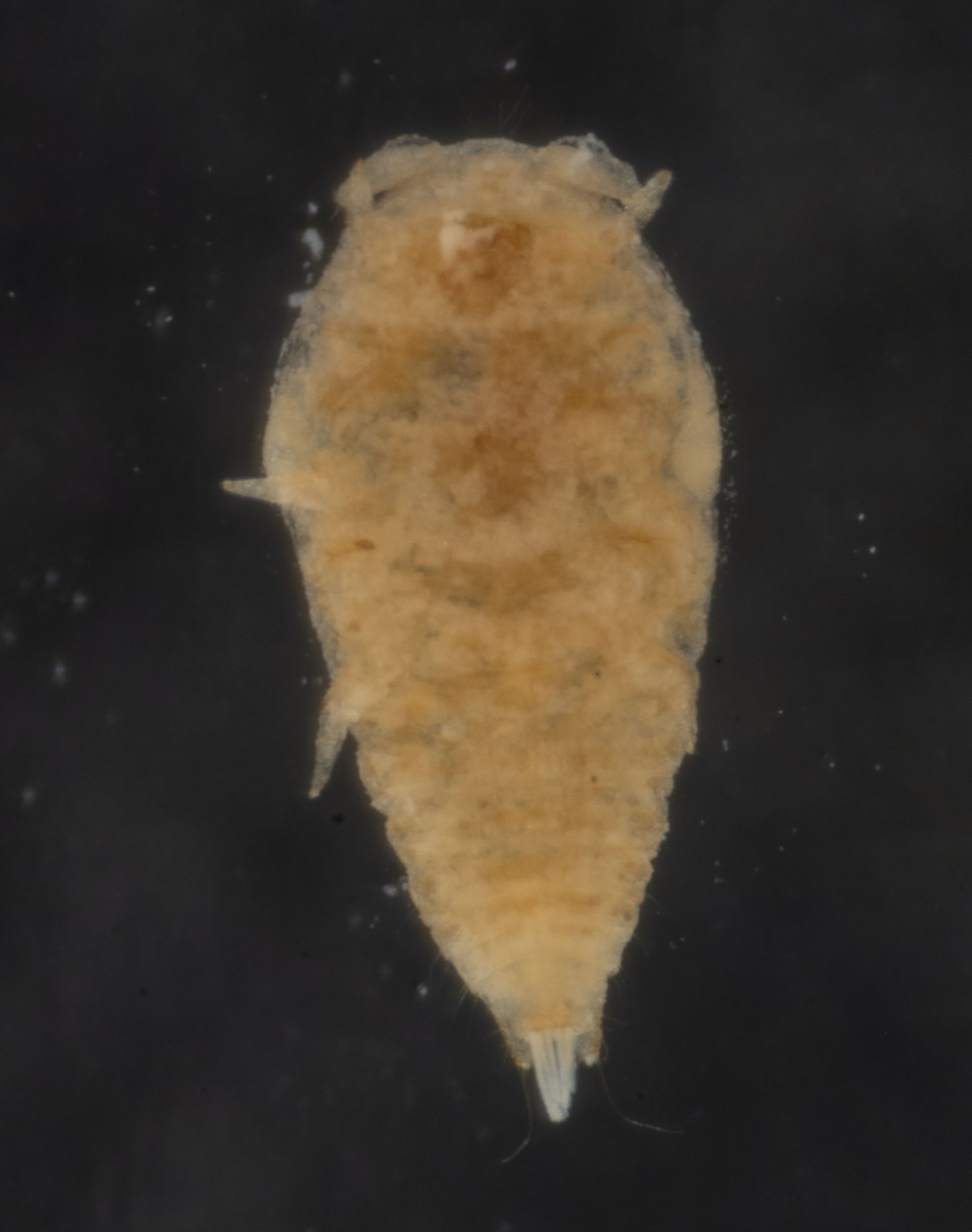
Female gall was dissected on 1st March. A living female was inside with many crawlers (1st instar nymphs). She was producing nymphs continuously. I imaged here alive together with many crawlers.


female removed from gall, continued to pump out nymphs.


I fixed female in ethanol and placed her in a vial. I removed most of the body wax by soaking in a household detergent solution for an hour or so.


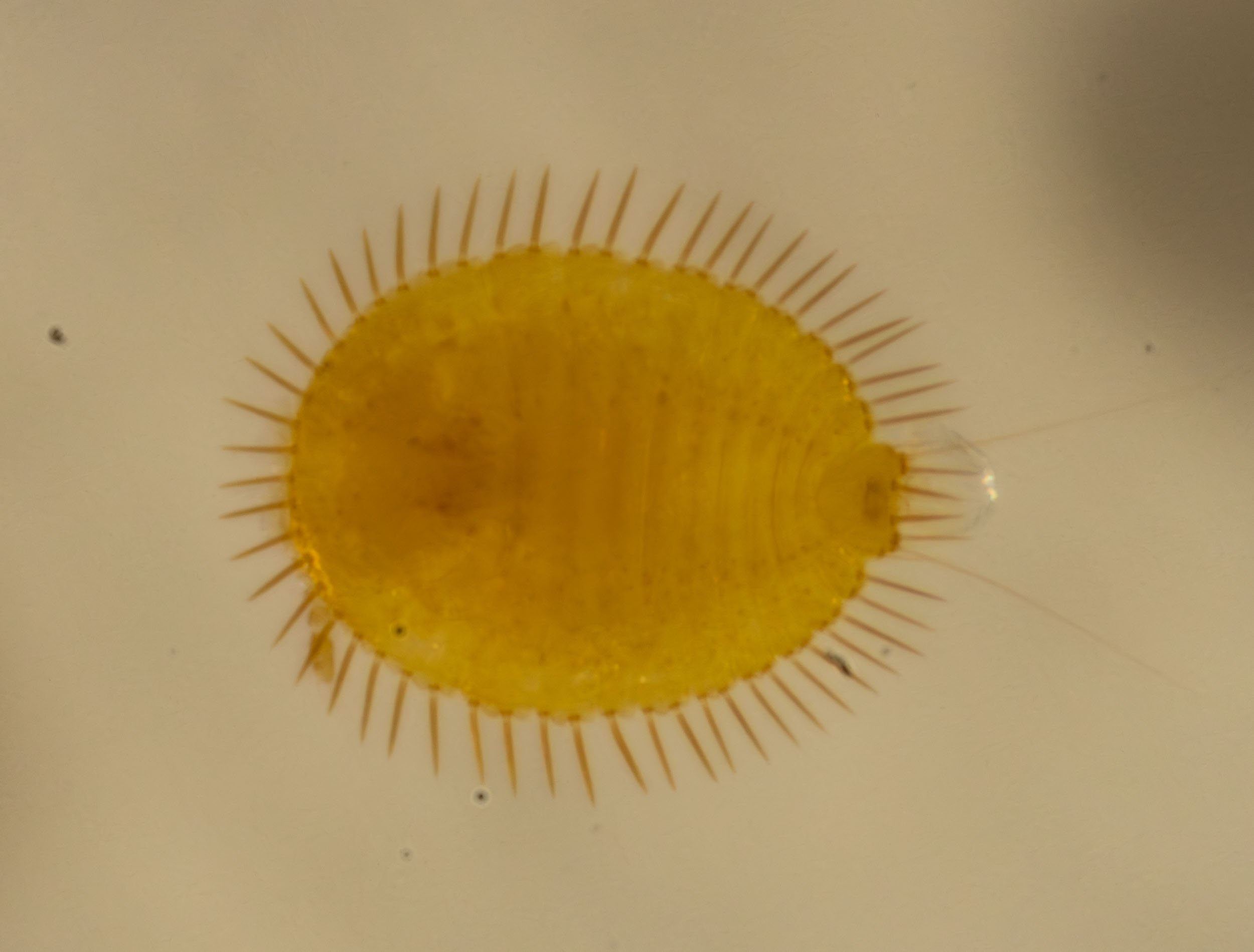
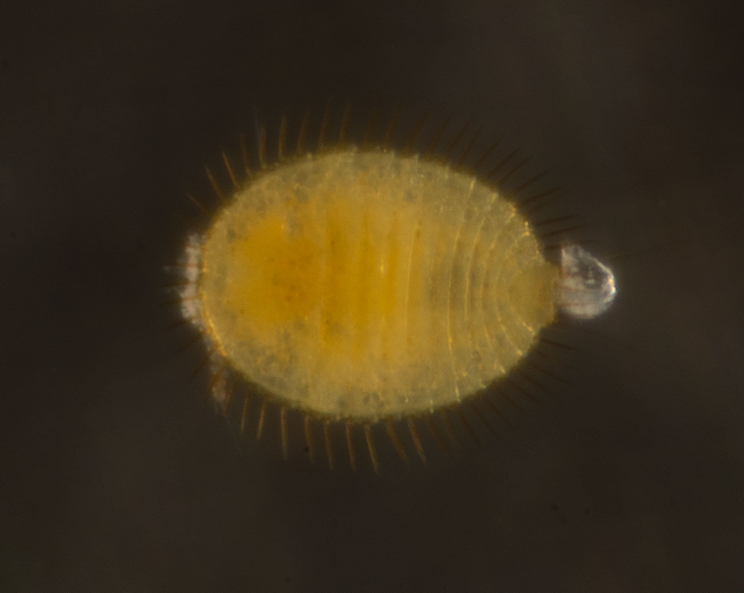
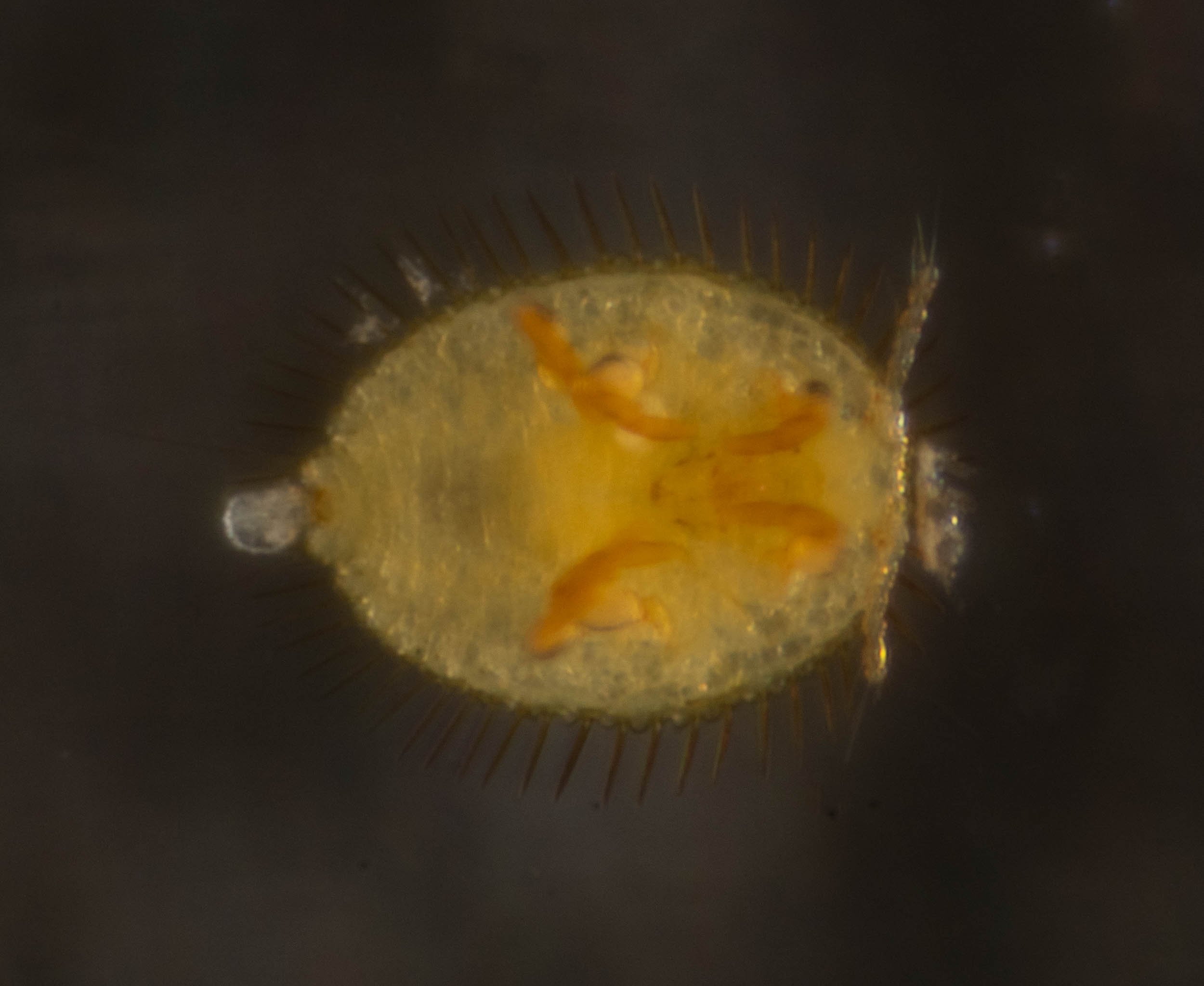
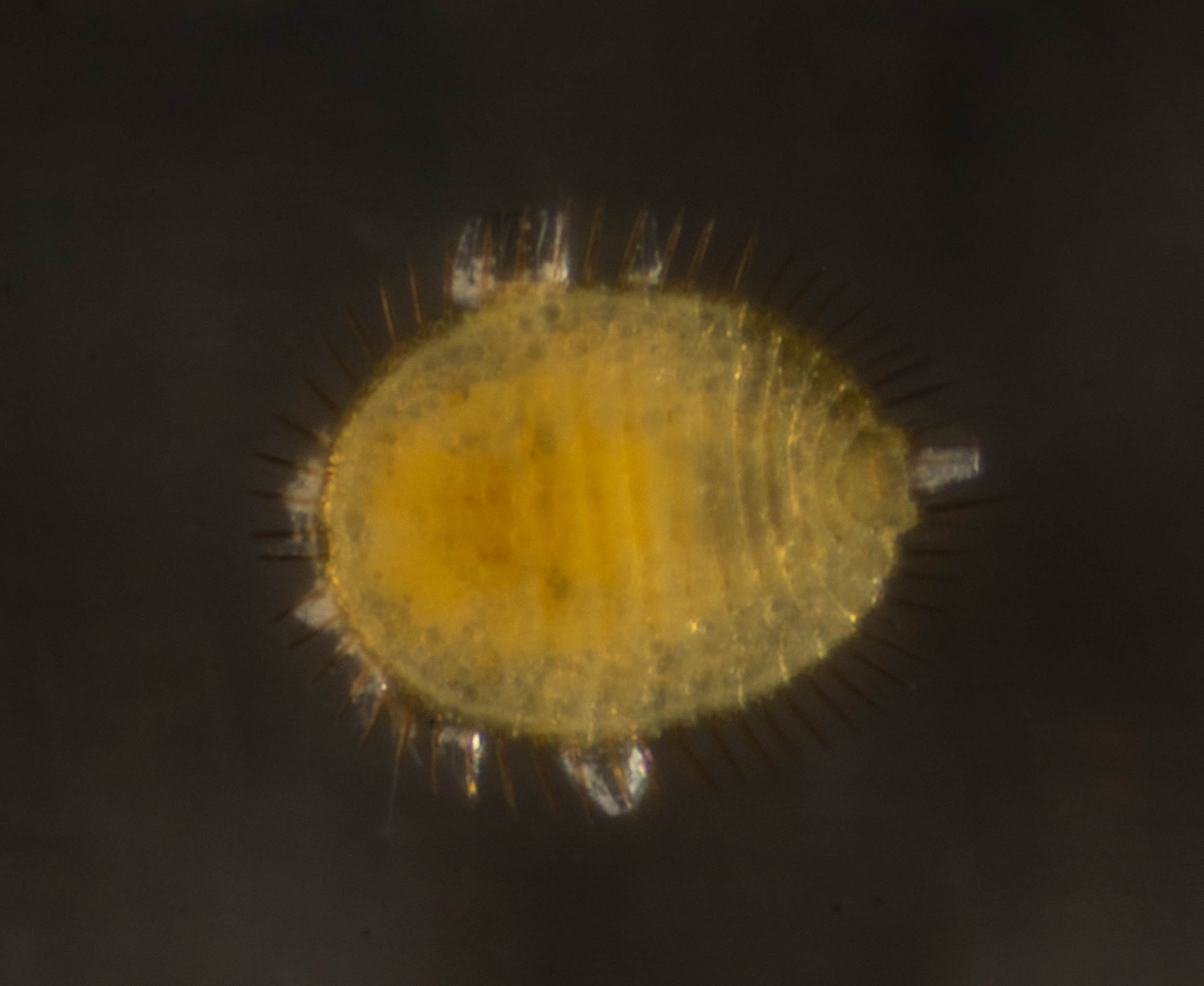
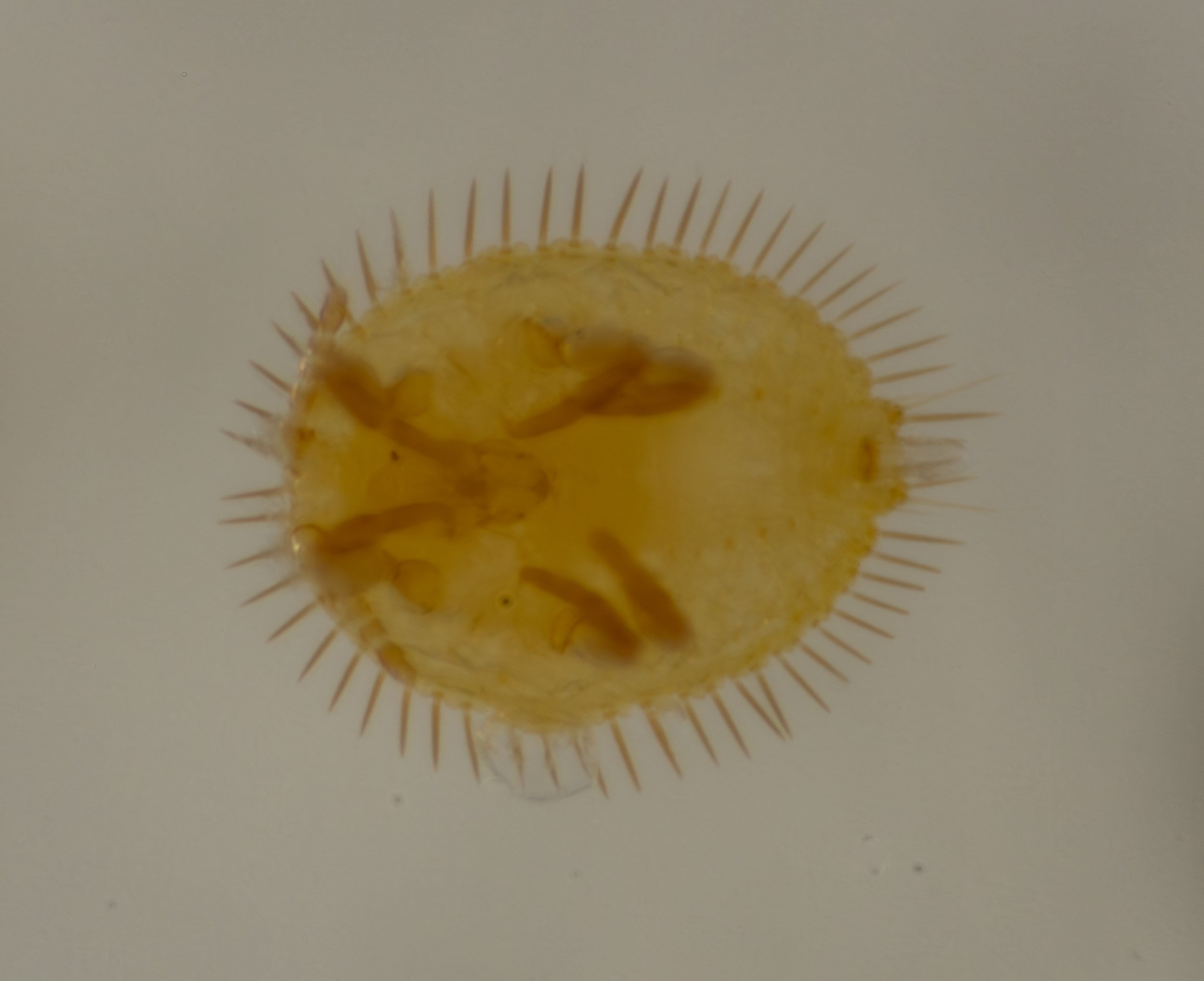
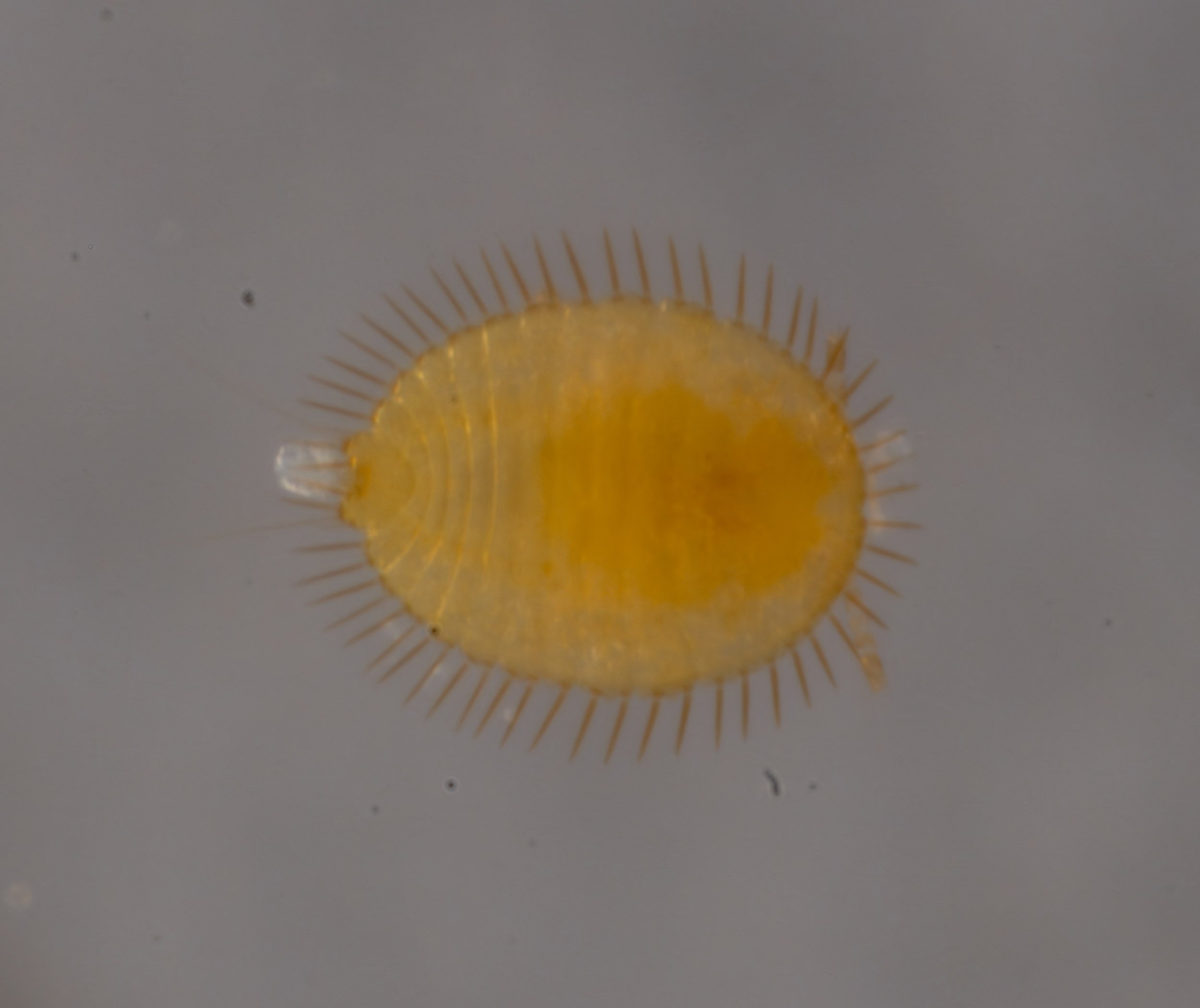
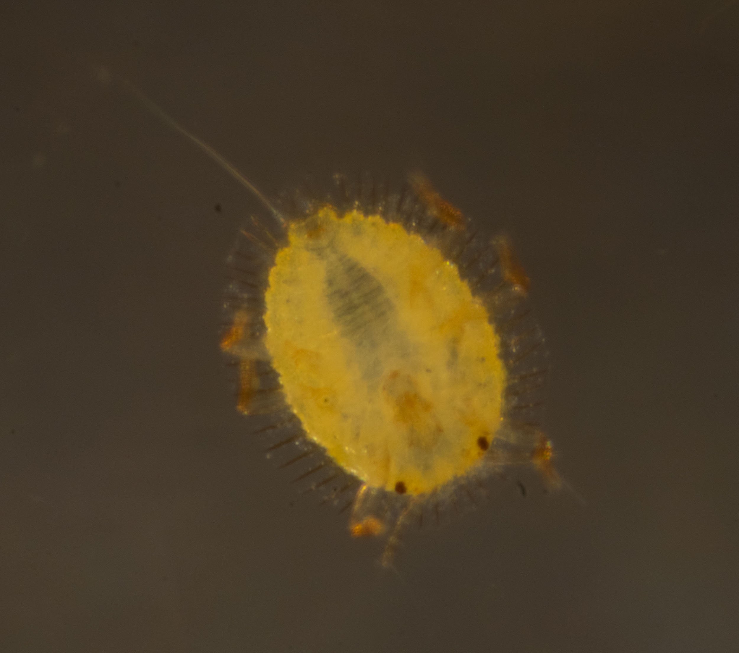
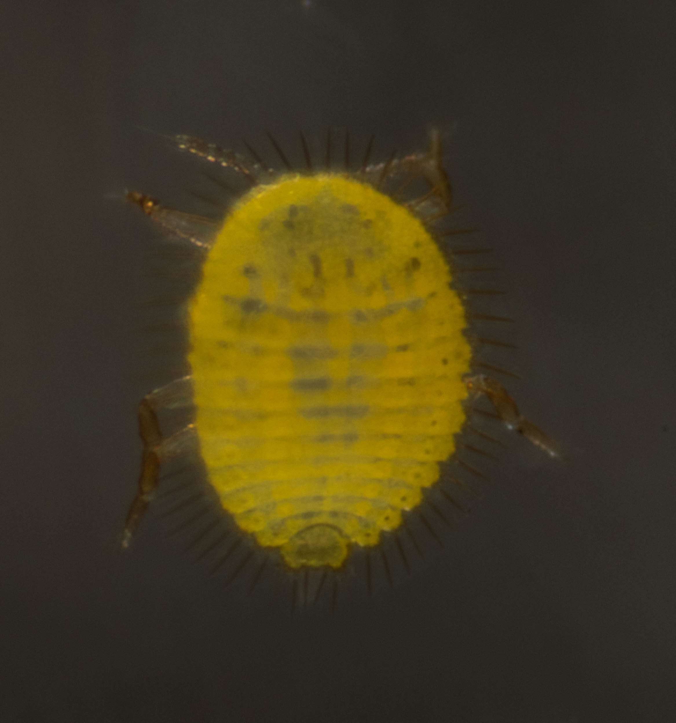
I imaged the living nymphs inside the gall, collected some and fixed them in ethanol before imaging under Tessovar.


I washed the ethanol treated nymphs with water, then transferred one to a drop of paraffin oil on a coverslip, soaked up the water with a kimwipe, coverslipped it and imaged it under DIC at 16x magnification in the Photomicroscope.
The following images show selected focal planes - focus stacking of multiple planes doesn’t work! The ethanol appears to remove the wings of the marginal setae.


These 1st instar nymphs were mounted in the same way as above, but imaged in the Tessovar under maximum magnification. I think this is the best way to get images of these nymphs. Quick, gives some 3-D information, colour and can invert slide to image other side of nymph.
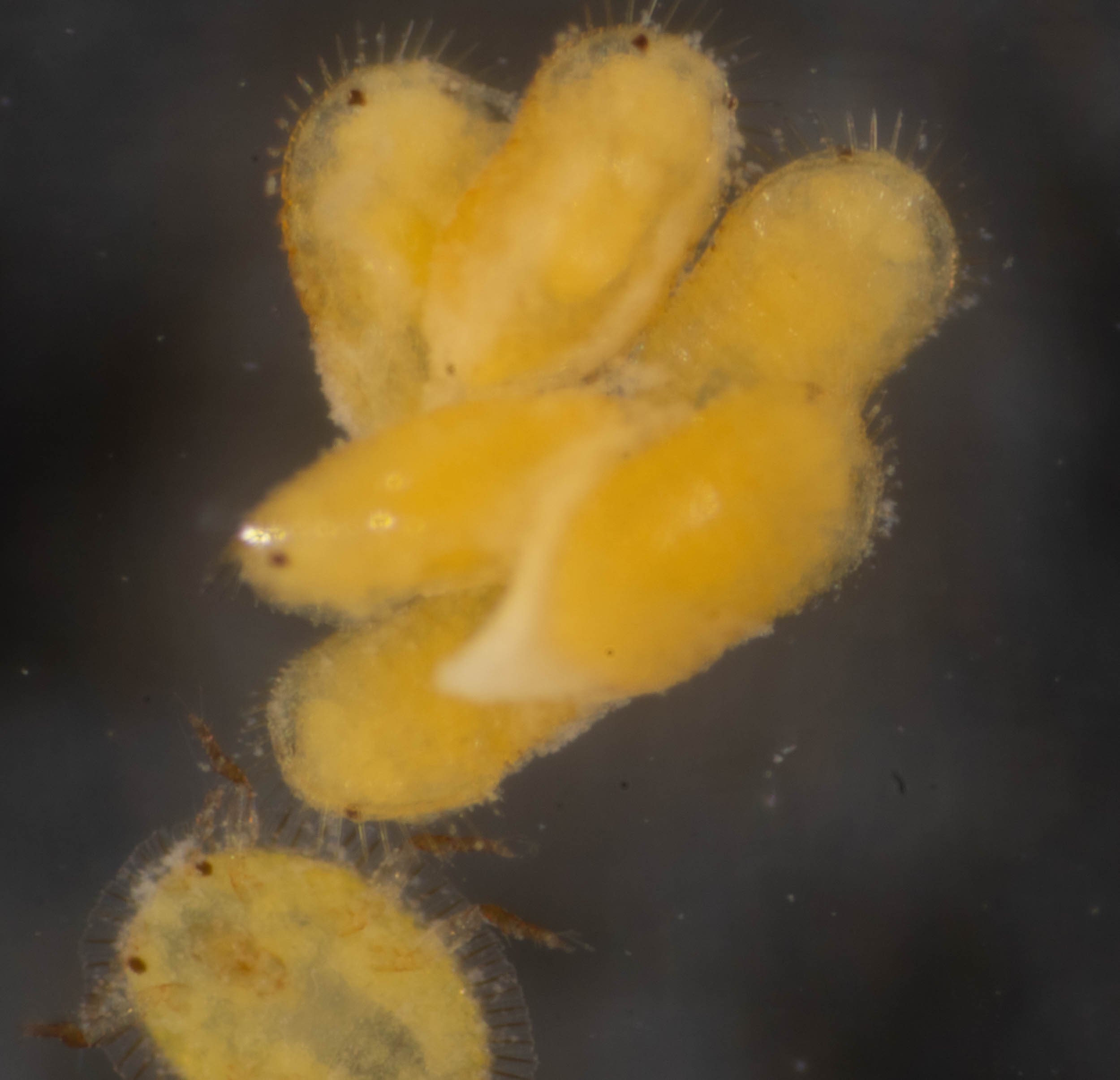
I split open a number of male galls and found adults about to eclose, together with some early stage nymphs.
Several of these were imaged, under the Tessovar and Photomicroscope.


Apiomorpha pharetrata #4
gall on small eucalypt (either E. globoidea or E. sieberi - probably former) on Lot 6 track, near junction with xantho loop.


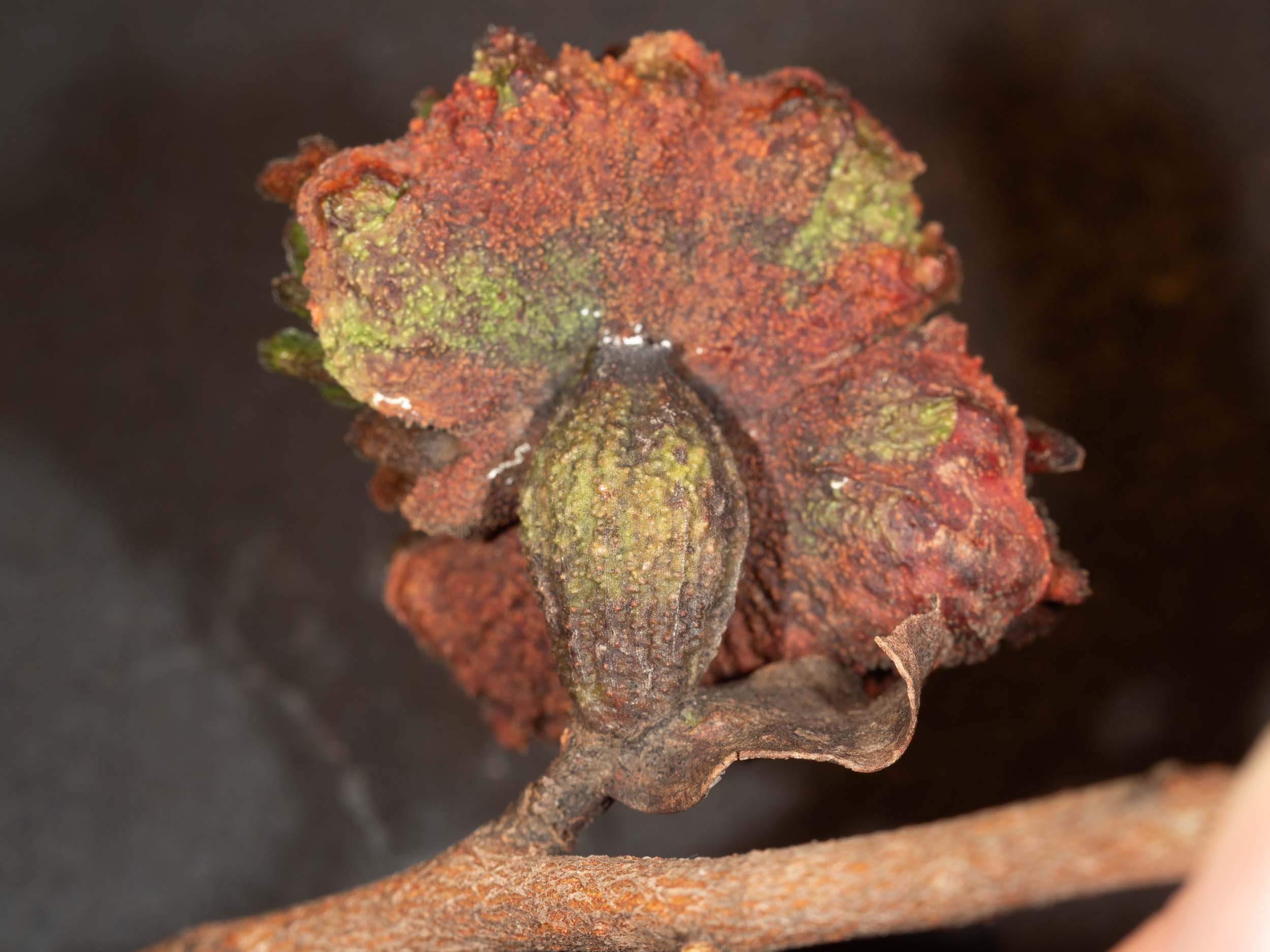
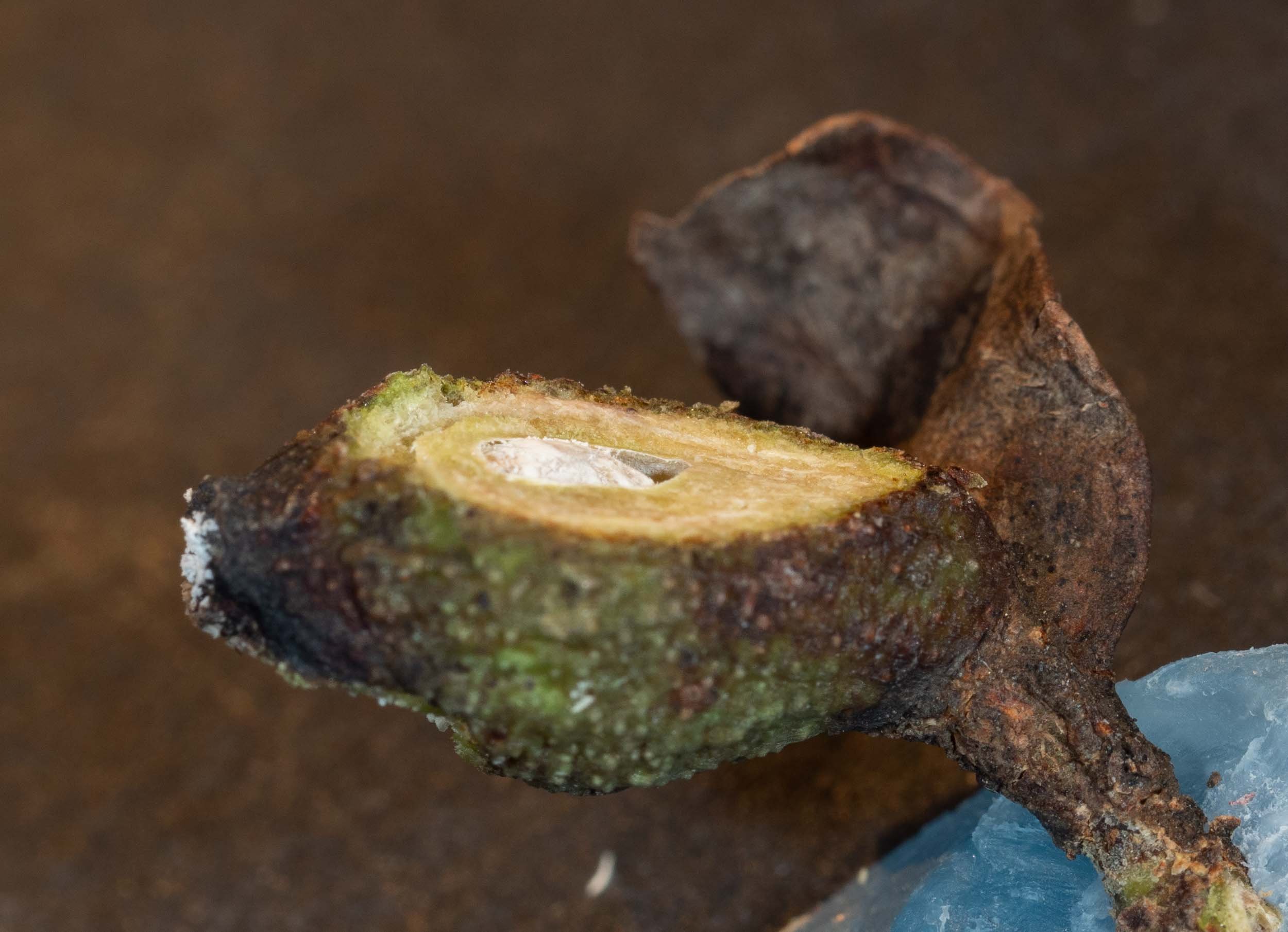
The gall was removed from the tree with its surrounding vegetation on 29/2/2024. The female gall is at base of petiole of a leaf, which has died. The compound male gall is attached to the female gall near the apical pore.


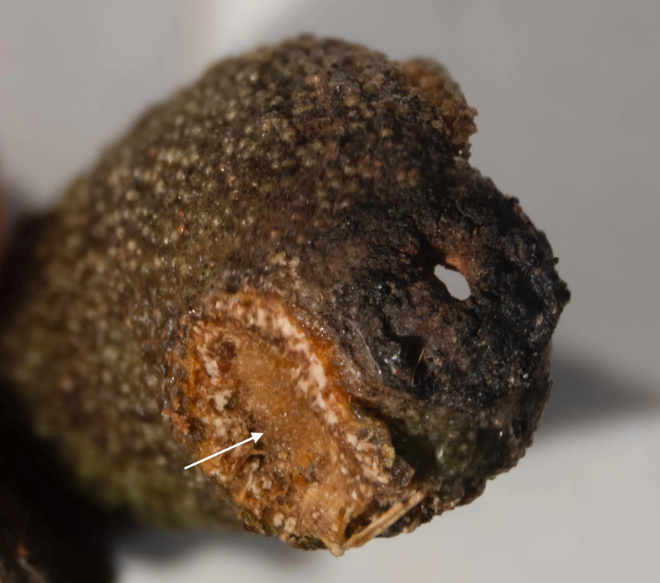
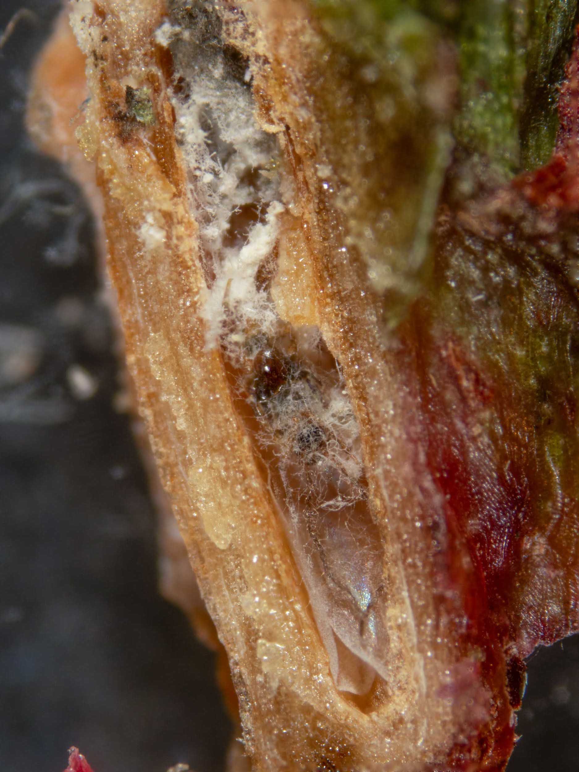
I sliced off the top of the female gall with a scalpel blade, to expose the female coccoid within. She had been parasitised by dipteran larvae, several of which were evident inside the gall, and was a husk.
The gall was placed in a covered dish. On 5/3/24 two pupae were found on the floor of the dish outside the gall. These have been placed into a labelled dish to see what adults emerge.
14/3/24 An adult fly same species (subfamily Oscellinae) to adults seen in #1 eclosed.


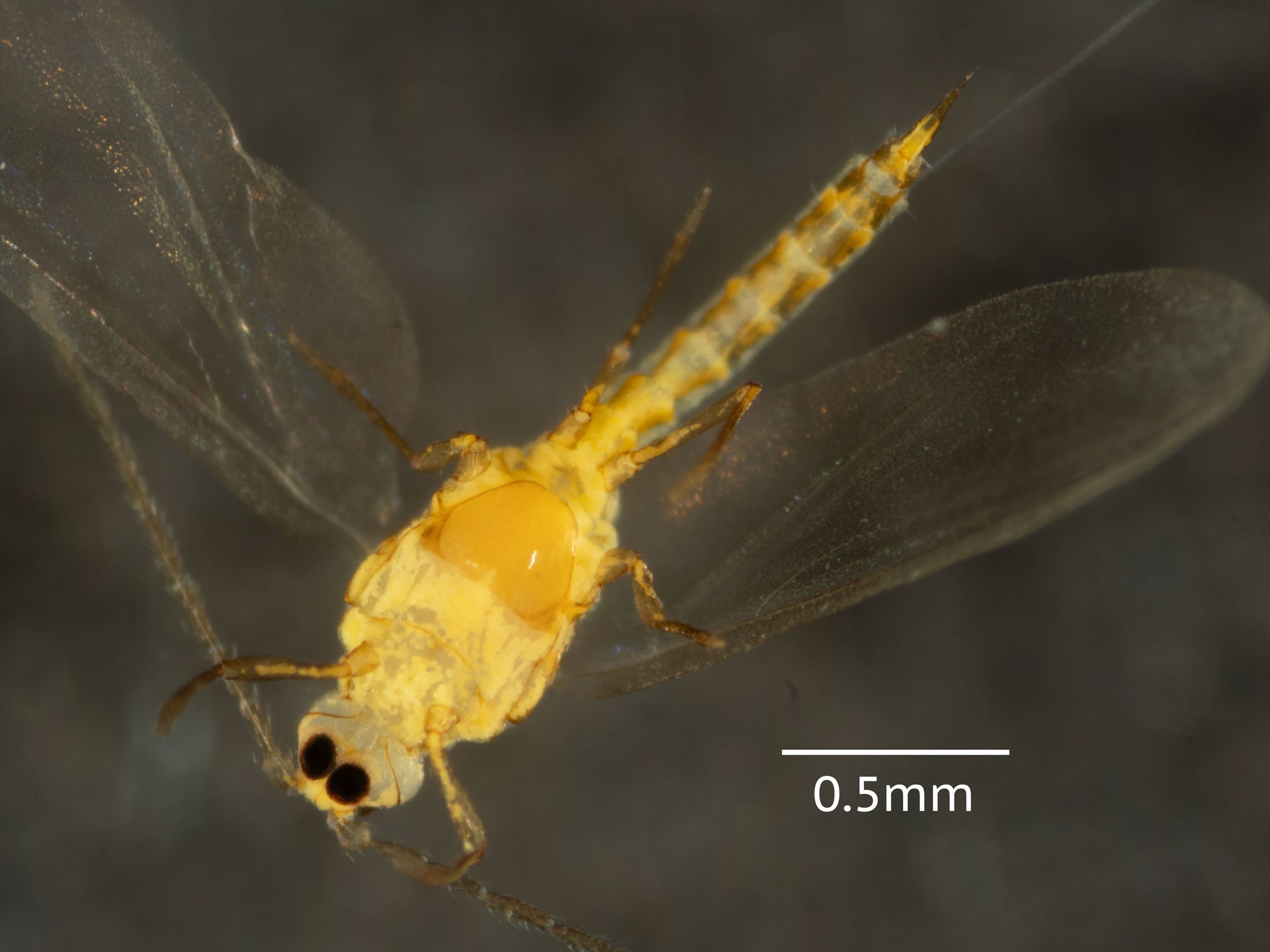
I photographed the upper surface of the male galls to show some male alates with their apical setae extended. An alate was exposed within its gall by slicing away the side wall. This individual was removed and photographed - first alive and then after being frozen. Placed in insect collection PW001.


Apiomorpha pharetrata #7
6/3/24 collected by Kerri on eucalypt, probably globoidea on home trail. She photographed the tree. I photographed the intact gall.
female gall on leaf rather than petiole, but two brown strands of tissue running across leaf surface connect gall to petiole.


An empty corpse of female coccoid found in the female gall on opening. There were 4 fly pupae in/on/around husk of female coccoid, several late stage larvae inside husk together with an active beetle larva. These were photographed. I kept these in covered petri dish to see what ecloses. 9/3/24 a single adult fly - same species as in #1 (subfamily Oscellinae) has eclosed - photographed.


After opening cells, several adult males were found, photographed, one kept in vial alive, another fixed in ethanol.


This video shows the movements of the legs and moutparts of a mite which has been restrained for viewing under the microscope.
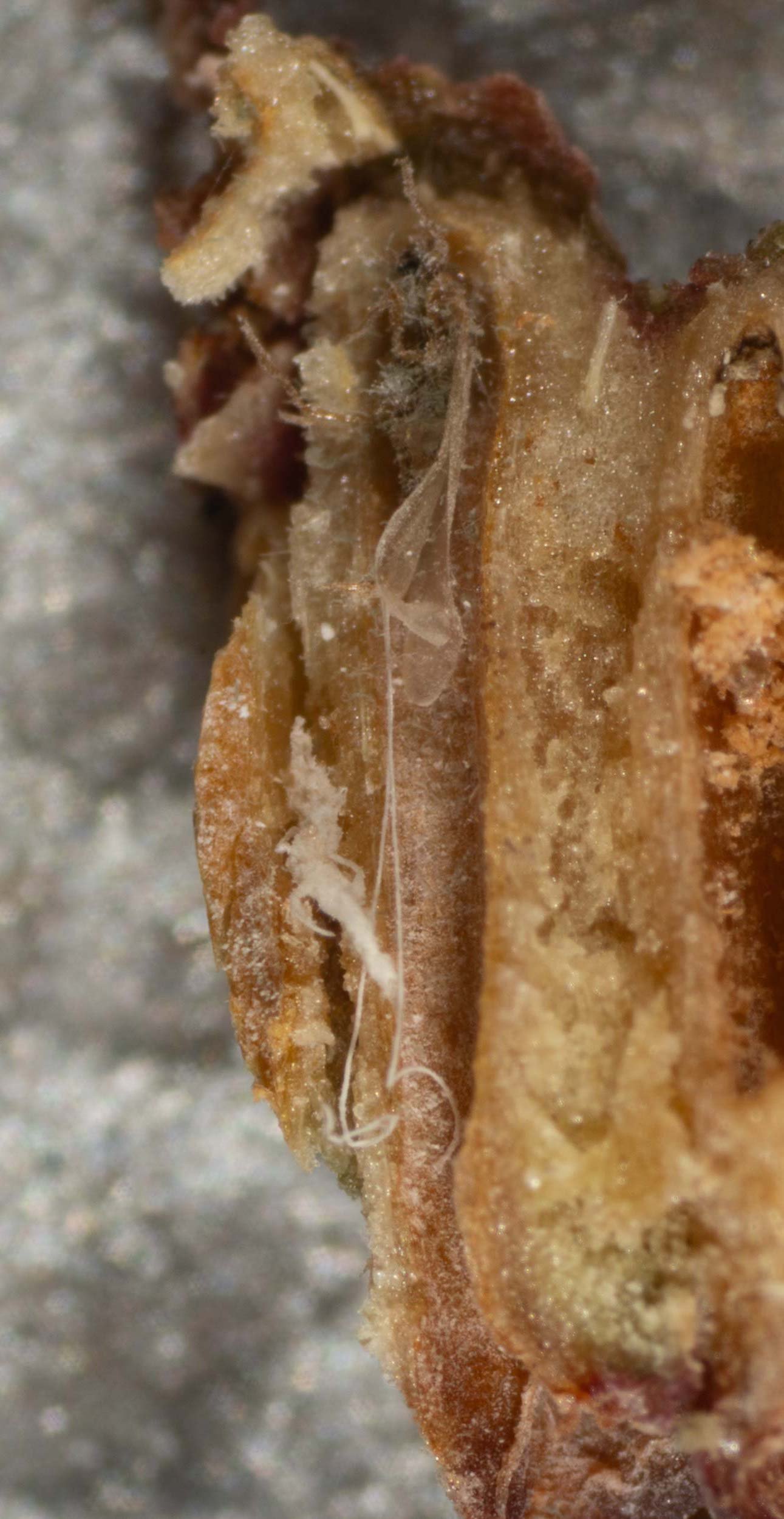
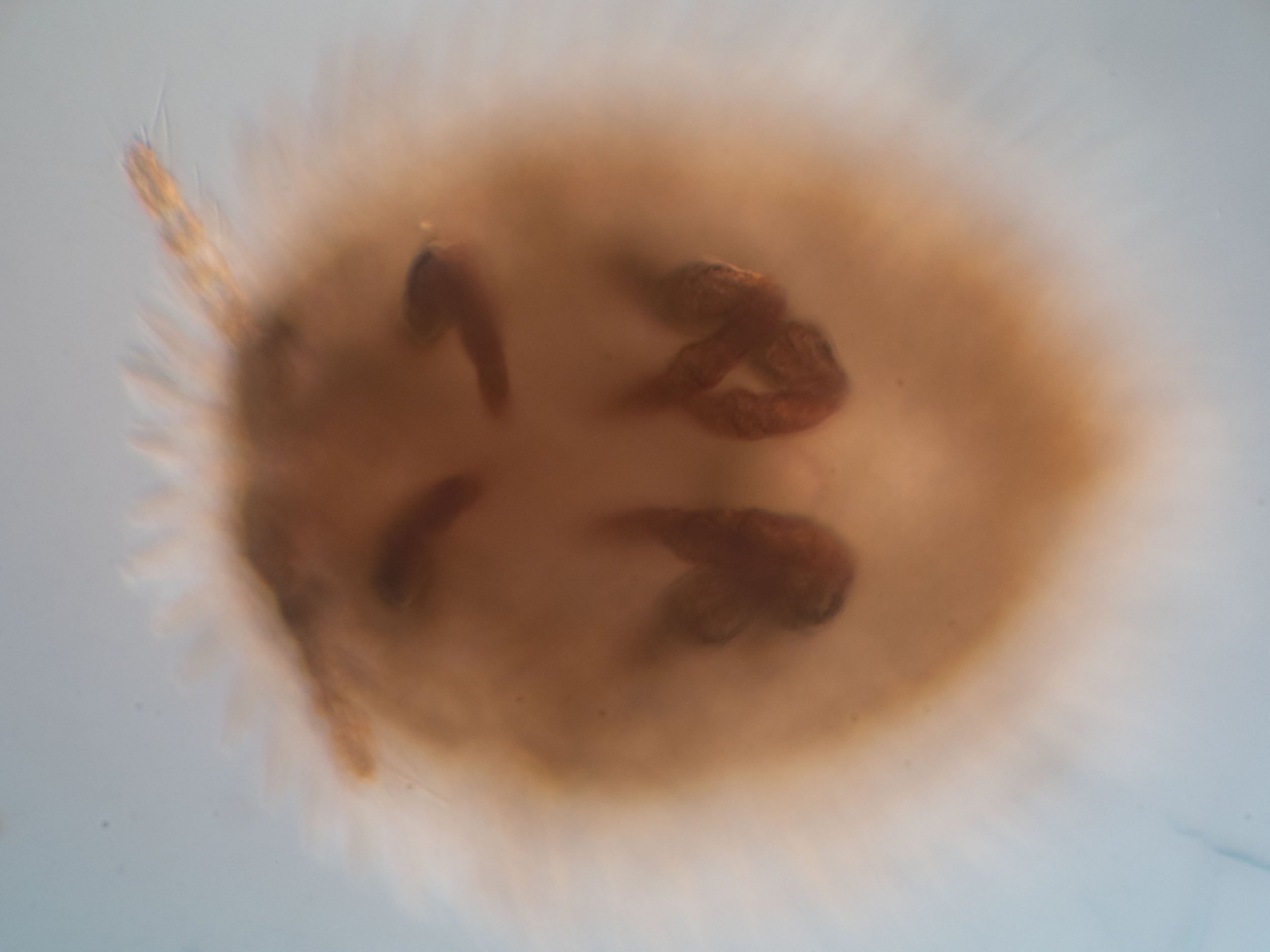
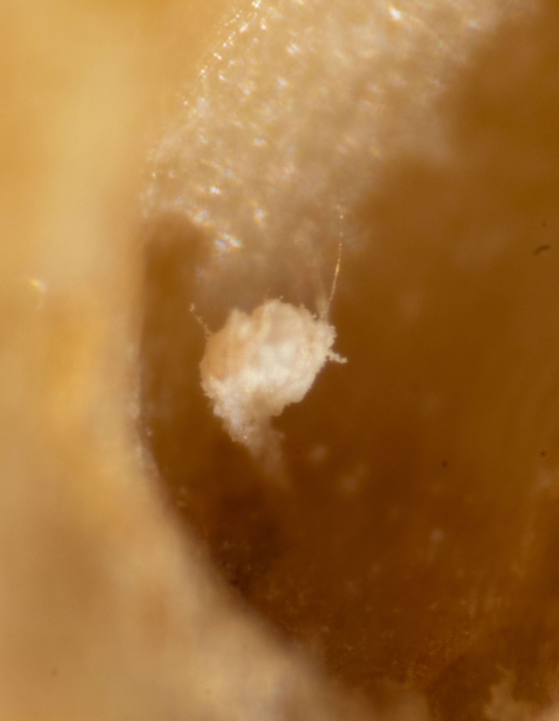
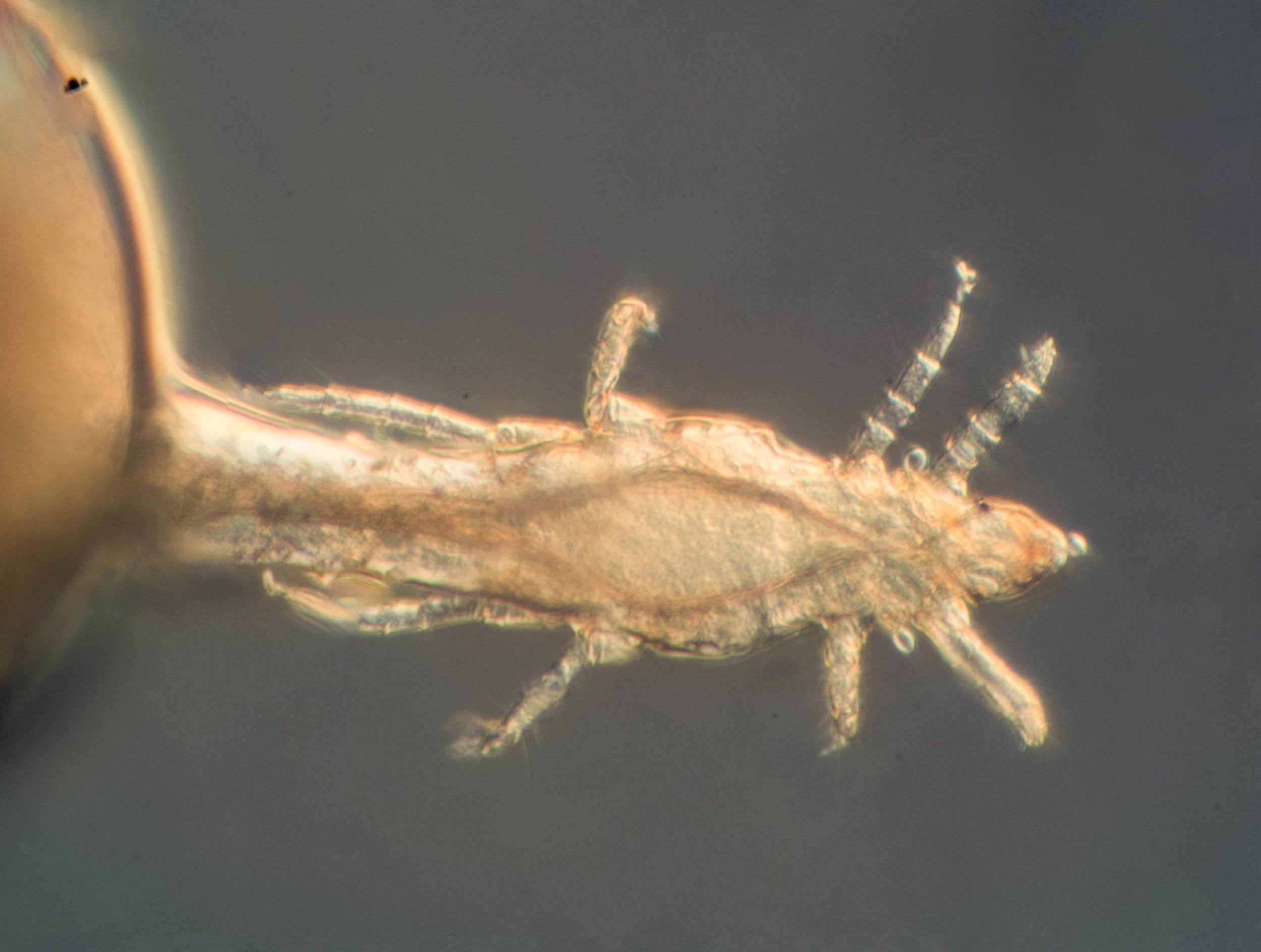
Almost all male cells had been parasitised. Several wasp pupae and adult wasps were found, but most of these were dead, parasitised by a strange “honeypot” mite. Several of these were fixed. Identified as Pyemotes sp. by Owen Seeman (iNaturalist observation).
(from Prendergast 2018) - Species in the Pyemotes group parasitise a range of hosts including bees, parasitising not only the adults but also larvae and pupae. Once a female mite attaches to the host it injects saliva containing neurotoxic compounds into it, leading to paralysis and eventual death (Klimov et al. 2016). Globally, seven species of Pyemotes have been described as being associated with bees (O’Connor and Klimov 2012). In Australia, five species of Pyemotes have been documented, of which Pyemotes ventricosus Newport was found on the non-Australian apid bee Anthophora retusa (L.) in laboratory cultures and infesting honeybee (Apis mellifera L.) colonies in Queensland (Halliday 2003b). Distinguishing Pyemotes species is very difficult (Halliday pers. comm.); none of the five species in Australia can be distinguished with confidence (Halliday 2003b). Also, the original descriptions of most species are poor (Halliday 2003b).
(from Lehrbuch der Speziellen Zoologie. Band I: Wirbellose Tiere. 4. Teil : Arthropoda)
Famiy Pyemotidae. Predators of insects and their developing young. Pyemotes. Females suck the larvae of Homoptera, Hymenoptera, Coleoptera and Lepidoptera. P. herfsi. Male 0.13-0.23mm, young females before the physiogastric stage 0.2-0.32mm long. The female feeds mainly on larvae of clothes moths, which become lame about a quarter of an hour after being injected by the chelicerae, then moving only with difficulty or not at all. The toxic effect is caused by a substance which is similar to spider venom and when isolated also affects humans. The mite kills the caterpillar by injecting the toxin and then sucks it up. Its opithosoma swells to a sphere in 7-11 days and attains the size of a small pin head (0.6-1.0mm), as the ovaries also grow massively. Embryonic and postembryonic development takes place within the mother’s body and the young are born alive. This occurs as soon as 9-12 days after the mother begins feeding on her prey. The number of offspring varies between 7 and 284, while in extreme cases over 40 can emerge from the birth canal in a single day. The females only reach their maximal size when few of them are feeding on a single caterpillar. The male do not leave their mother after birth, but remain living as parasites on her until they die (only within a week, seldom as long as a month). They feed by tapping into the mother’s abdomen and suck her body juices. Usually three or four cluster around the genital opening. If the front end of a young female appears here, one of the males turns around and grabs his sister with his pincer-shaped rear legs. It uses these to progressively lever her out of the birth canal - a forceps delivery! The males use this method only to extract females - never other males. This assistance is not required, because the first born are often females. The male does not release the female, but immediately mates with her. The young female leaves her mother after approximately 4 minutes and seeks prey. She must achieve this within 1.5-2 days, which is as long as her nutrient store lasts.
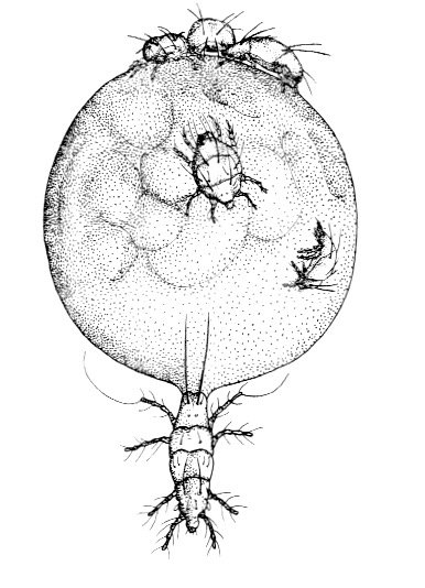
Physiogastric female of Pyemotes herfsi. Several males on her rear end. Diameter of abdomen 0.8mm. Fig. 247 Lehrbuch der Speziellen Zoologie.
Apiomorpha pharetrata #8
6/3/24 collected by Kerri on broken E. globoidea sapling behind water tank on driveway.
Only female gall present on petiole, no male galls.
empty corpse of female coccoid in gall together with 3 fly pupae and an empty puparium
Apiomorpha pharetrata #9
6/3/24 collected by Kerri on broken E. globoidea sapling behind water tank on driveway.
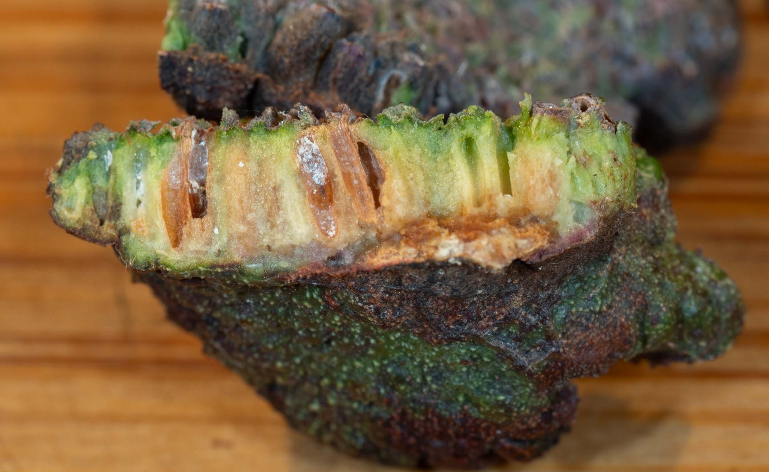
The female gall lay midway along the leaf on the midvein which is thickened proximal to gall. The large male compound gall was attached to the female gall in the usual position. There was a large amount of wax around the apical opening to the female gall. When slicing off the top section of the female gall, it was evident that the gall tissue was moist.


A living female coccoid was present within gall with many living crawlers. The female is very responsive to light touch. Crawlers began crawling out off the gall immediately. Some of these were fixed in 70% ethanol and transferred to paraffin oil after drying with a tissue and imaged under the Tessovar.


The female coccoid was imaged after removal from the gall, then placed in the killing jar for an hour. She was then treated with a detergent solution to remove the body wax and fixed in 70% ethanol. No parasites were evident in the gall after removal of the female.


Description of female coccoid from Gullan 1984
Body. C. twice as long as wide, 3.9-10.8 (7.4)µm long, 1.2-5.9 (3.7±0.9)µm wide.
Integument. Membranous except for sclerotization on anal lobes, posterior half of abdominal segment IX and posterodorsal margin of abdominal segment VIII.
Antennae. 4-segmented, 100-180 (143±16) µm long. Apex with 5 stout, long (26-40µm) fleshy setae and usually 2 slender, shorter (13-27µm) fleshy setae. 5-7 hair-like setae, 20-50 µm long, distributed on segments as follows: 2 or 3 (mostly 3) on I, 1-3 (mostly 2) on 11, 0-2 on III (mostly 0), 0 on IV.
Labium. 110-150 (131±14)µm long, 150-190 (175±11)µm wide.
Legs. With few hair-like setae on coxa and femur. Forelegs 330-500 (390±30) µm long; tarsal claw prominent. Middle legs 410-630 (480±50) µm long; tarsal claw distinct. Hind legs 450-750 (550±60) µm long; coxa and femur almost completely covered with pustules which are large in relation to leg size; tarsal claw distinct.
Spiracles. Of synlabiate type with an elongate peritreme. Mesothoracic spiracles 100-150 (120±12) µm long, 50-85 (64±9) µm wide. Metathoracic spiracles 90-160 (122±14) µm long, 50-100 (67±10) µm wide.
Abdominal segment IX. Lightly sclerotized posteriorly, slightly longer than preceding abdominal segment, 260-600 (460±70) µm wide, with posterior margin truncate ventrally, merging with anal lobes dorsally.
Anal lobes. Lightly to moderately sclerotized, 360-550 (420±50) µm long, subcylindrical, slightly attenuate subapically, rarely divergent apically. Each apex bifurcate, terminating in 2 prominent, spine-like processes, 30-65 µm long, with inner process longer than outer process. Hair-like setae, 65-250 µm long, longest on outer margins of lobes, with 1 longer (250-320µm), more robust seta originating ventrally near each apex.
Anal ring. 100-180 (126±16)µm in diameter, invaginated but clearly visible, with c. 16 ring setae, 120-450 µm long.
Venter. Without spine-like setae. Hair-like setae on all body segments, 40-350 µm long. Multilocular disc pores sparsely distributed on head and thorax, confined to anterior half of abdominal segments III-VII, rare on abdominal segment VIII, absent from IX and apex of head; pores of 6-13 locules present, 9-locular pores predominant.
Dorsum. With spine-like setae, 30-70 µm long, of similar size and in regular to slightly irregular row on segment, except rare on head and thoracic segments I and III, absent from abdominal segment IX; distribute on head and thoracic segments as follows: 0-5 (40-60) µm long on head and I; 0-14 (30-60 µm long) on II; 0-13 (30-70 µm long) with medial gap in row on III; and on abdominal segments: 0-11 (35-70µm long) with medial gap in row on II; 0-14 (30-70µm long) with medial gap in row on III; 4-15 (30-70 µm long) on IV; 7-16 (30-70µm long) on V; 9-16 (30-70µm long) on VI; 6-14 (35-70 µm long) on VII; 5-12 (30-70 µm long) on VI; 6-14 (35-70µm long) on VII; 5-12 (30-70µm long) on VIII; 0 on IX. Hair-like setae on all body segments, 50-400µm long, longest near spine-like setae on posterior abdominal segments excluding IX. Multilocular disc pores rare. confined to anteriolateral margins of thoracic segment III and abdominal segments II-VII; pores of 7-11 locules present, 9-locular poreses predominant.
several late stage male nymphs present and a few early stage ones. collected and imaged several. several late stage nymphs present and a few early stage ones. collected and imaged several.
>90% of male cells were parasitised by frit flies.
Apiomorpha pharetrata #11
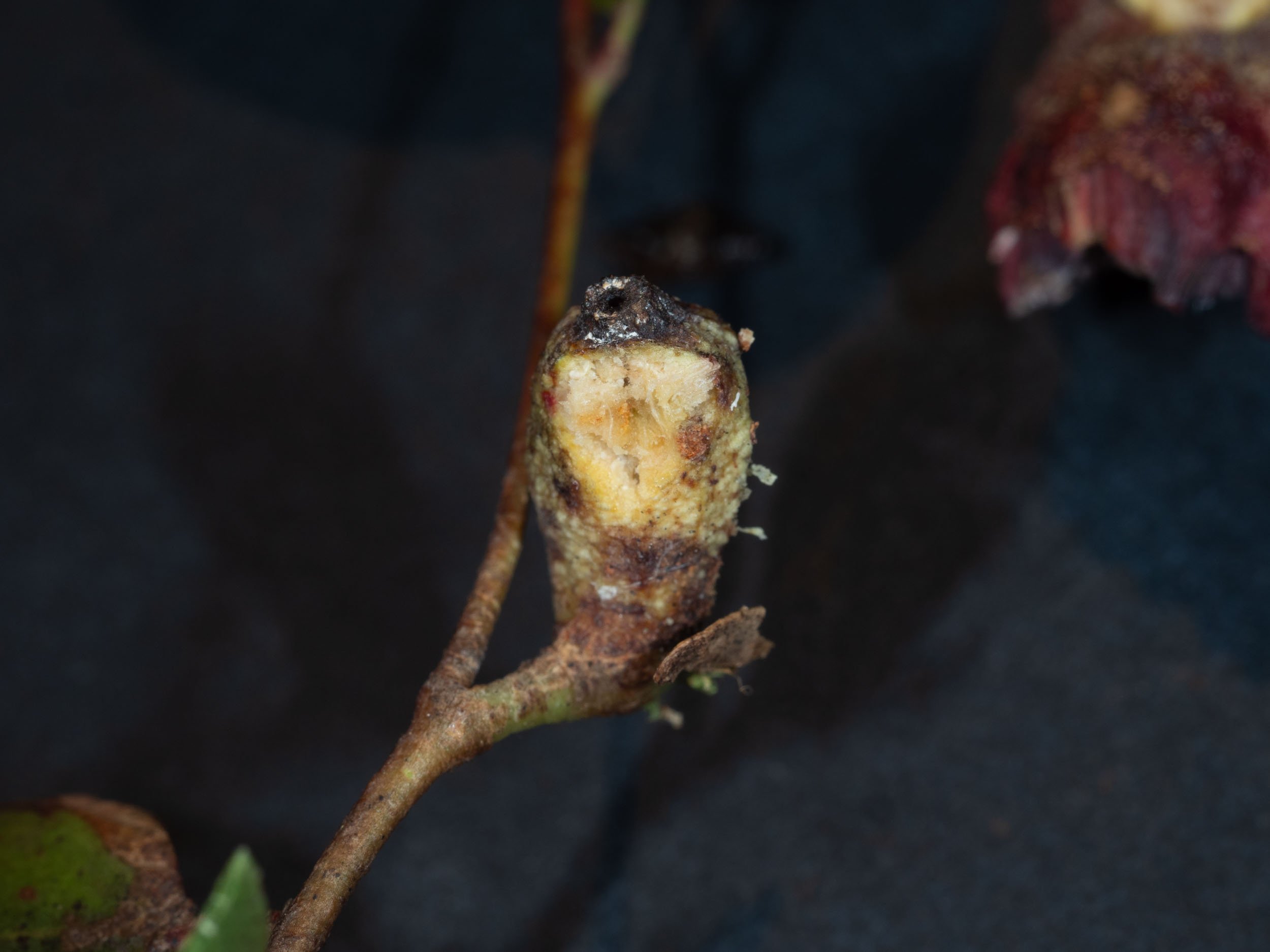
9/3/24 a compound gall was collected from an epicormic branch on broken E. globoidea on driveway behind water tank. It was in the usual position, close to the petiole of the leaf.


I sliced off the top portion of the female gall. A shrunken but not dessicted dead female was found inside. She was surrounded by fluid from her body in which several active fly larvae were found. The coccoid was fixed in ethanol, then washed in a detergent solution.


Most of the cells of the male compound gall were empty or contained fungus. Dipteran? eggs and freshly hatched larvae were found in a number. No adult wasps were present. A living mature male coccoid was found in one cell and a 2nd instar male nymph in another. The latter was removed, fixed in ethanol, transferred to a drop of paraffin oil on a slide and imaged under the photomicroscope.


Key references:
Gullan, P.J. (1984) A Revision of the Gall-Forming Coccoid Genus Apiomorpha Rubsaamen (Homoptera : Eriococcidae : Apiomorphinae) Aust. J. Zool. Suppl. Ser. 97: 1-203
Cook, L.G. & Gullan, P.J. (2008) Insect, not plant, determines gall morphology in the Apiomorpha pharetrata species-group (Hemiptera: Coccoidea). Aust. J. Entomol. 47: 51-57
Cook, L.G. , Gullan, P.J. & Stewart, A.C. (2000) First-instar morphology and sexual dimorphism in the gall-inducing scale insect Apiomorpha Rubsaamen (Hemiptera: Coccoidea: Eriococcidae). J. Nat. Hist. 34:6, 879-894
This is a workbook page … a part of our website where we record the observations and references used in making species identifications. The notes will not necessarily be complete. They are a record for our own use, but we are happy to share this information with others.