
Perginae larva breeding biology
Workbook
This page deals with the breeding biology of Perginae sawflies.
It draws on information in the published literature and iNaturalist observations.
There are 8 genera in the subfamily Pterygophorinae - Perga, Pseudoperga, Pergagrapta, Xyloperga, Cerealces, Acanthoperga, Antiperga and Paraperga.
Click the name of each genus to view a page for species identification of adults.
Larval food plants
This table from Schmidt et al. (2006) summarises the host plants of Perginae larvae. As indicated, almost all species use a eucalypt (Tribe Eucalypteae) - either a Corymbia, Eucalyptus or Angophora species.
The exceptions are Acanthoperga marlatti and Antiperga enslini, for which Acmena (Syzgium) smithii (Lilly Pilly) and Melaleuca sp. (probably), respectively are listed as host plants.
This iNaturalist observation shows eggs deposited by Acanthoperga marlatti or A. cameronii on Lophostemon suaveolens (Swamp Mahogany). Purcell & Goolsby (2005) describe how Acanthoperga cameronii oviposits on Melaleuca quinquenervia. So it appears that Acanthoperga may have a range of host plants.
Egg laying
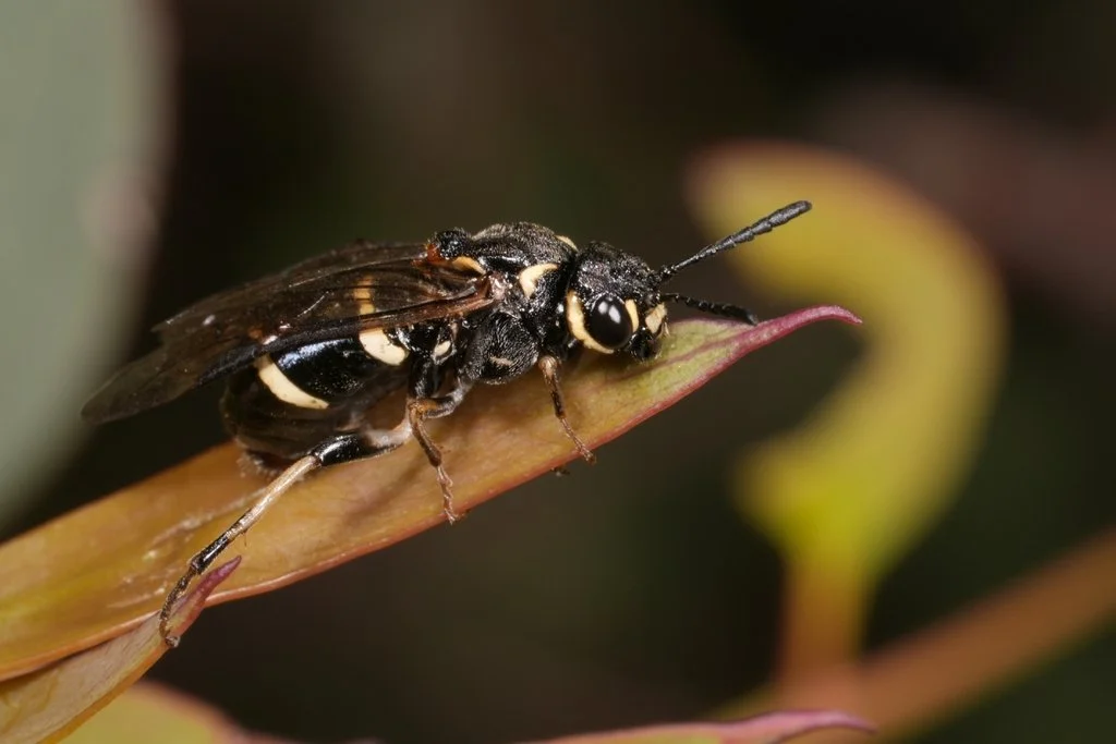
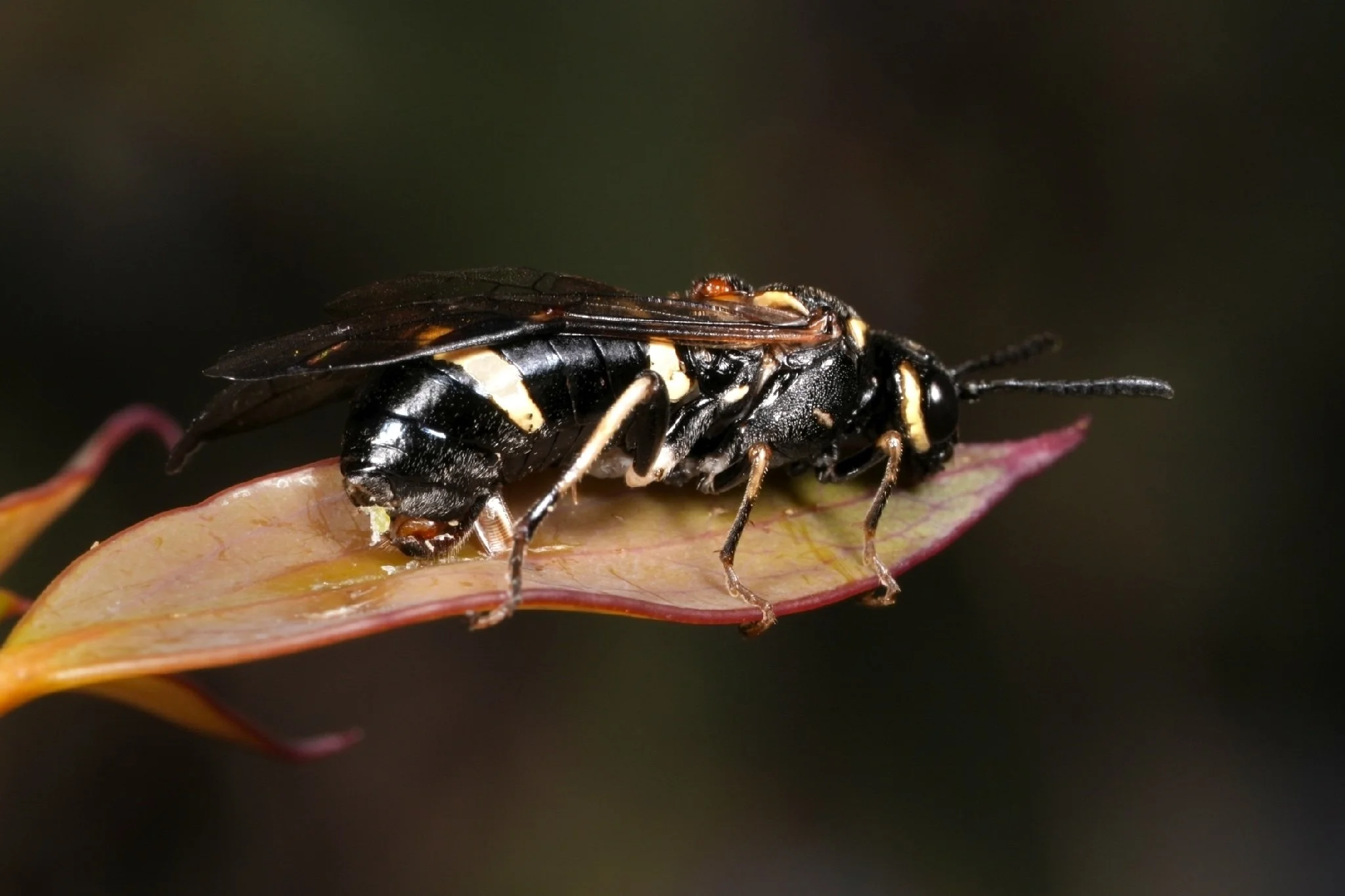
The following description of this process - by Carne (1962) - applies strictly to Perga affinis. However, iNaturalist observations show that other Perginae genera use similar modes of egg laying.
Removal of wax from leaf surface
“Most flight activity occurs in the early hours of warm autumn afternoons. After alighting on foliage, the female explores a number of adjacent leaves, walking rapidly over both surfaces, stopping occasionally, and appearing to test the surface in some way with her saw sheath. She may select one of the leaves for oviposition or fly off and inspect another group of leaves elsewhere in the same manner.
Preparation of the leaf for oviposition involves a complex behavioural pattern. Once the female begins this process she becomes quite unresponsive to any external stimuli and continues to behave according to this pattern, even if subjected to quite violent disturbance. Thus it is possible to remove shoots bearing females from a tree and to observe oviposition in the laboratory, or to take photographs involving the use of strong flood lighting.
The wasp first moves, head downward, to the leaf apex and begins to abrade the surface over a median zone some half to three-quarter of an inch in width. She grasps the leaf margins with her claws, and scrapes away the waxy surface layer by vigorous abdominal flexure, pressure being applied as the abdomen is extended”.
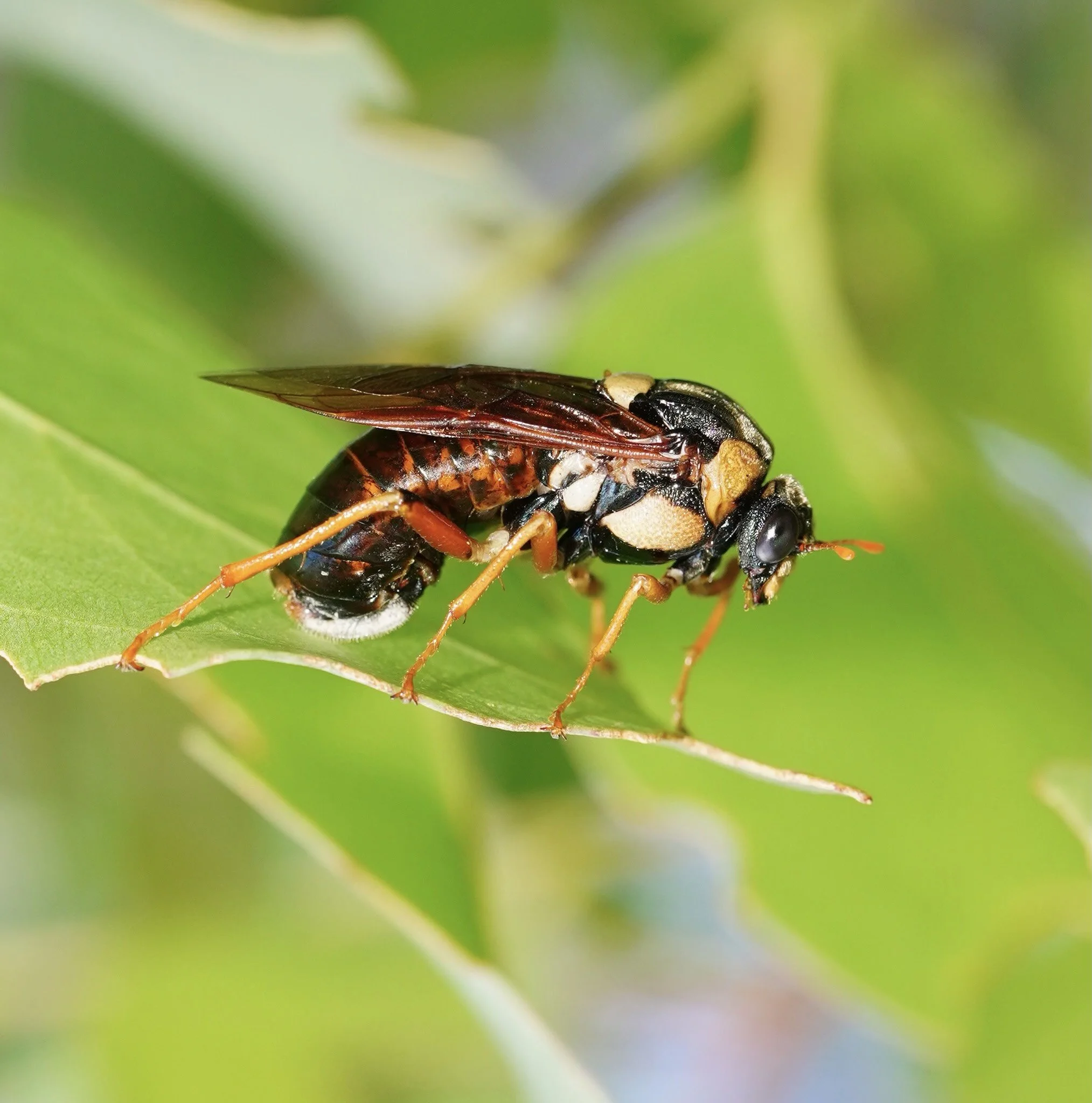
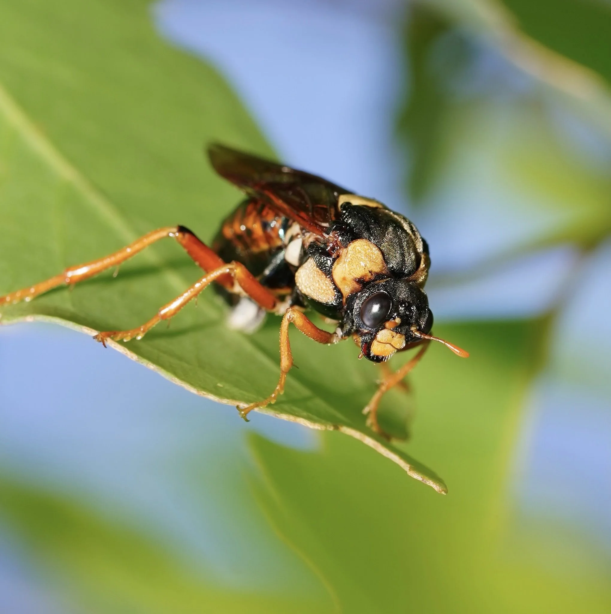
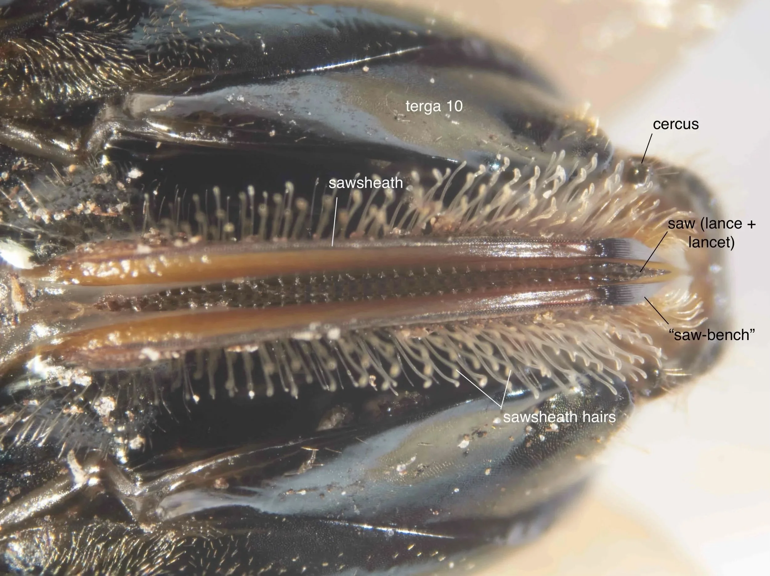
“The abrading instrument is the finely-grooved ‘saw-bench’ situated on the apical third of the sawsheath (also called ovipositor valves or valvula 3) - see Figs. 3-7 below.
The ovipositor valves bear laterally dense arrays of spatulate hairs, while their margins are transversely serrated for most of their length. Distally, these serrations are replaced by the longitudinal ridges of the ‘saw-bench’, which plays an important role in pre-ovipositional activities.
Two distinct actions are involved in the scraping process. A series of powerful outward thrusts of the abdomen produces an audible high-frequency rasping sound, as the saw-bench tears the leaf surface. These then give way to more delicate polishing motions, barely audible, during which the abraded fragments are collected by the hairs lying on either side of the saw-sheath. This comb of hairs eventually becomes thickly charged with abraded tissue fragments”.
Fig. 8 shows Perga affinis scraping the leaf surface.
This video from zosterops99 of Pergagrapta bella and this one from bigwigger of an undescribed Perga sp. from WA (probably a melanistic form of Perga schiodtei) show these species carrying out the same activity.
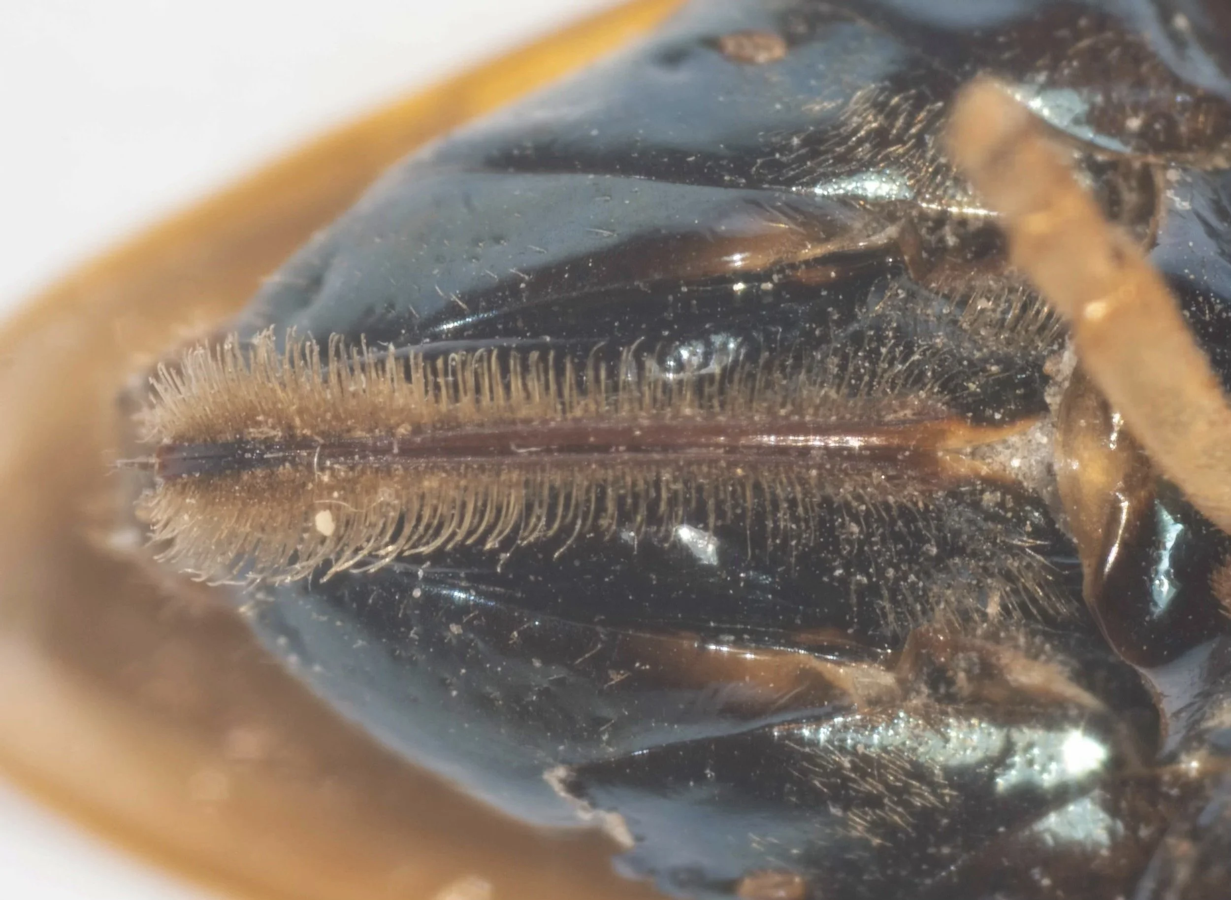
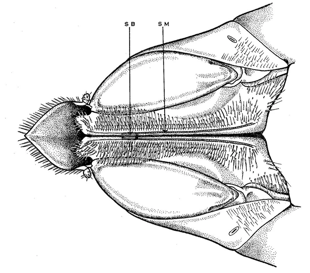
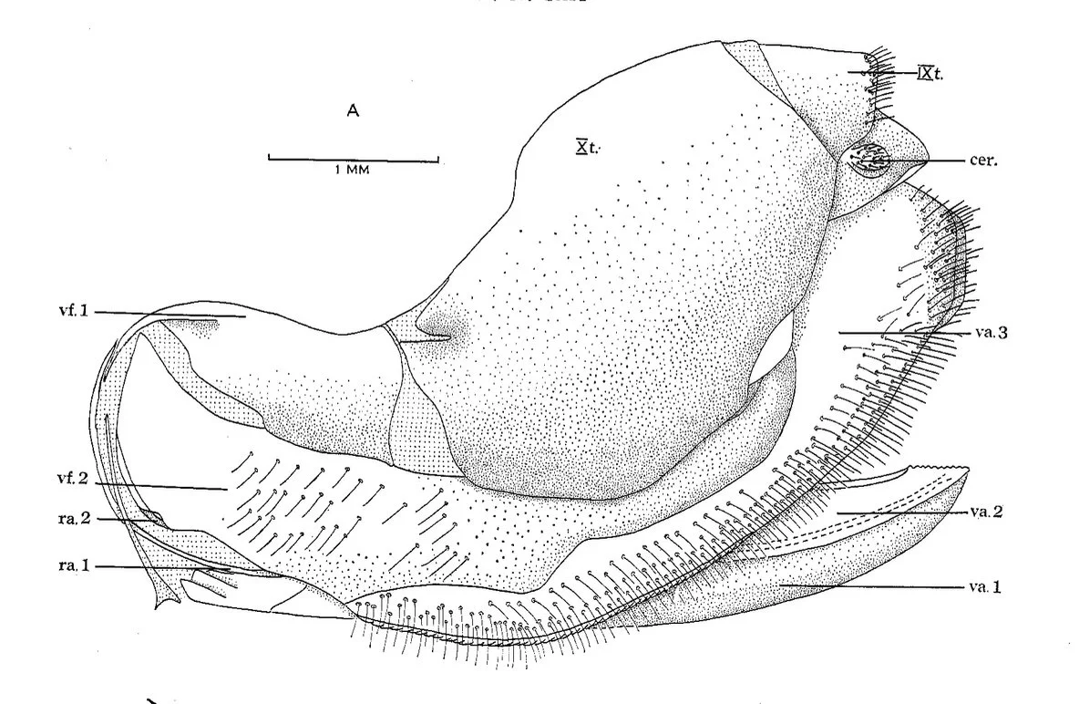
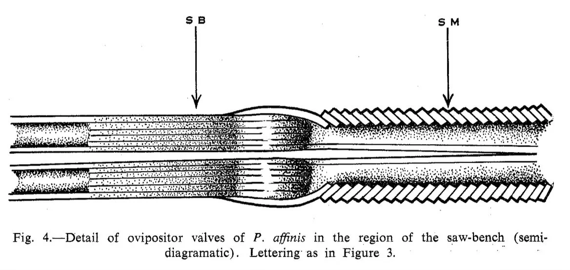
“As part of the leaf is cleared of surface wax, the female moves backwards up the leaf and begins to scrape a fresh area slightly overlapping that already treated. She continues this process to the base of the leaf, or to a point at which the width of the leaf blade exceeds her leg span. After remaining motionless for 0-15 min, the female moves down to the leaf tip, and treats the other surface in the same manner. Having worked over both sides of the leaf, the female repeats the process several times.
The whole operation, including resting periods, varies in duration from 2-4 hr. As the peak of daily flight activity occurs in the early afternoon, oviposition rarely begins before 4-5 p.m. Some females are not ready to oviposit until dusk, when decreasing temperatures bring about the cessation of all activity. Such individuals do not resume activity until early the following morning. Thus the process of oviposition is most frequently observed in the early morning or late afternoon”.
2. Oviposition
Perga affinis - Carne (1962)
“The female moves backwards up the leaf to the basal limit of the abraded area. The ovipositor is withdrawn and held at right angles to the main body axis, the margins of the leaf are usually grasped by the female as in the preparative stage, the point of the ovipositor being placed against the leaf surface beside the midrib, and inserted.
The ovipositor is extended and held at right angles to the main body axis during oviposition. It consists essentially of two pairs of valvulae, the outer pair being fused ventrally and formed distally into a serrated lance. The inner pair (the lancets) are separate, each being attached to an inner face of the lance by means of a virga; their dorsal margins are provided with a series of fine teeth.
When the ovipositor has penetrated the upper surface, the lancets begin to operate and the whole apparatus is bent through 90°. The ovipositor cuts through the midrib and passes into the tissues on the opposite side of the leaf.
By means of powerful muscles, the lancets may be caused to reciprocate within the virgae. The ovipositor is thus well adapted to its function of inserting eggs into the tissues of leaves. The lance facilitates the initial entry of the ovipositor into the leaf; the sawing action of the lancets permits an appropriate cavity to be prepared for the reception of the egg which subsequently passes down a duct formed by the two pairs of valvulae.
When cutting the vascular bundles, the lancets make a distinctly audible vibration which becomes louder and of lower frequency when they encounter the midrib. The motion of the lancets then slows down and an egg is placed in the cavity formed; the lance is then gently withdrawn, the tissues of the midrib close, and the egg lies in a virtually sealed cavity.
The wasp then moves downward a very short distance and repeats the process. As the ovipositor opens up a cavity wider than the distance between each incision, the eggs lie freely in a continuous row, or "pod", and are not isolated by portions of uncut tissue..
During oviposition, large drops of a viscous greenish yellow fluid accumulate on either side of the abdomen near the base of the lance (see Fig. 10 of Pseudoperga guerinii). These subsequently harden to a resin-like substance and become detached. The fluid, a product of the accessory glands, may serve to lubricate both the lancets and the egg duct, and possibly to seal the leaf incisions.”
Oviposition by other Perginae
Fig. 11 by flynnprall1 and this movie by rewildingsuburbia show Perga schiodtei ovipositing. The saw can be seen inserted into the leaf on one side of the midrib. Mel Logozzo reports that a female Perga schiodtei commenced laying at 13:30 and finished at 16:45.
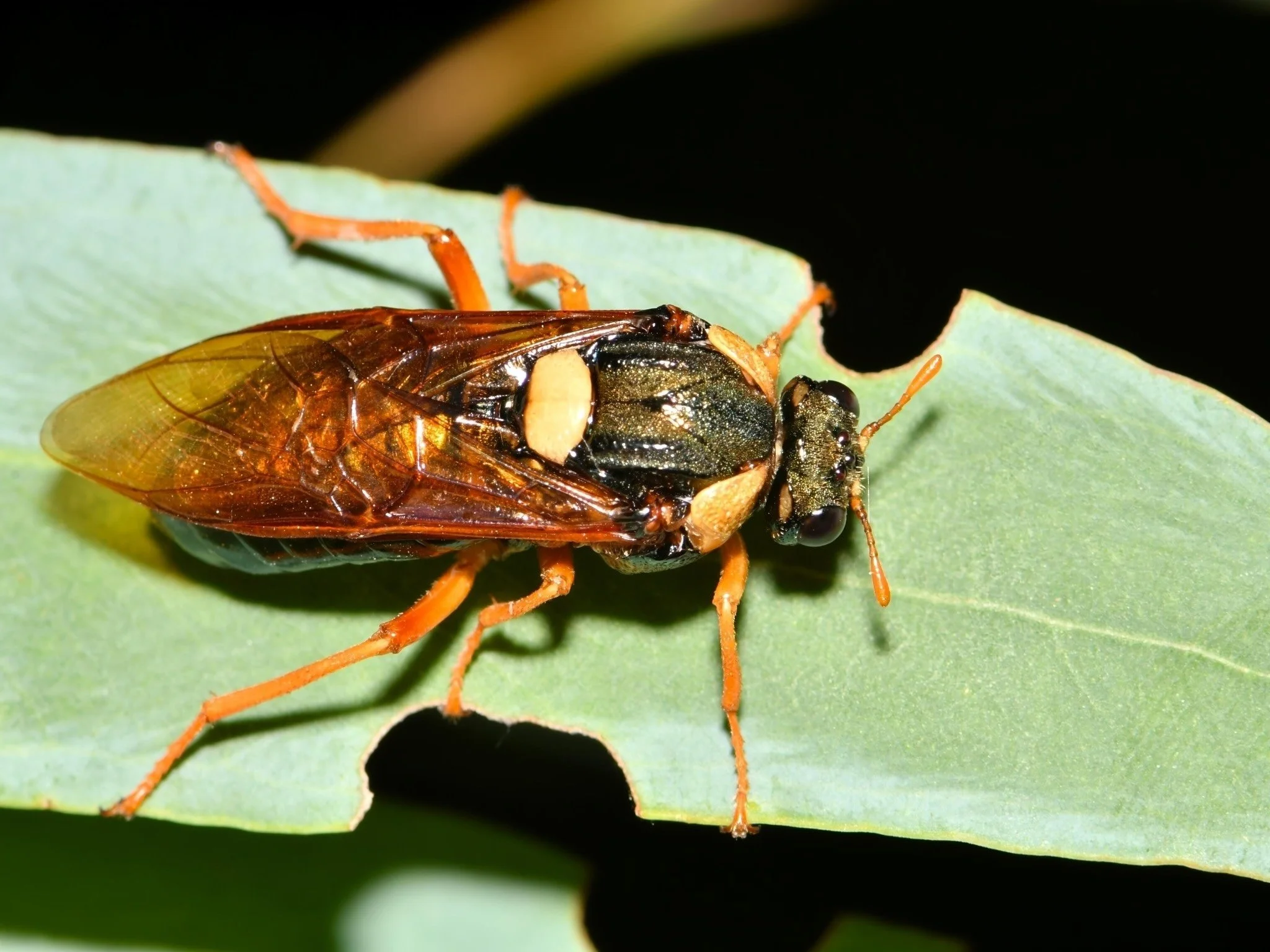
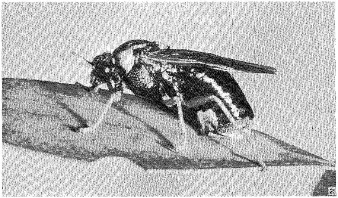
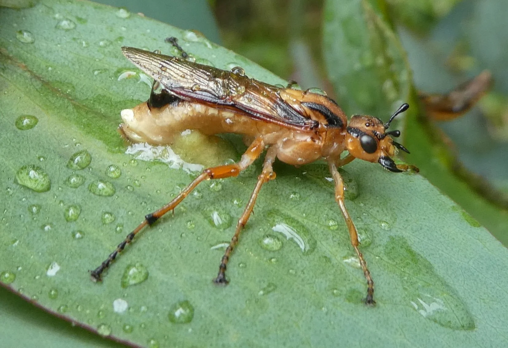
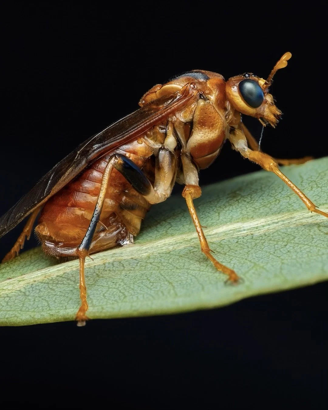
Xyloperga amenaida uses a variation of this method when ovipositing on Eucalyptus pauciflora (Snow Gum) leaves.
In this case, the female does not appear to abrade the leaf surface. Rather she grasps the leaf margin with her legs and then advances along the leaf, scoring the surface with her saw to make a line parallel to the leaf edge (Fig. 12, 13).
She then moves along that line from its starting point, inserting her ovipositor beneath the cuticle to deposit a row of eggs (Figs. 14-16).
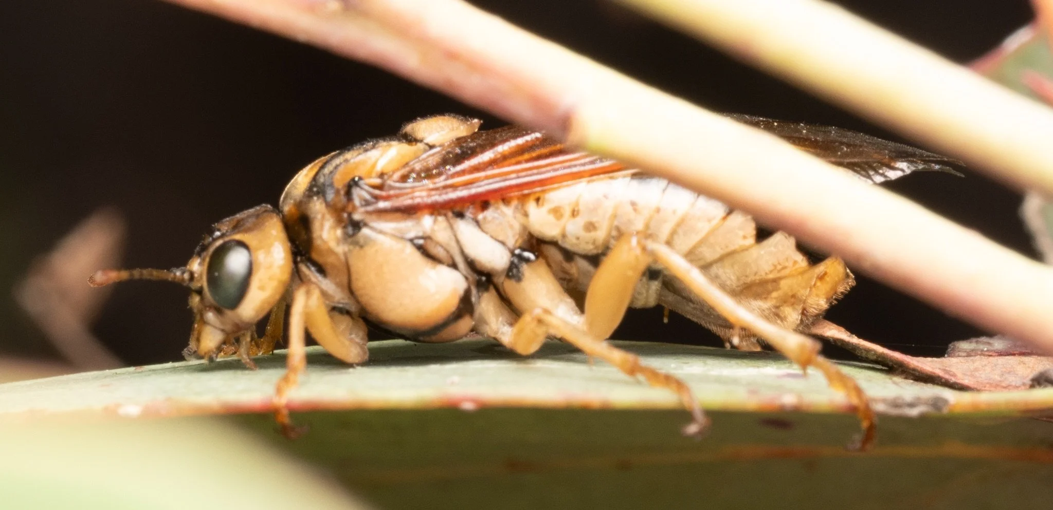
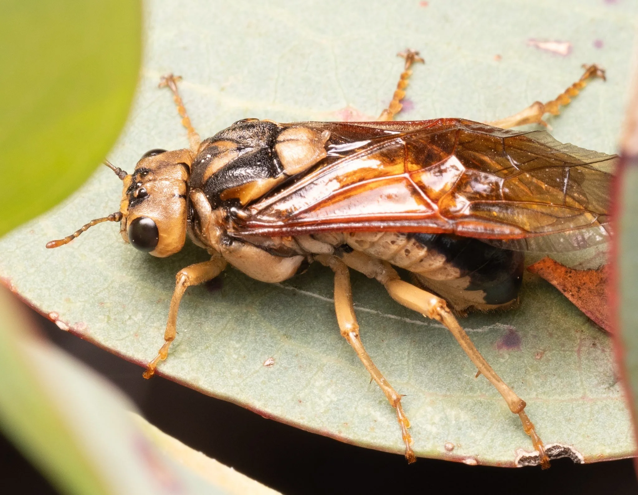
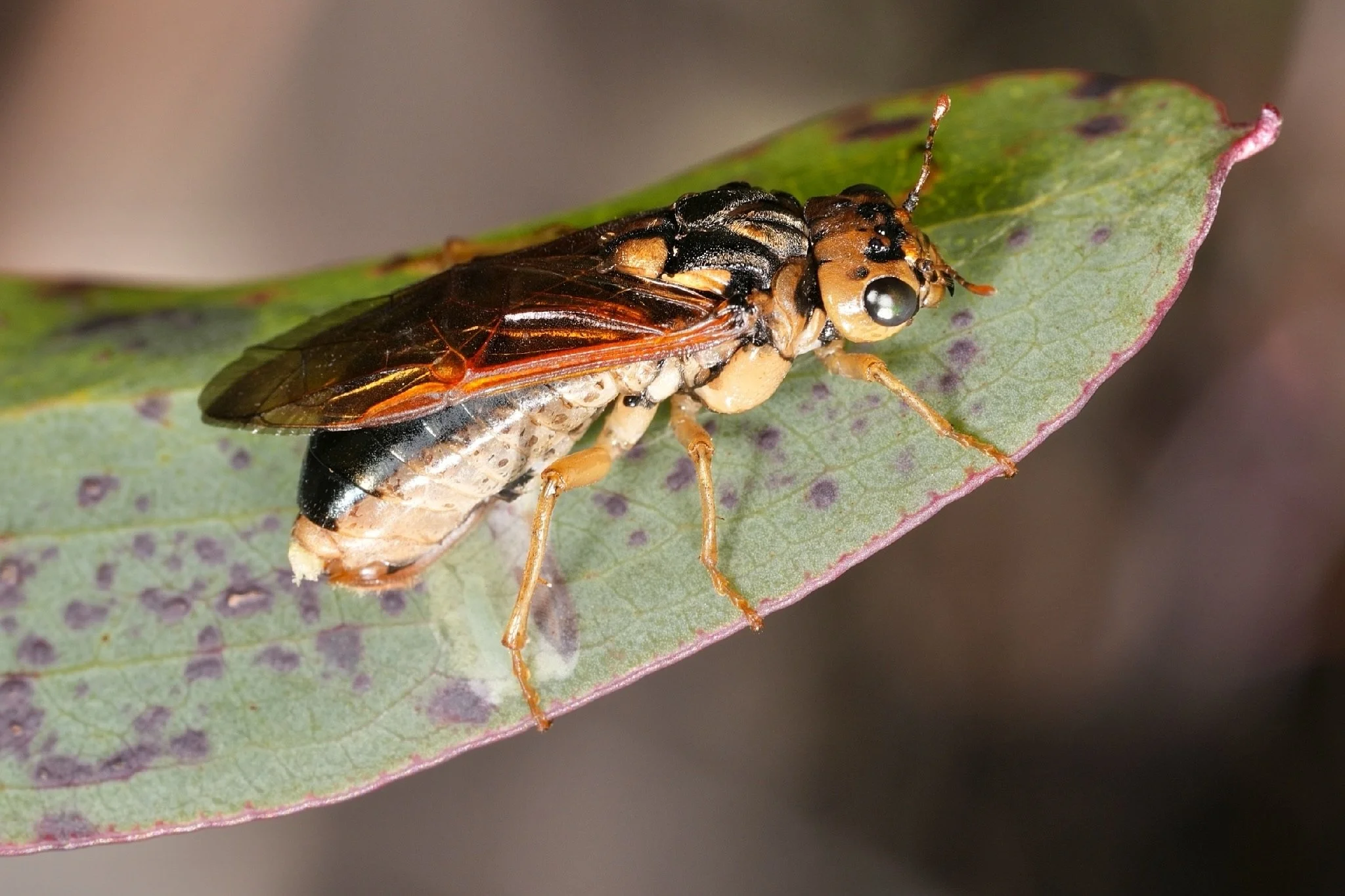
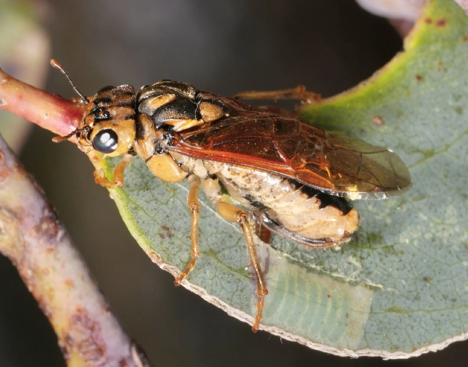
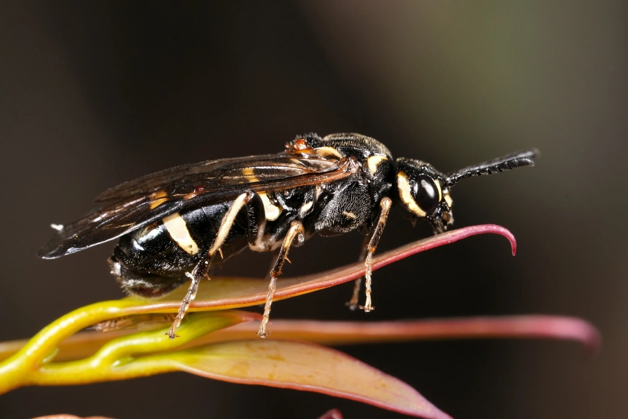
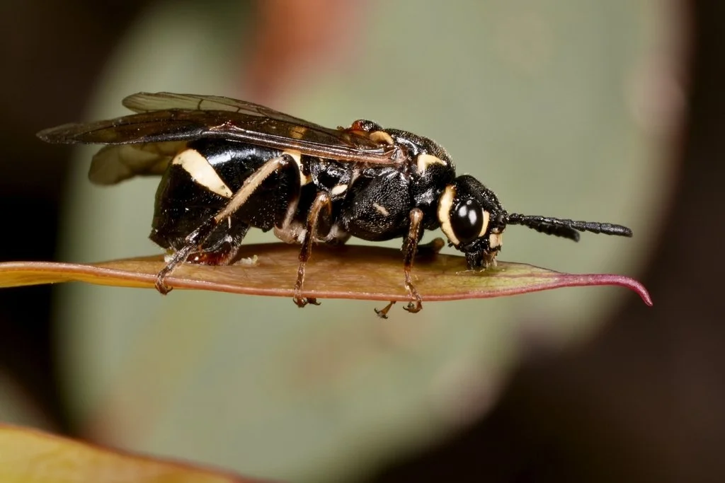
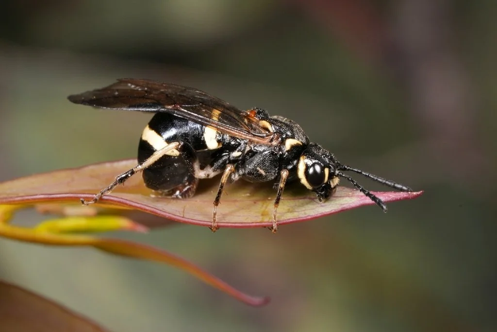
Cerealces scutellata females use a similar method to Xyloperga amenaida when ovipositing into Eucalyptus pauciflora leaves, as seen in the following panel of images. However, younger, fresh leaves are favoured.
The female grasps the edges of the leaf as she moves forward towards its tip, making a cut in the surface (Fig. 18-20). The narrow width of the young leaf results in that cut line being placed close to its midline.
She then returns to the start of the cut line and pushes her ovipositor beneath the epidermis to begin the process of egg laying (Fig. 21).
Egg arrangements in Perginae
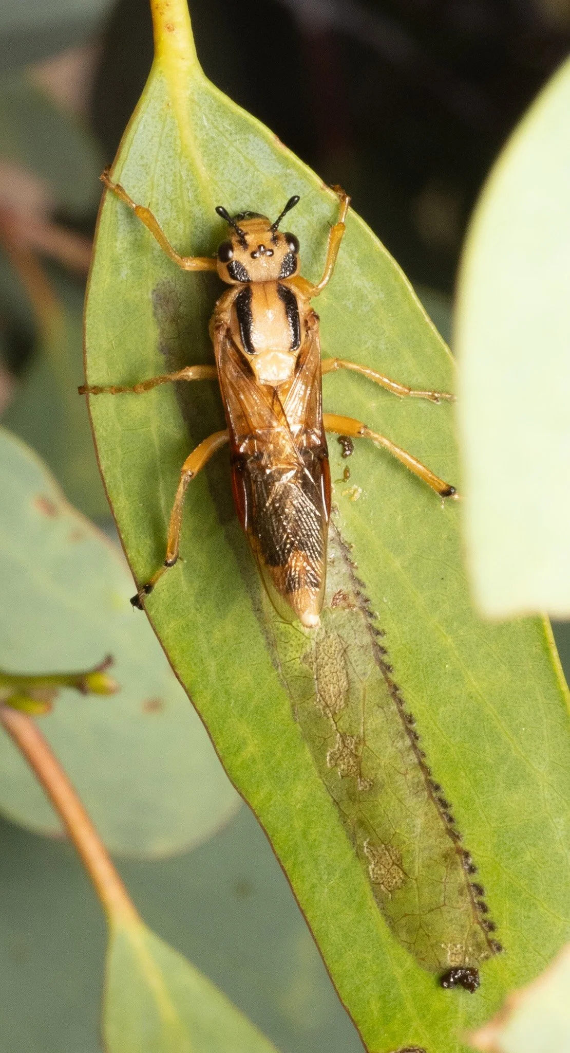
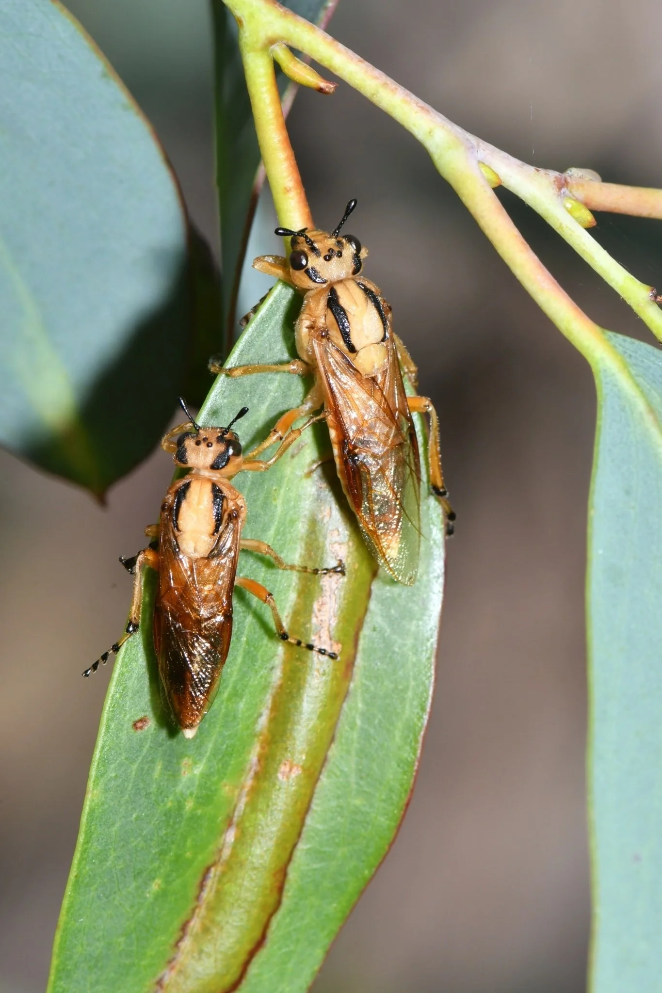
iNaturalist observations of Pseudoperga guerinii, Pseudoperga lewisii and Pseudoperga belinda females suggest that these species generally lay a single row on one side of the midrib (Figs. 22-26, 30). The initial points of incision are evident as a regular series of dark marks angled at 30° to the midrib.
On the other hand, Lewis reports (Westwood, 1836) that P. lewisii “places eggs transversely in a double series” and Macdonald and Ohmart (1993) state that “females of Pseudoperga lay eggs on both sides of the midrib in a single oviposition site.” Furthermore, iNaturalist observations show Pseudoperga ferruginea females laying a row of eggs on each side of the midrib (Figs. 27-29).
This aspect of oviposition behaviour thus appears to vary both within a genus and species.
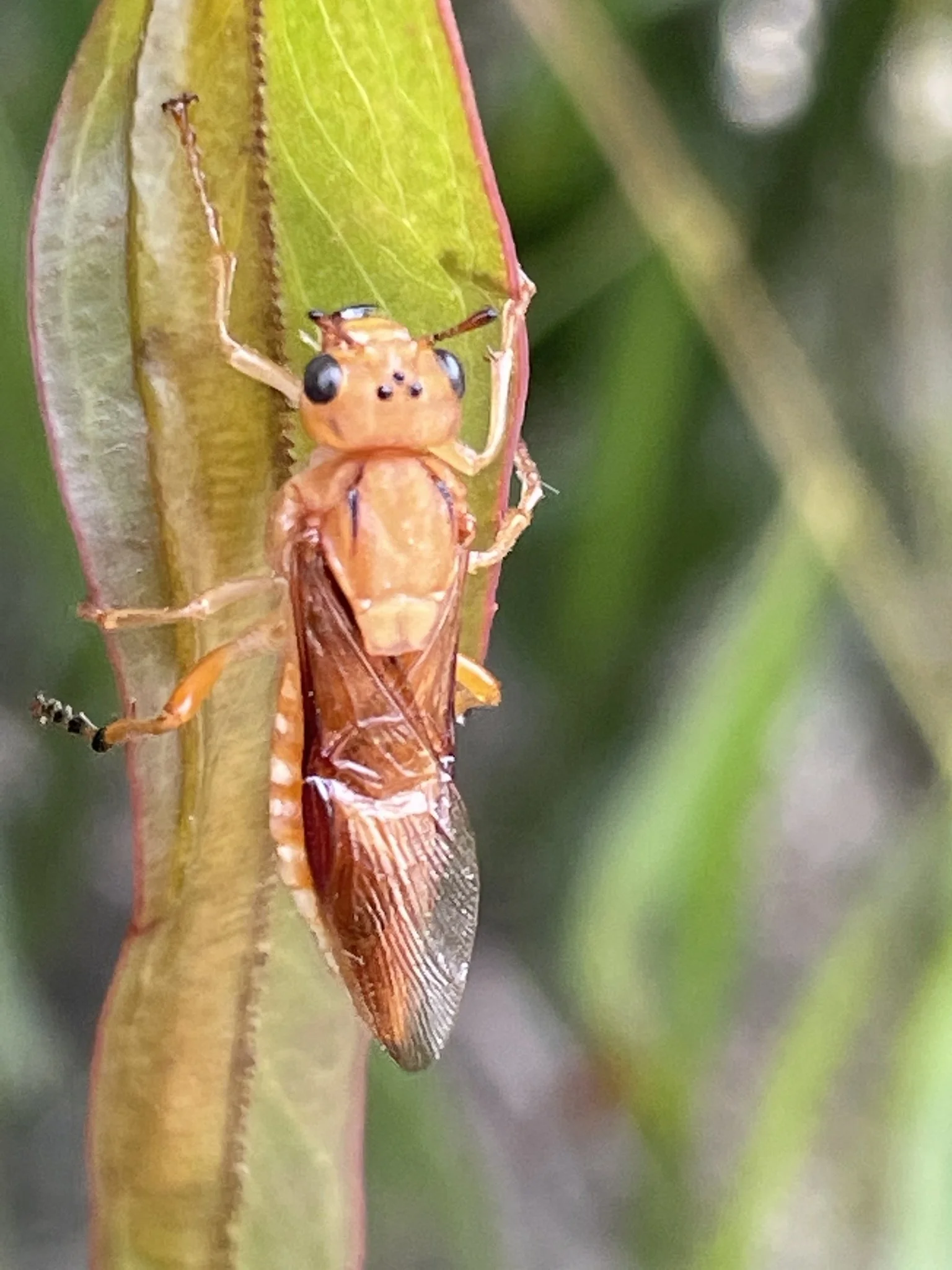
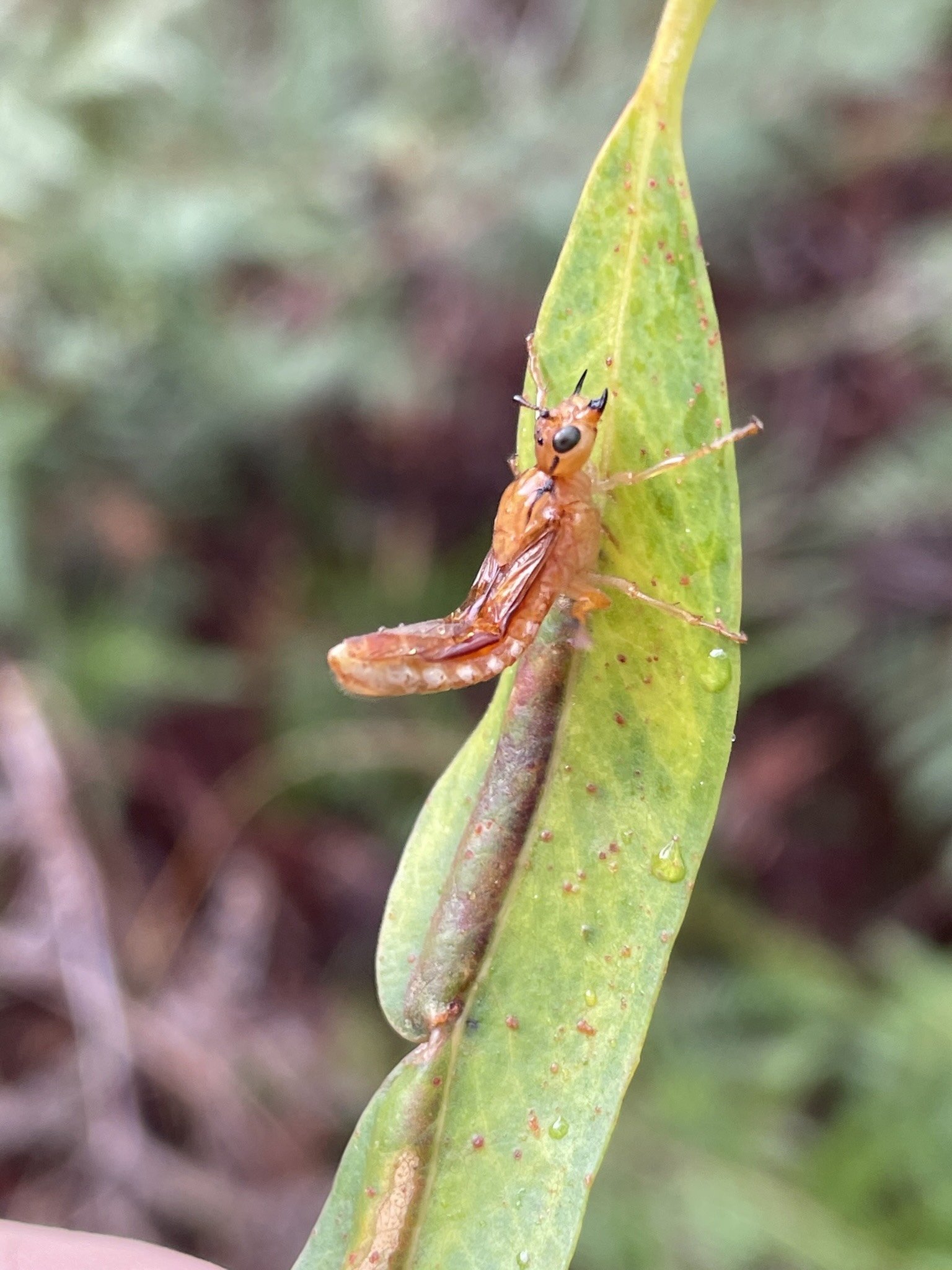
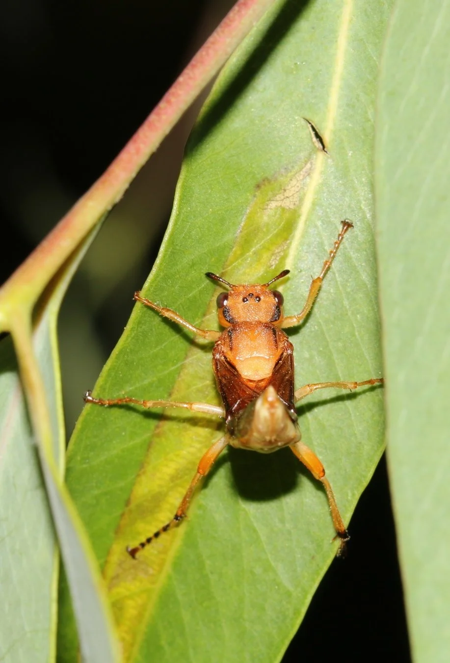
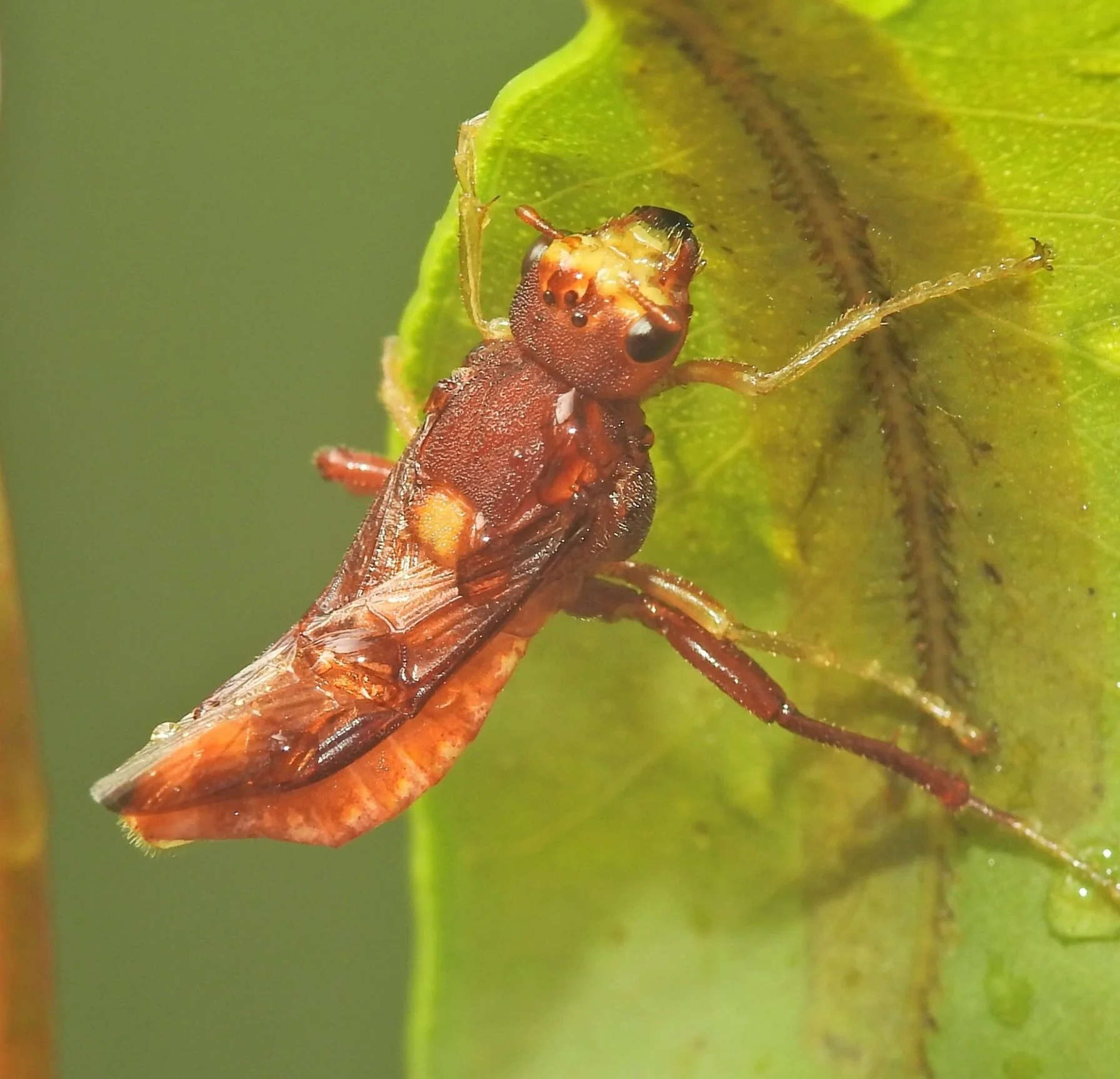
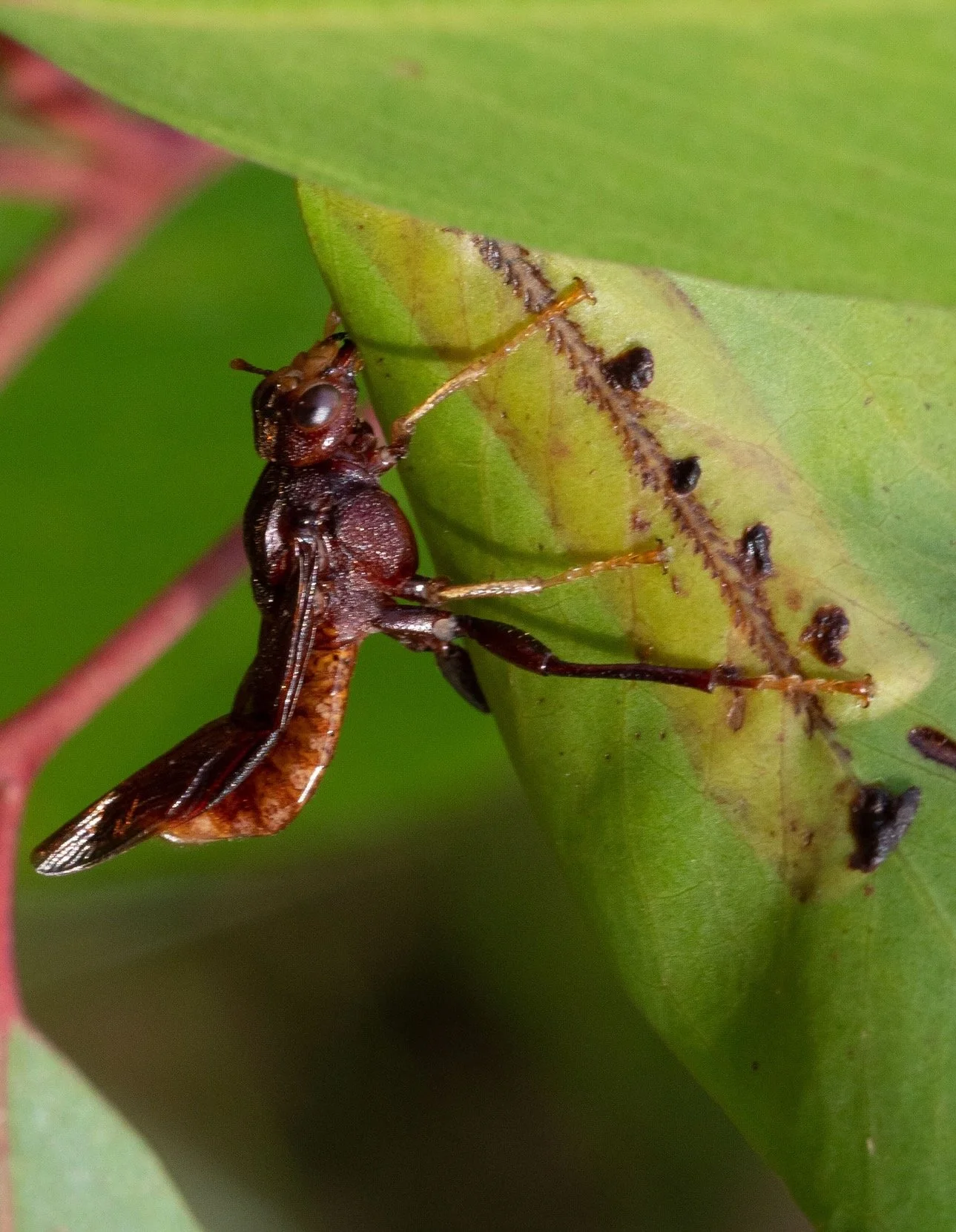
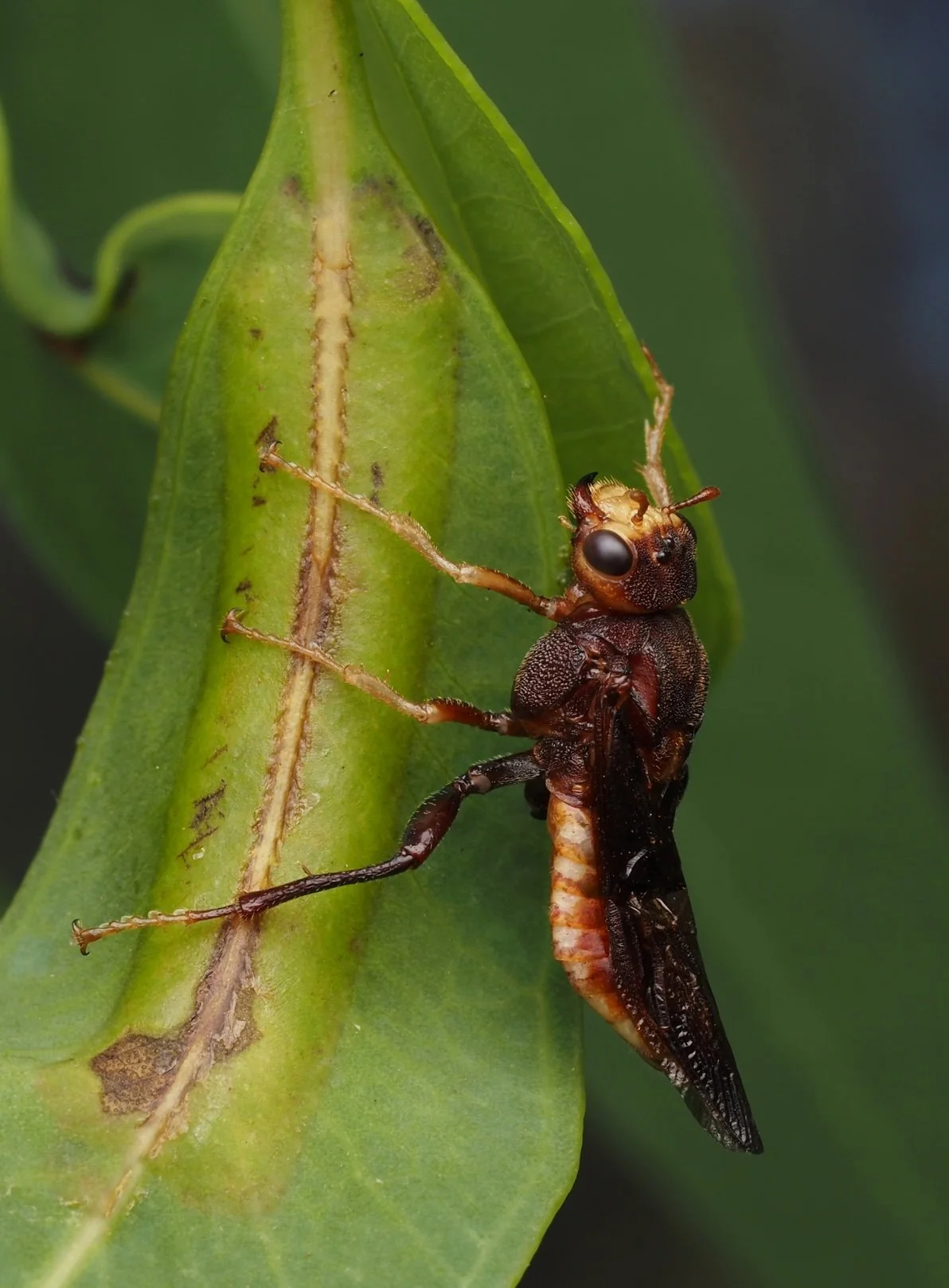
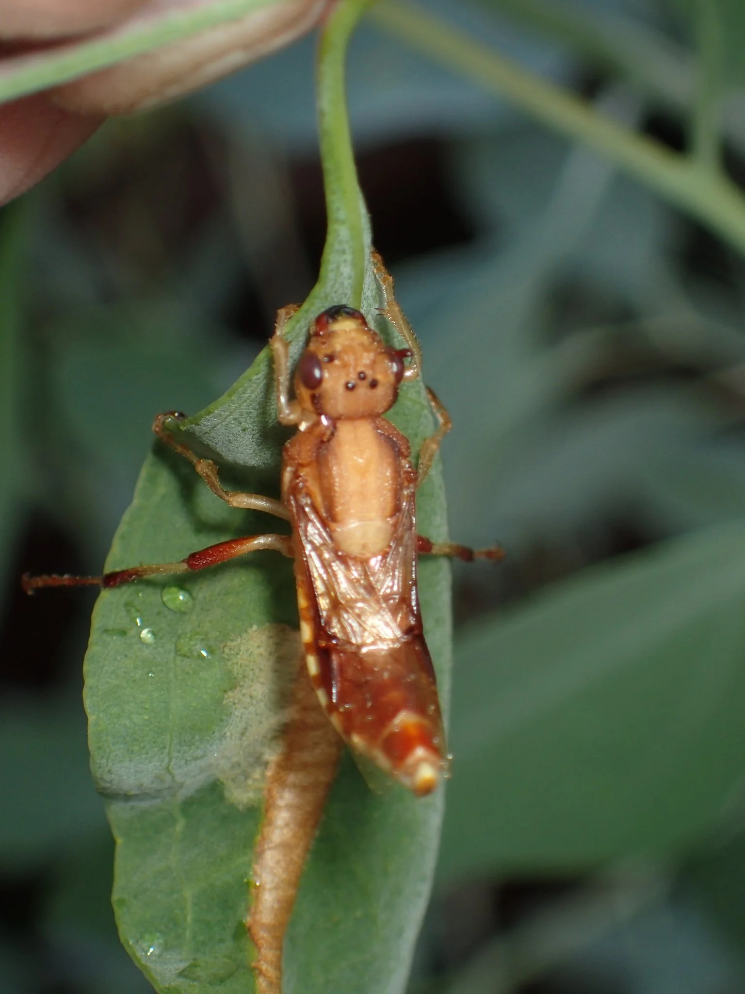
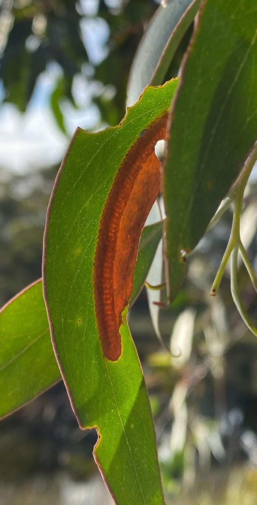
Pergagrapta bella (Figs. 31-32), Acanthoperga cameronii (Fig. 33) and Perga schiodtei (Fig. 34) lay a single row of eggs.
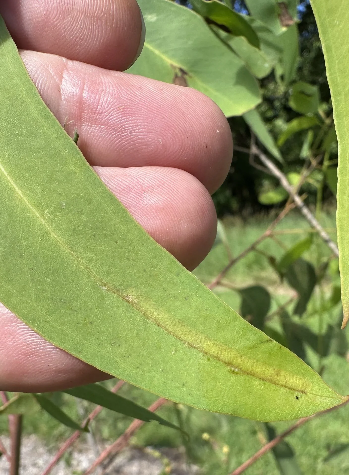
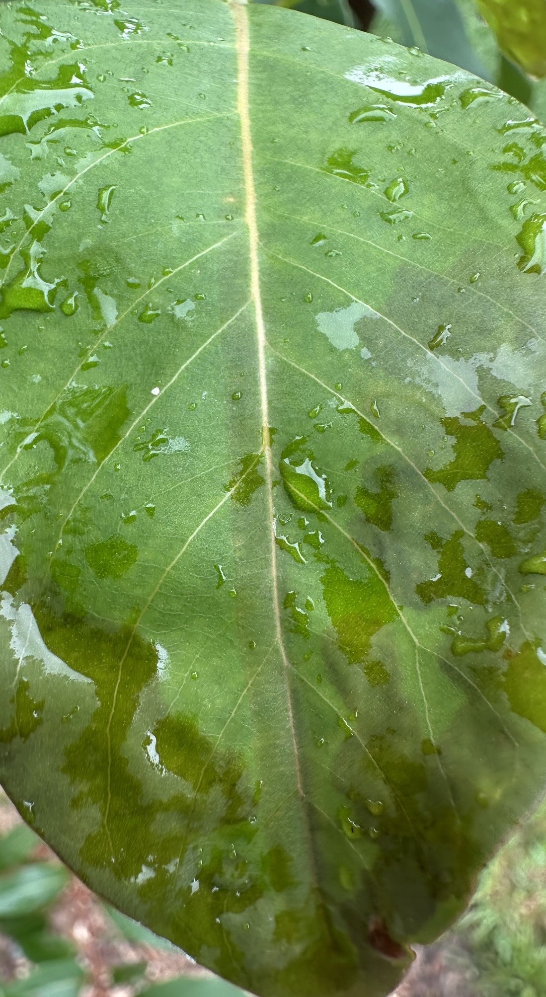
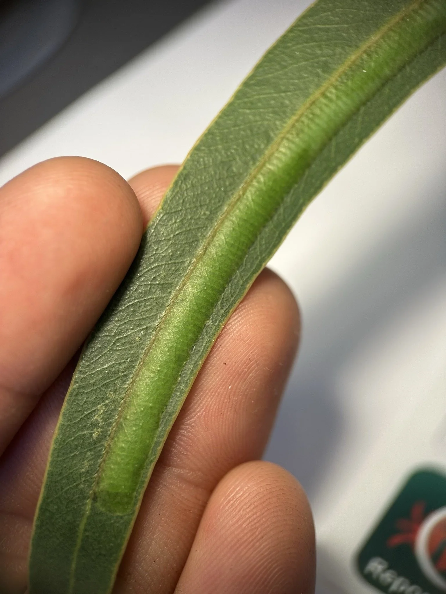
Inside the egg pod
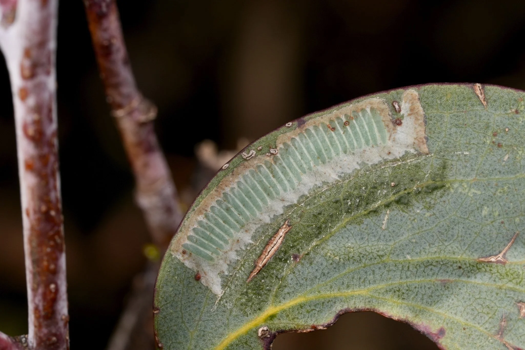
Carne (1962) “The eggs of Perga affinis are long, slender, slightly sigmoidal in shape, with a thin, unornamented chorion. They are laid at very regular intervals along the pod (i.e. the portion of the leaf adjacent to the midrib where eggs have been laid and which is visibly distended by their presence - Fig. 35). The eggs lie freely in the pod cavity and, as they develop, they increase in diameter and come to occupy fully the available space.”
This same arrangement is seen in other Perginae egg pods (Figs. 36, 37).
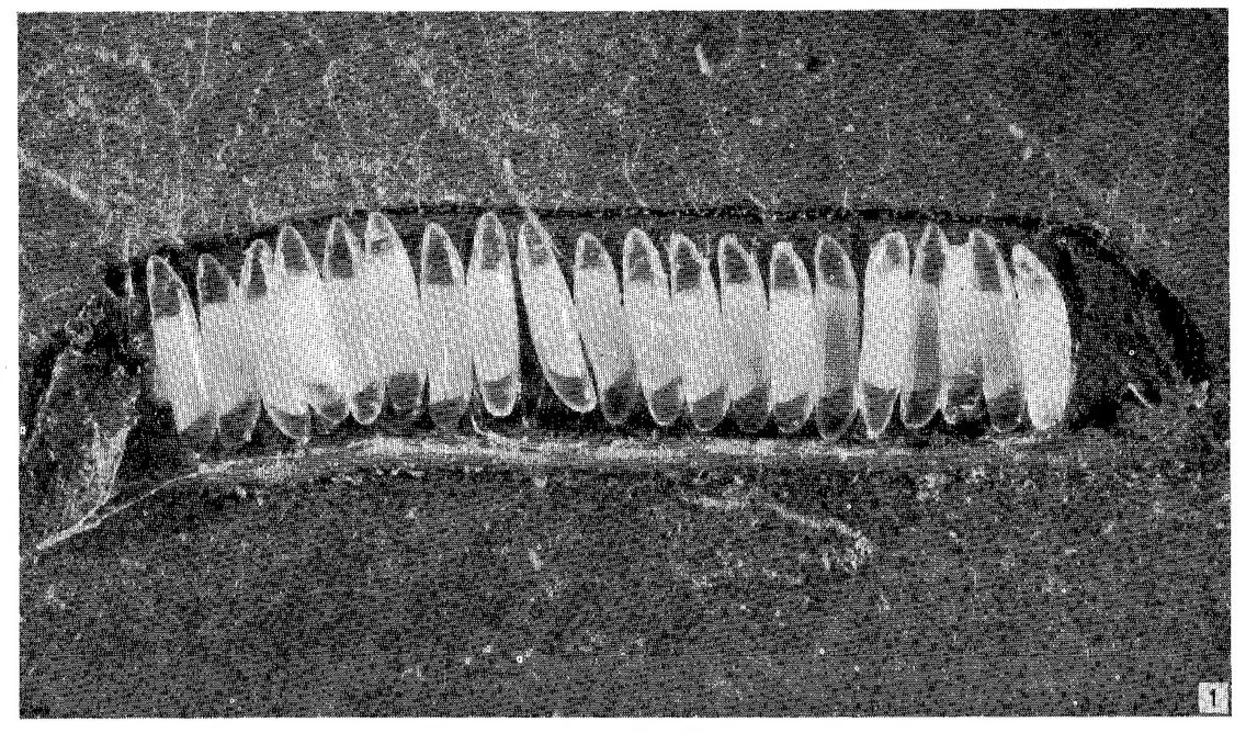
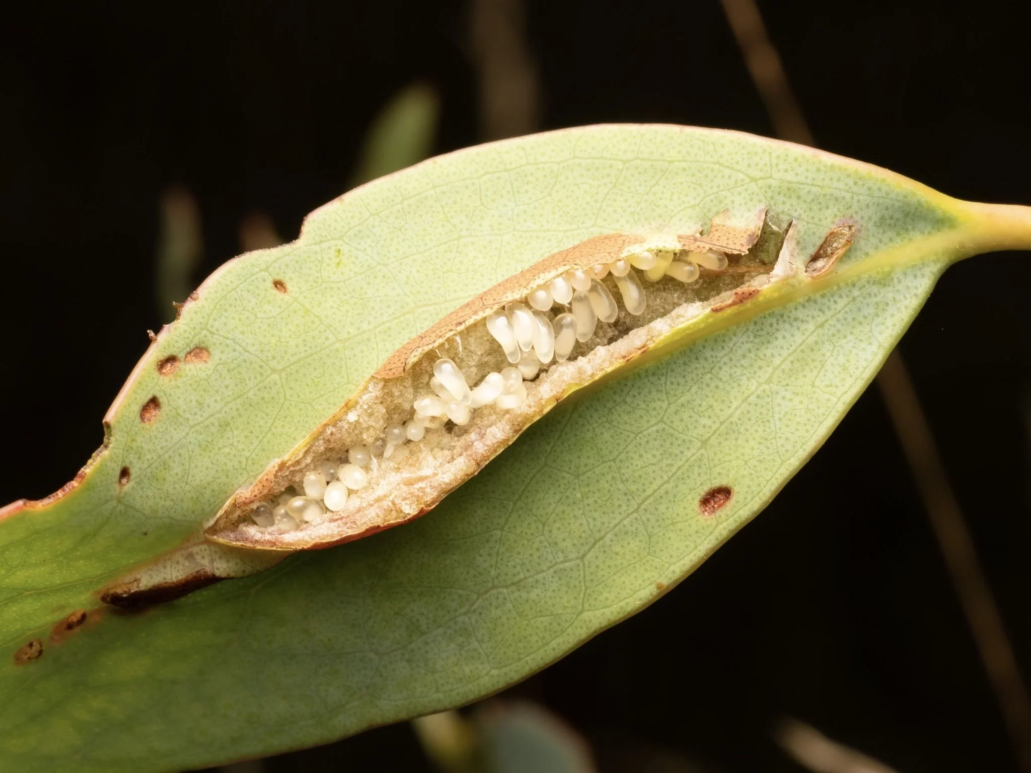
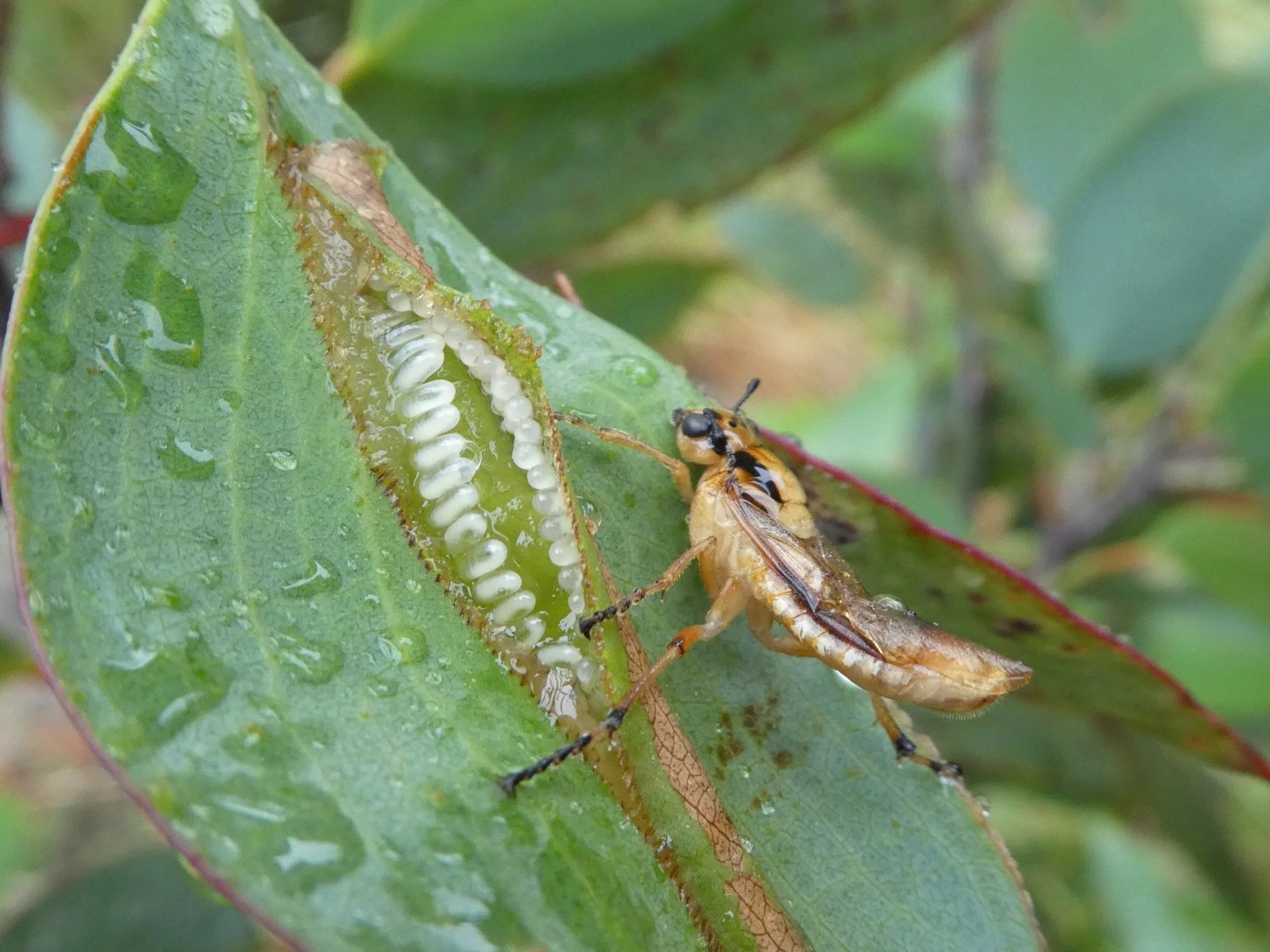
Perga affinis - Carne (1962)
“Oviposition ceases when the wasp reaches a point at which surface abrasion began. Within the limits of the female's egg complement, the size of pod is related to the dimensions of the leaf, being of greater average size on elongate narrow leaves than on broader leaves (cf. Fig. 38 a,f,g). If her egg supply becomes exhausted before the available oviposition space is occupied, a female will usually continue to make incisions. Indeed, once the behaviour pattern associated with oviposition has been established, it continues until death. Consequently, towards the end of the flight season, most of the females scraping or making incisions are found to contain few or no eggs, the resulting abortive pods (Fig. 38 b) being distinguishable from functional pods on close inspection. Some leaves are found abraded but not incised (Fig. 38 c) owing to the death of females.
The great majority of pods are aligned against the midrib. However, the female does not seek out the midrib when making incisions, but, owing to her habit of grasping the leaf margins, and to the central position of the midrib in most leaves, such an alignment occurs automatically. Occasionally leaves damaged by other insects are selected for oviposition; the alignment of the pod then follows the approximate new median line of the leaf, and may cross the actual midrib at an appreciable angle (Fig. 38 h).
Females never attempt to oviposit in the same region as others. However, several females may utilize the same leaf (Fig. 38 e), but only when practically all of the surrounding leaves have received eggs.
Although the wasps have been seen to drink from drops of water trapped in leaf axils, they do not feed and are dependent for all muscular activity upon energy reserves derived from the larval fat-body; absorption of ovaries does not occur. The average longevity of females caged at room temperature with fresh foliage is 7-9 days. In the field, unless taken by predators, the female dies of exhaustion a few days after laying the last of her eggs. Dead or moribund females were never found to contain more than 2-3 unlaid eggs, the oviducts of the majority being completely empty.”
Fig.38. Carne (1962) Fig. 13 Diagrams illustrating the types of egg pods formed by Perga affinis, and the influence of leaf geometry on pod length and position.
Details of saw structure and mechanics of oviposition - from Tait (1962)
Figs. 39-40.
First Valvula (Lancet) - The lancet consists of a blade along the edge of which are tooth-like sclerotizations resembling the teeth of a saw. A basal radix (rad.) and blade-like lamnion (lam.) is further subdivided horizontally into segments by alternating bands of intense and weak sclerotization. The lamnion is also divided longitudinally by a fold, the crespidium (crp.). This causes the ventral half of the lancet to be inclined inwards towards the mid-line. Within the less sclerotized regions on the outer surface are various projections. On the upper surface of the blade is the ala (al.), which is produced into a number of spines, the alaspinulae. These terminate near the crespidium in wing-like projections, the alaspiculae (als.). On the ventral margin of the saw are spurs, the spiculellae (spl.). Between these and the alaspiculae is a row of spines, the subalar spines (s.sp.). The more heavily sclerotized regions are produced ventrally into tooth-like projections along the edge of which are serrations forming a serrula (ser.). The upper half of the inner surface of the lance is densely covered with hairs. On the inner ventral surface, flap-like sclerotizations extend from the crespidium half way to the ventral margin.
Second Valvula (Lance) -The lance is similarly divided into zones. The regions of low sclerotization, however, gradually diminish anteriorly towards the ventral margin. The distal tip of the lance is highly sclerotized and is provided with a toothed edge along the upper surface. This region makes the first incision into the tissues of the leaf. The two lances are joined along their dorsal edges by a membrane. The proximal end of the lance forms a hump, the basal process to which muscles are attached. The lances and lancets are joined by means of sliding grooves called virgae (vir.). The virga of the lance is situated along the mid-longitudinal line while that of the lancet is situated on the dorsal surface. These virgae allow the lancet to slide back and forth against the lance. The insect is thus able to make a cavity between the upper and lower epidermis of the leaf in which it deposits an egg.
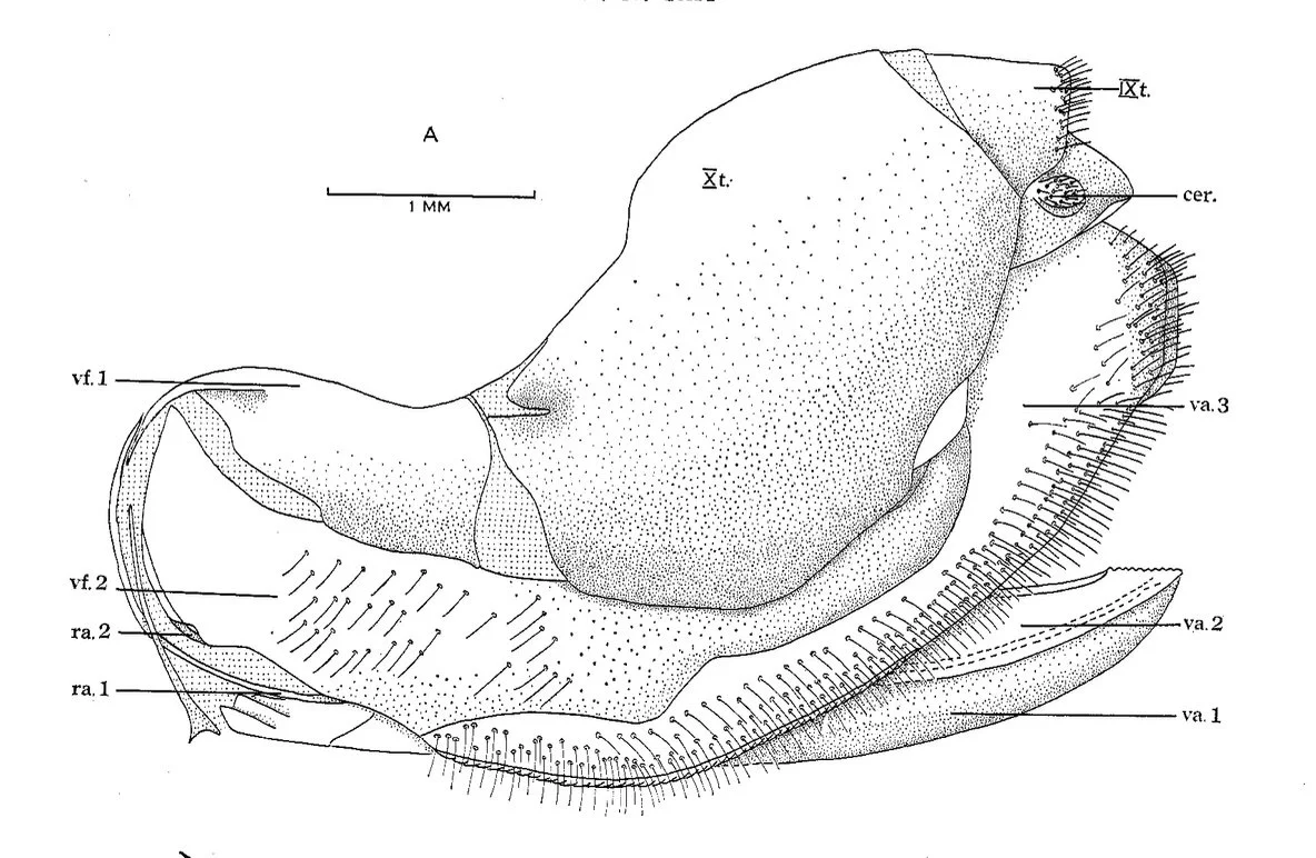
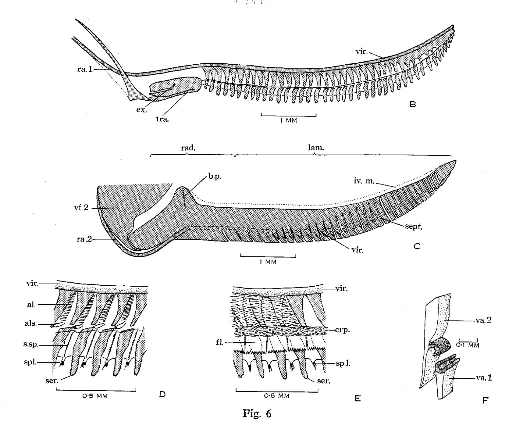
Third valvula (sawsheath) - The third valvula is densely covered along its ventral lateral border with stiff bristles arranged in 3 or 4 rows. Along the ventral border of the third valvula is a heavily chitinized rasp-like cord. There is also a region of longitudinal corrugations termed the "saw-bench" by Riek (1961). This is used to remove wax from the surface of the leaf prior to ovipositing. The posterior tip of the sheath is produced into a cap that protects the tips of the lance and lancet when these are not in use. The third valvulae are joined dorsally by a bridge to which several muscles are attached.
Fig.41.
There are two separate actions in ovipositing: firstly, the protrusion of the ovipositor, and secondly the sawing motion itself. Contraction of muscle 30, which arises on the suture dividing the second valvifer and the third valvula and is inserted on the basal process of the lance, causes the protrusion of the ovipositor from its sheath so that it is at right angles to the abdomen. This action is opposed by the contraction of muscle 31, which is attached to the ramus of the second valvula and to the basal process of the lance, causing the return of the ovipositor to the sheath.
Contraction of muscle 28 causes the protraction of the lancet and retraction of the lance. The first movement is brought about by muscle 28b through the action of the second valvifer on the first valvifer relayed to the lancet by ramus 1. The second movement is brought about by the action of muscle 28a on the ramus of the second valvifer. Muscle 37 could also be of importance in the retraction of the saw. Muscle 29 opposes this action.
The lancet is retracted by the bending of the quadrate plate being relayed to the first valvula and thence via ramus 1 to the lancet. Protraction of the lance is brought about by contraction of muscle 29, attached to the second valvifer. Muscles 32 and 33 are antagonistic muscles protracting the third valvulae which act as a brace when the insect is ovipositing.
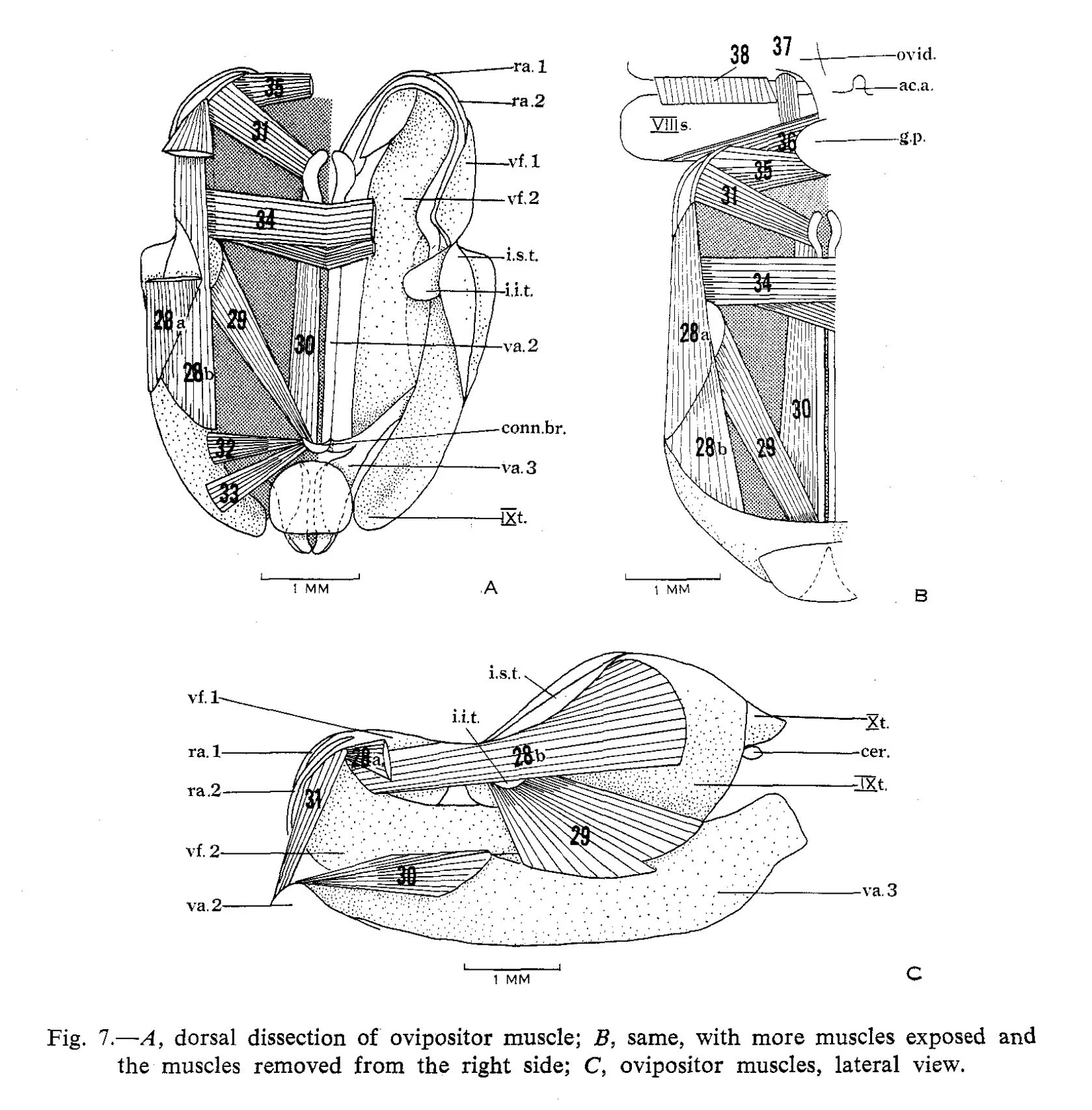
Fig. 41. Tait (1962) Fig. 7
References
Carne, P.B. (1962) The characteristics and behaviour of the saw-fly Perga affinis affinis (Hymenoptera). Australian Journal of Zoology 10: 1-34.
Macdonald, J. & Ohmart, C.P. 1993. Life history strategies of Australian pergid sawflies and their interactions with host plants. pp. 485-502 in Wagner, M. & Raffa, K.F. (eds) Sawfly Life History Adaptations to Woody Plants. San Diego: Academic Press.
Riek, E.F. (1961) The distribution and inter-relationships of Perga affinis Kirby and Perga dorsalis Leach (Hymenoptera, Symphyta). Proceedings of the Linnean Society of New South Wales 86: 237-240.
Tait, N.N. (1962) The anatomy of the sawfly Perga affinis affinis Kirby (Hymenoptera: Symphyta). Australian Journal of Zoology 10: 652-683.
Westwood, J.O. (1836) P. 234 in postscript to Lewis, R.H., Case of maternal attendance on the larva by an insect of the tribe of Terebrantia, belonging to the genus Perga, observed at Hobarton, Tasmania. Transactions of the Entomological Society of London 1: 232-234.
This is a workbook page … a part of our website where we record the observations and references used in making species identifications. The notes will not necessarily be complete. They are a record for our own use, but we are happy to share this information with others.