
Elatichrosis (Elateridae: Denticollinae)
Workbook
Beetle captured around 14/12/23 at home in Wonboyn. I used Andrew Calder’s “Genera of the Australian Elateridae” CSIRO Publishing 1996 to identify it.
Which subfamily?
Denticollinae
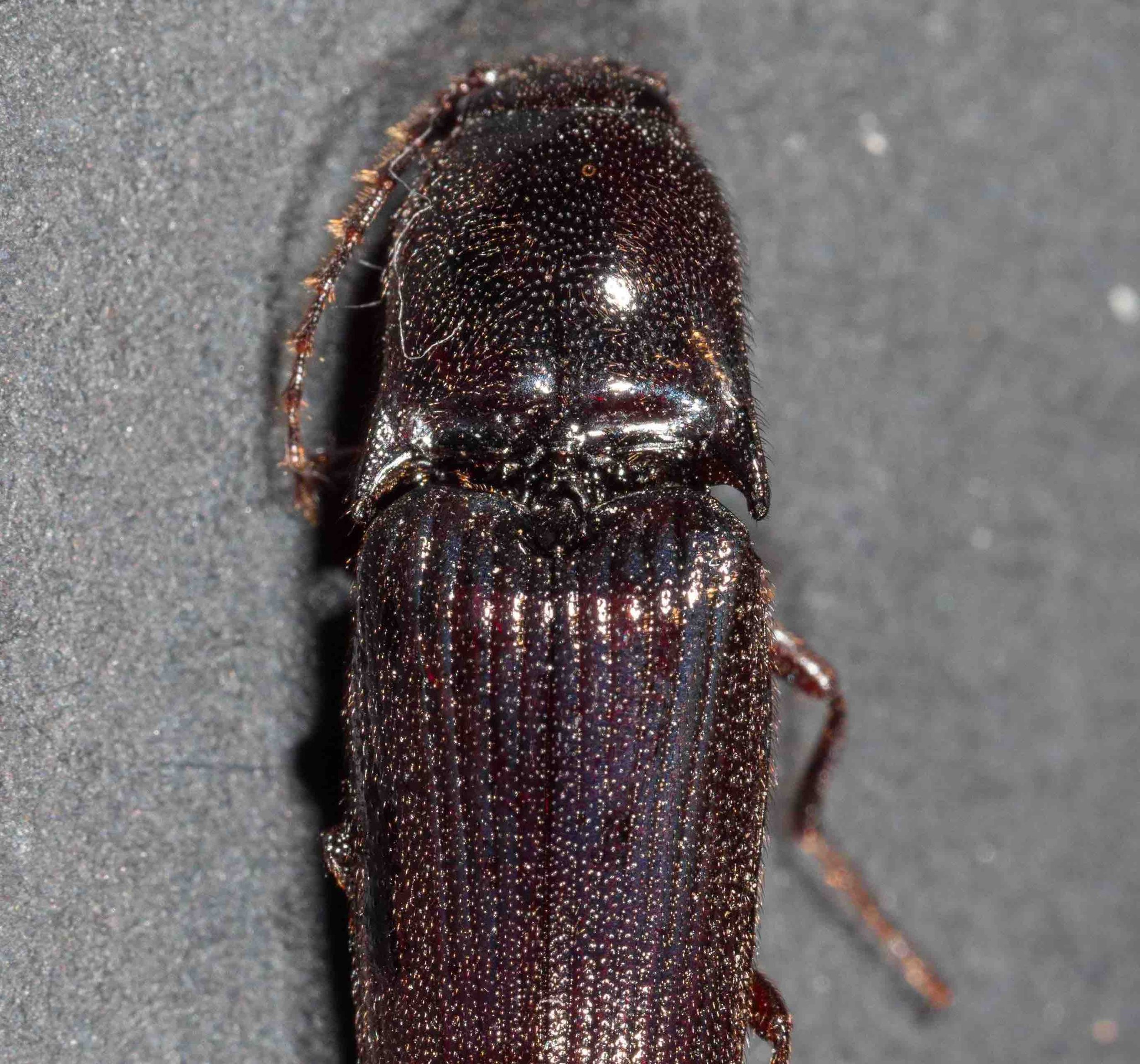
Calder states that “in the Denticollinae the anterior portion of the head capsule is flattened dorso-ventrally causing the mandibles to be directed more anteriorly, giving such elaterids a characteristic prognathous appearance”.
This can be seen in this frontal view of the head of the beetle.
This image shows that the frontal carina is incomplete medially, another general feature of the subfamily.
frontal view of head
Other features of this subfamily evident in our beetle include:
ventral view of head and prothorax
prosternum produced anteriorly to form ‘chin piece’
marginate edge of hypomeron inclined meso-dorsally
ventral view of left side of mesothorax
mesocoxal cavity open to both mesepimeron and mesepisternum
hind wing with short radial cell; vein MP4 with apparent crossvein to CuA2; wedge cell present; apex of wing membrane (without venation) occupying at least 0.2X total length of wing membrane; apex with two sclerotisations at acute angle to each other.


hind tarsus - lacking spongiose pads or lamella (i.e. simple)
tarsomeres usually with spongiose pads apico-ventrally, but occasionally simple as in our beetle
hind tarsus showing claws without basal setae
tarsal claws simple without basal setae on outer flat portion
Which genus in subfamily Denticollinae?
The key in Calder leads to genus Elatichrosis by the following steps:
1.Tarsal claws without basal setae (see previous image)
15. Mesocoxae open to both mesepimeron and mesepisternum (see third image above)
26. Anterior margin of scutellum well defined, sharply angulate and steeply declivous to prescutum (left image below); labrum fully exposed at base (right image below).


27. Tarsomeres simple (see tarsus image above)
28. Mandibles bidentate (upper and lower teeth indicated by 1 and 2 in this image)
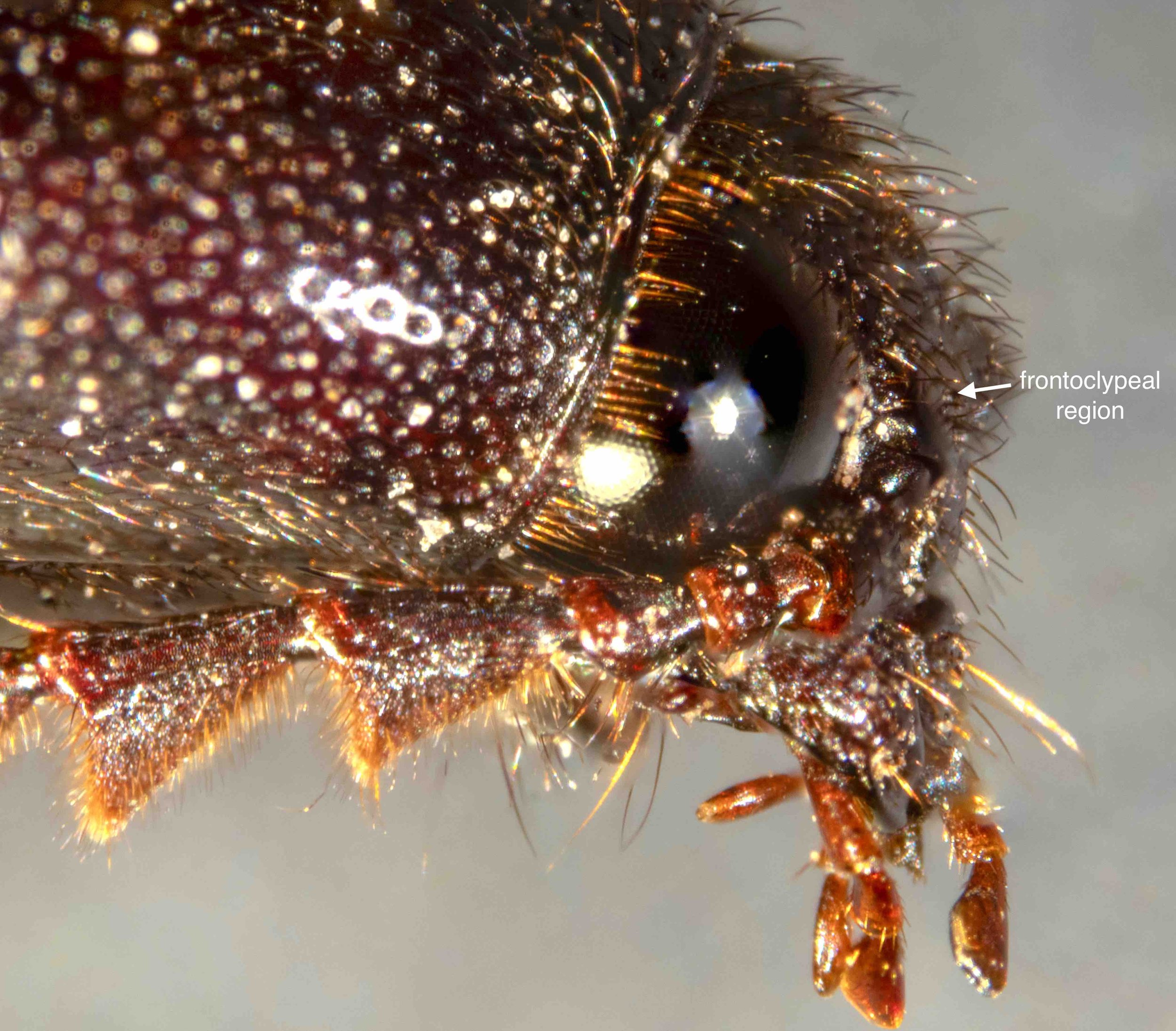
35. Frontoclypeal region gradually sloping to base of labrum

36. Hind tarsomere 1 longer than hind tarsomere 5
37. maxillary palp elongate, widened distally


38.
Pronotum longer than wide (left hand image below);
Large 15.2-32.8mm in length (18mm - middle image below);
Hind tarsus longer than hind tibia (middle image),
Lateral margins of prothorax broadly rounded not carinate ——> Elatichrosis
As shown in the right hand image below, the lateral margin of the prothorax is carinate in our beetle, but it extends to only about halfway along the margin. Calder’s description of Elatichrosis states that some species have no trace of the carina, while others have a carina which is incomplete anteriorly, as shown here.
I conclude that the beetle belongs to genus Elatichrosis.


Does our beetle match Calder’s description of Elatichrosis?
Summary of defining features of this genus.
Adult
3.8-4.4 X as long as wide, moderately convex. (a bit less - 3.3X)
Colouration. Dark brown; black; orange brown or reddish-brown with black markings (dark brown)
Length 15.2-32.8mm (18mm)
Images of whole beetle above are consistent with this.
Head
Flattened anteriorly - (Yes see first image in subfamily diagnosis above)
Frontal carina incomplete medially (carinate over antennal bases) - (Yes, see first image in subfamily diagnosis above)
Frontoclypeal region gradually sloping to base of labrum - (Yes, see step 35 in key above).
Eyes of male small, protuberant, 0.2-0.3 X interocular distance (Yes, 0.3)
Mouthparts issuing from front of head capsule, directed forwards (Yes, see first image in subfamilly classification)
Mandibles bidentate (Yes, see step 28 in key above)
Labrum fully exposed at base; semicircular, wider than long (Yes, see RH image in step 35 in key above)
Maxillary palp elongate, hatched-shaped; segment 3 shorter than segment 2 (Yes, see images in step 37 in key above)
Antennae serrate from antennomere 4; with 11 antennomeres; antennomere 3 longer than antennomere 2; antennomere 4 longer than anntenomere 3; (Yes, see LH image below).
Antennae short, not reaching apex of hind angles of pronotum or long, exceeding hind angles (almost reaching apex of hind angles, see RH image below)


antennal insertions small, separated by greater than two diameters (Yes, see RH image at step 37 of key above).
Prothorax
Longer than wide (PI 125-157) - (Only just longer than wide PI=106)
With strongly impressed median longitudinal depression - (No, only a week depression evident - see LH image in step 26 of key above)
Anterior angles strongly produced to cover half of eye - (Not sure - somewhat less than half eye covered in photos in last panel)
Lateral margins not entirely carinate (Yes - see RH image in step 38 of key above)
Base of pronotum produced medially but notched in front of scutellum (Yes - see LH image at step 26 and RH image in step 38 of key above)
Hind angles of pronotum not carinate or unicarinate (Yes - unicarinate, see LH image at step 26 of key above), narrow, elongate, strongly divergent (Not sure, but these look similar to hind angles in the habitus image of Elatichrosis trisulcatus in Calder)
Protonotosternal suture straight (but distinctly curved outwards posteriorly at procoxa); marginate along hypomeral border; anterior portion of polished band along inner margin of hypomeron inclined meso-dorsally (Yes, see second photo in Which Subfamily? section and LH photo in ‘Head’ section above)
Prosternum without longitudinal carinae; marginate around procoxal cavities; well developed anteriorly to form ‘chin piece’ (Yes, see second photo in Which Subfamily? section)
Prosternal spine more or less horizontal without ledge immediately behind procoxae (Yes); with strongly developed subapical tooth (Not clear); without median longitudinal groove (Yes); portion of spine posterior to coxae long (1.6 X coxa diameter - 1.3X) - see photo below
ventral view of head/prothorax removed from rest of body to show prosternal spine
Mesothorax
scutellum pentagonal or tongue-shaped (tongue-shaped); apex broadly rounded, sides not notched (yes); without longitudinal carina (yes); anterior margin well-defined, sharply angulate and steeply declivous to prescutum (yes) - (see LH image at step 26 and RH image at step 38 of key above).
mesocoxae rounded, wide apart, separated by posterior margin of mesosternal cavity (0.4-0.7 X coxal diameter - yes 0.7 X) - (see LH image at step 26 and RH image at step 38 of key above).
mesocoxal cavity open to both mesepimeron and mesepisternum (yes - see third image in Which Subfamily? section)
mesosternum and metasternum separated at midline by distinct external suture (yes - see RH image in Head section above)
Hind wings
well-developed; 2.8-3.1 X as long as wide (yes, 2.9 X - see wing photos in part 4 of Which Subfamily? section and image below)
wing membrane not notched in anal area (yes - see wing photos in part 4 of Which Subfamily? section)
vein MP4 with apparent cross vein to CuA2, branching from MP3 proximal to apparent cross vein between veins MP4 and CuA2 (yes - see wing photos in part 4 of Which Subfamily? section and image below)
wedge cell present (yes - see wing photos in part 4 of Which Subfamily? section)
apex of wing membrane (without venation) occupying 0.1 X length of wing membrane; with 2 sclerotisations at acute angle to each other (yes - see wing photos in part 4 of Which Subfamily? section)
hindwings visible after removal of elytra
Elytra
2.4-2.9 X pronotal length (yes, 2.45 X - see first image in panel at step 38 of key)
striae impressed, usually strongly punctate-striate (yes - see first image in panel at step 38 of key)
epipleura very gradually narrowed at level of hind coxal plate (yes - see image below)
lateral view showing epipleura of right elytron
apex of elytra bluntly or narrowly rounded (yes, bluntly - see first image in panel at step 38 of key)
Legs
hind coxal plate with distal width less than half greatest proximal width nearest insertion of trochanter; outer posterior angle rounded; outer margin of hind coxal plate without elevated fold (yes - see image below)
ventral view of abdomen and metathorax
meso-trochantin fully visible (yes - see image alongside)
ventral view of LH side of mesothorax
hind tibia subcylindrical, not compressed laterally, very slightly widened towards apex; with 2 short, subequal apical spurs; without rows of erect, spiniform setae; hind tibia longer than hind femur; hind tarsus longer than hind tibia (yes, see images below and RH image in Head section).


tarsomeres simple, densely pilose ventrally, tarsomeres 1-3 with erect spiniform setae apico-ventrally, tarsomeres 1-4 decreasing in length distally, tarsal claws simple without basal setae (yes - see last two images in Which Subfamily? section)
Abdomen
Abdominal tergites not strongly sclerotised, thin, pale in colour (yes - see image below)
abdominal tergites visible after removal of elytra and hindwing
last visible abdominal sternite (ventrite 5) narrowed to broadly arcuate apex (yes, see image below)
ventral view of abdomen
Male genitalia
(refer to panel of 5 images below)
aedeagus 2.5-2.8 X as long as wide (3.0)
median lobe planar; apex narrowly rounded, exceeding apices of parameres (yes)
basal struts short to moderately long, 0.3-0.4 X (0.3) total length of median lobe
parameres articulated with median lobe; each with lateral subapical barb; apex mostly sclerotised with inner apical angle membranous, sparsely setose (yes)
base of parameres well developed ventrally, extending beyond dorsal base, not fused together medially (yes)
basal piece 0.3-0.4 X (0.3) total length of aedeagus, distinctly separate (yes)


Summary
This beetle displays almost all of the features in Calder’s (1996) description of Elatichrosis.
The one feature that does not match is the absence of a “strongly impressed median longitudinal depression” in the pronotum. My beetle shows only a shallow depression in the posterior-most region of the pronotum.
However, I cannot find a better match in any of the other genera in the subfamily Denticollinae. I therefore conclude that it is very likely a member of that genus.
I have consulted the review by Neboiss of Elatichrosis “The genera Elatichrosis Hyslop and Lingana Gen. Nov. (Coleoptera: Elateridae)” Aust. J. Zool. 8: 289-306. His distribution map shows that Elatichrosis is found in south-eastern Australia. As all of the species he describes have a prominent impressed median longitudinal depression in the pronotum, I conclude that my beetle is likely to be an undescribed species in that genus.
This is a workbook page … a part of our website where we record the observations and references used in making species identifications. The notes will not necessarily be complete. They are a record for our own use, but we are happy to share this information with others.