
A second clutch of microgastrine cocoons
Workbook
A clutch of Cotesia-like micrograstine wasps as well as several ichneumonid wasps emerge from cocoons in one caterpillar
20 May 2021
I found another cluster of microgastrine cocoons, this time attached to a leaf on an Acacia longifolia seedling. There was no sign of a caterpillar beneath the cocoons. Its dessicated husk had presumably fallen away from the cocoons.


28 May 2021
A large number (24) of wasps had eclosed sometime during the night and were moving around actively in the jar covering the cocoons.


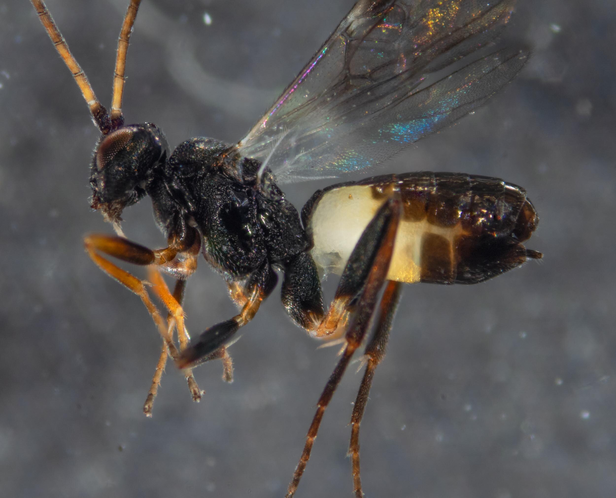
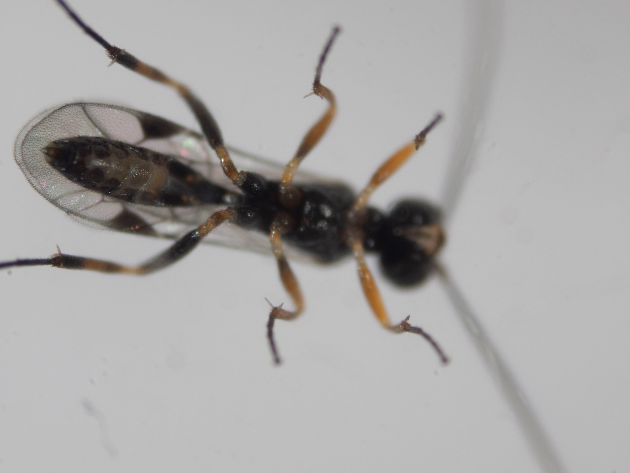
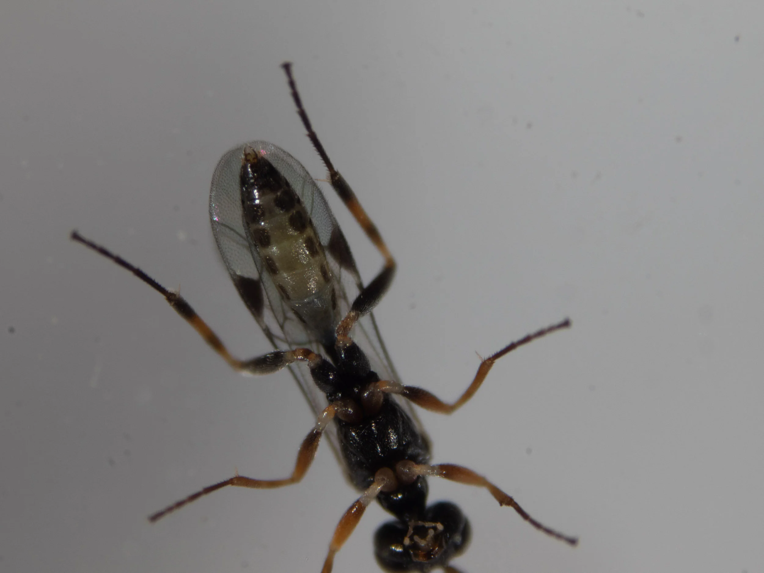
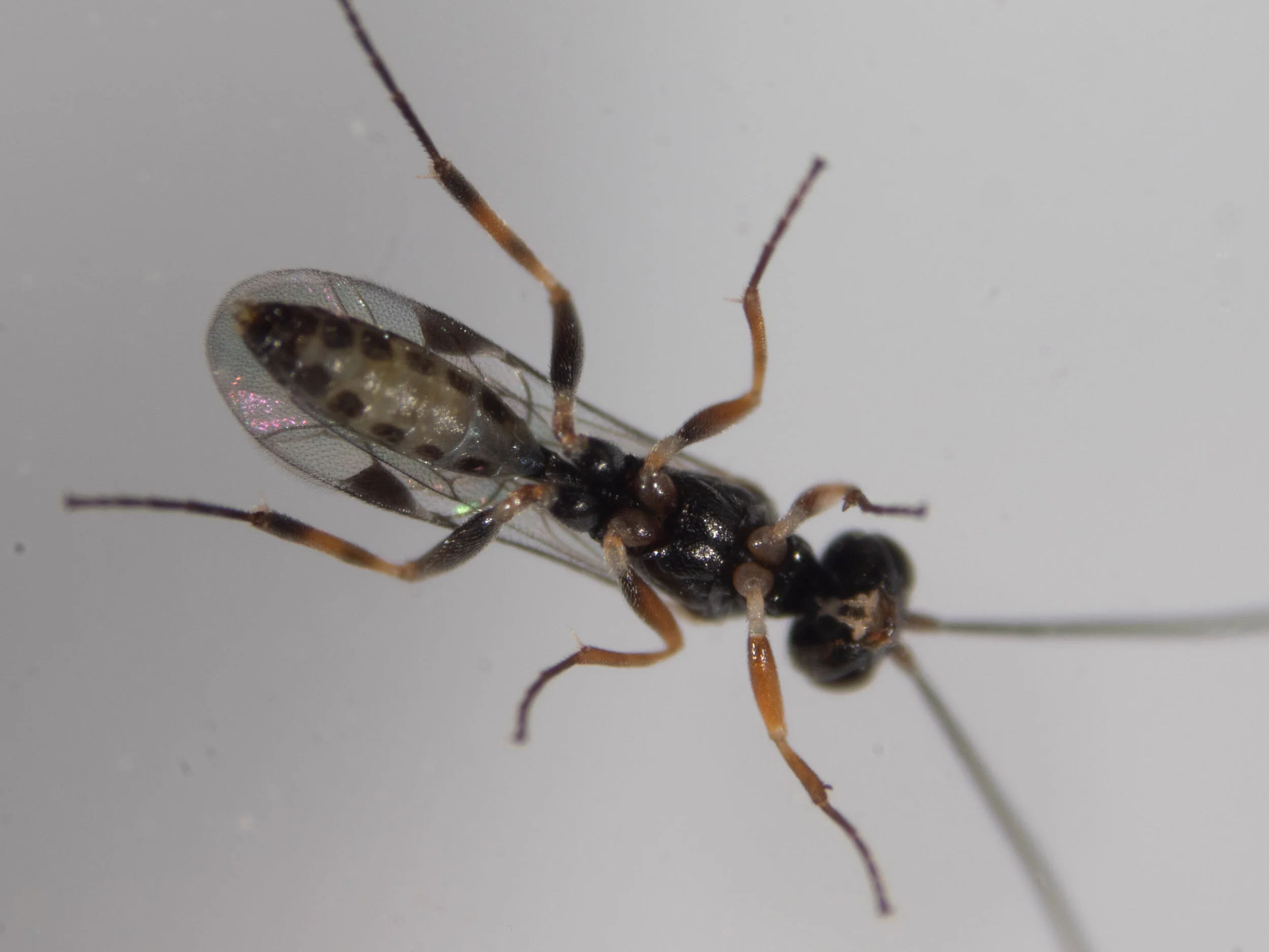
A single wasp (#1) was removed from the collection after it had been cooled for several minutes in the freezer. The following gallery shows this individual.
Dorsal views


Ventral views


This wasp is a male. I placed it in the freezer for a couple of hours and then put it in ethanol. All of the eclosed wasps were put in the freezer for several hours and then placed in ethanol.
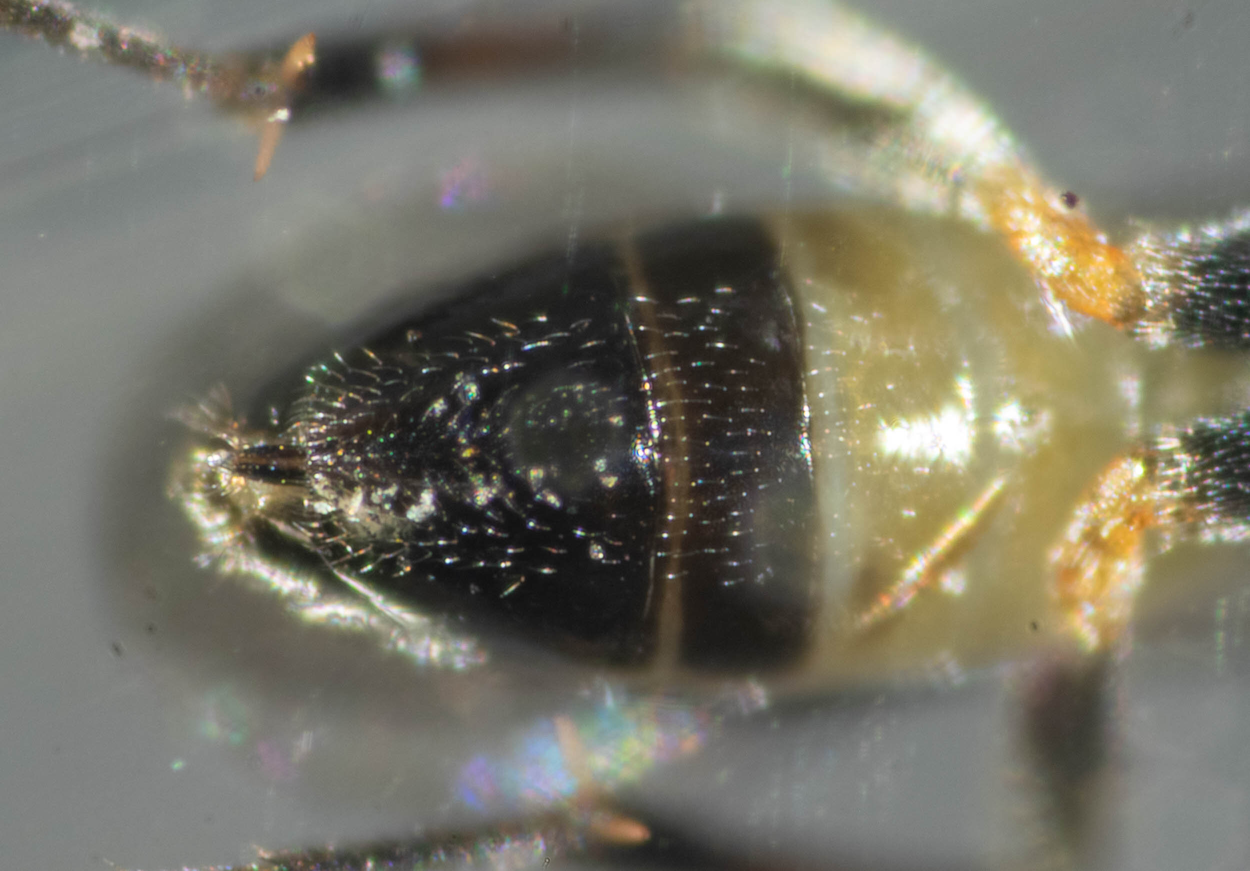
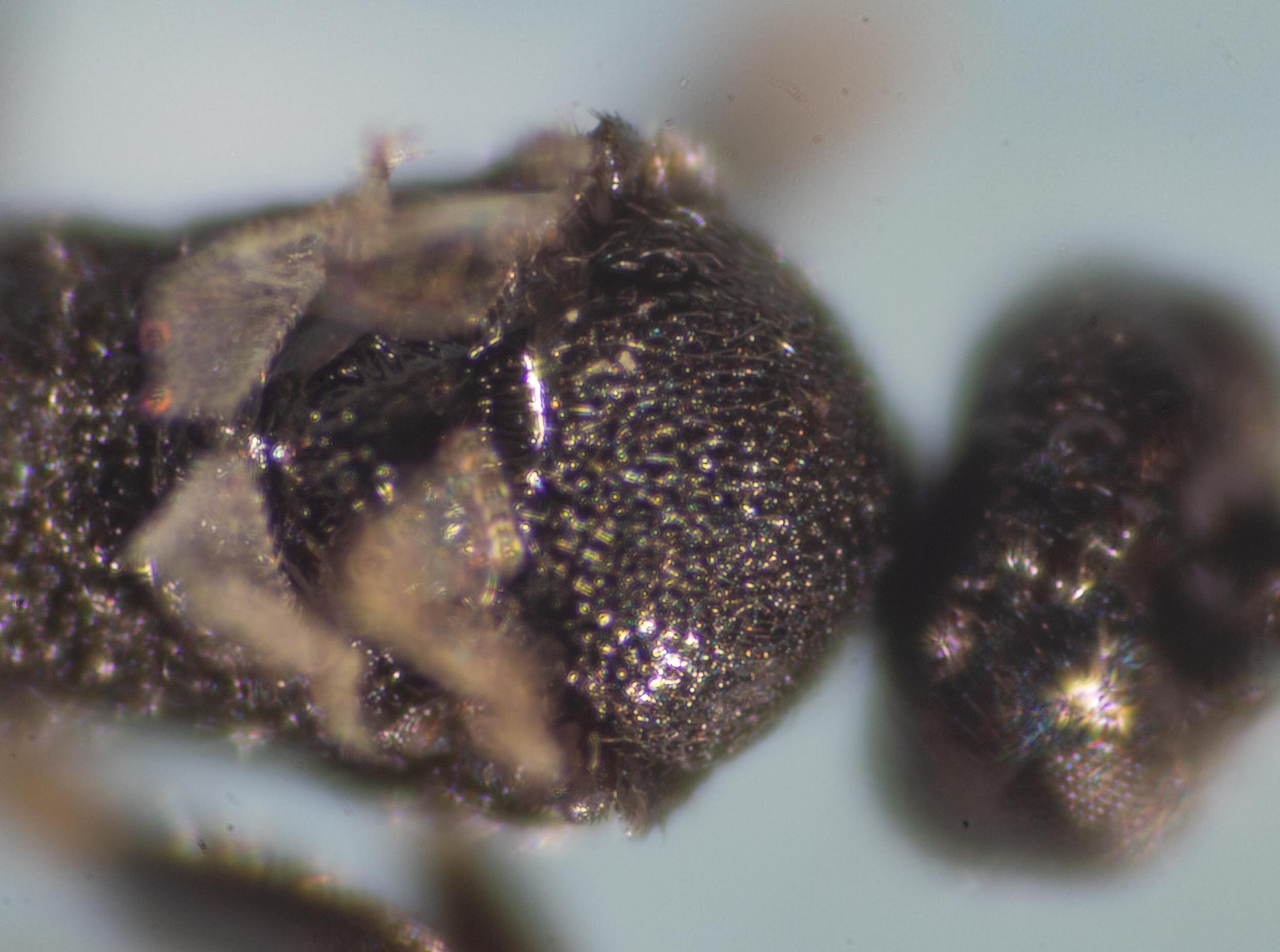
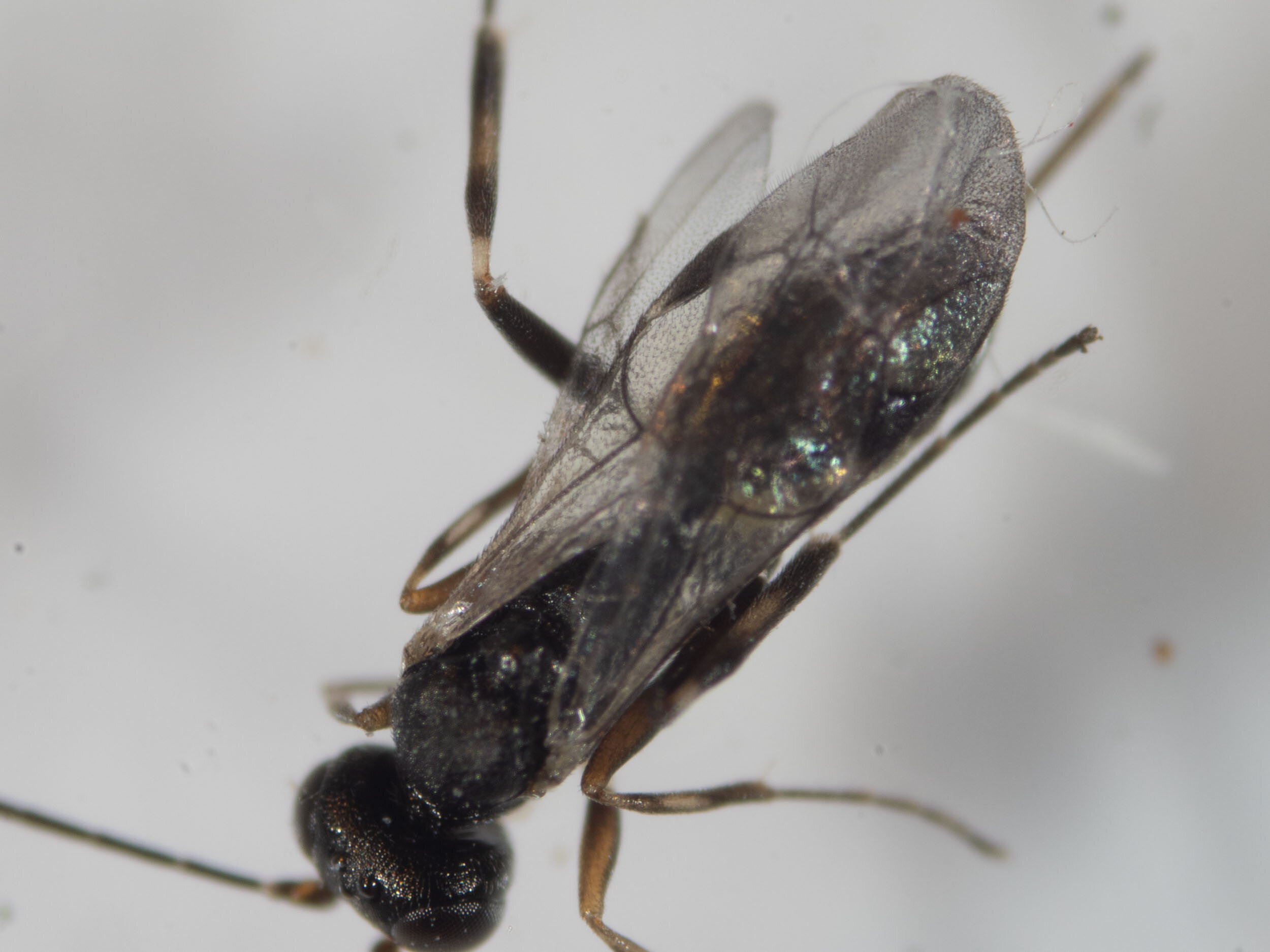
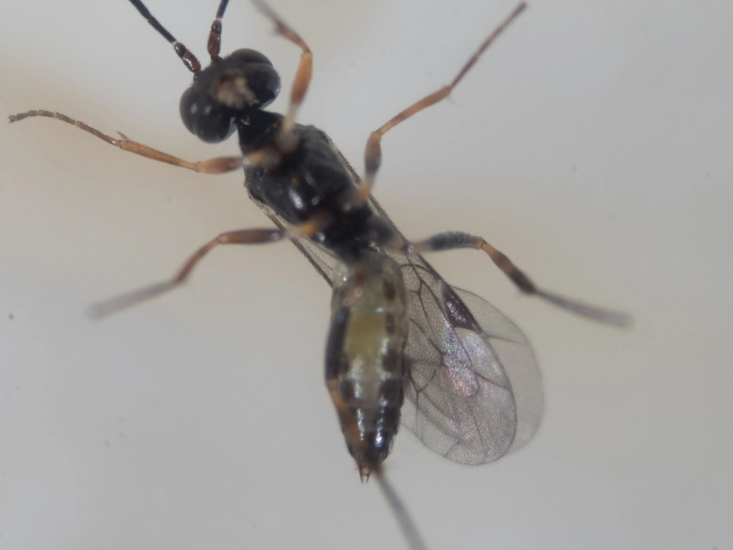
I selected another wasp (#2), a female, and removed the wings to give a clear view of the dorsal body wall.


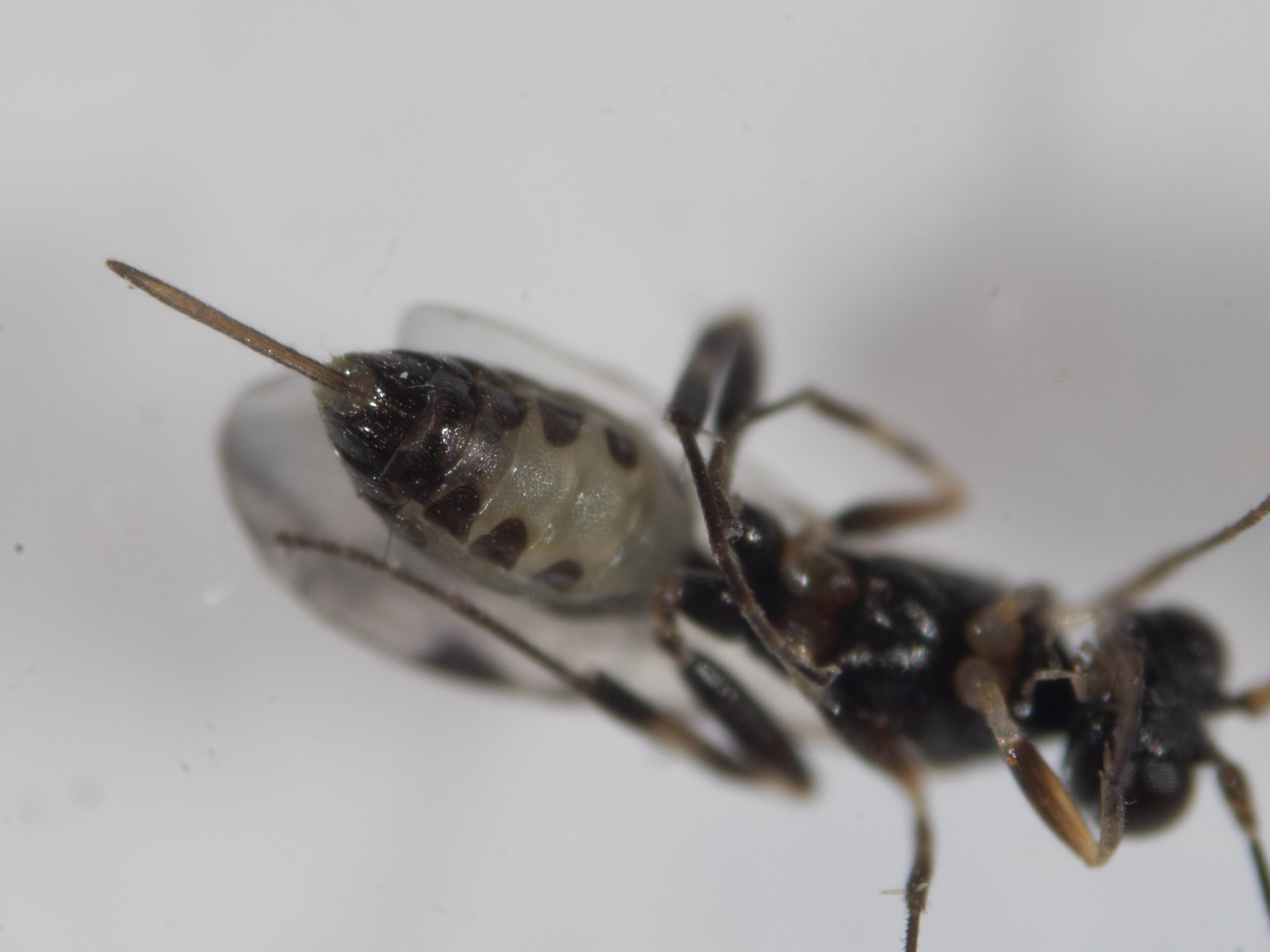
I imaged a number of the other wasps in ethanol. These are all identical in appearance to the microgastrine wasps that eclosed on 2 March, 2021.


All of the eclosed wasps were put in the freezer for several hours and then placed in ethanol. There were 13 females and 11 males. Several were set aside for Erinn.
There are more cocoons (approx. 40) than wasps. On the afternoon of 28 May I examined the cocoons and discovered that only 24 were opened (circular cap removed), while the rest were unopened.
I took the cap off one of these unopened cocoons and another wasp emerged. This was placed in ethanol. The unopened cocoons were placed under the glass to see if additional wasps would emerge.
29 May 2021
In the morning another wasp (female) had eclosed. It was photographed then placed in ethanol.


I dissected open one of the remaining cocoons and found a dipteran? larva inside with parts of an adult wasp body. This hyperparasite larva had apparently been feeding on the wasp pupa. The larva was actively moving and appeared to have begun building a pupal case.


I placed the rest of the wasp pupae into a petri dish to try to identify the presumed hyperparasite.
9 June 2021 - Ichneumonid wasps emerge
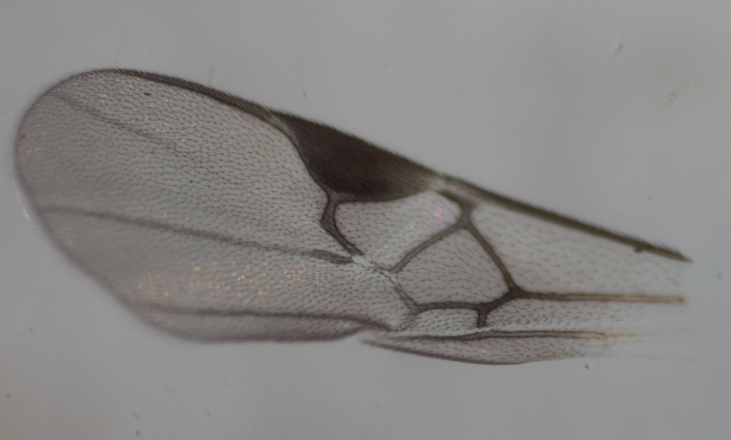
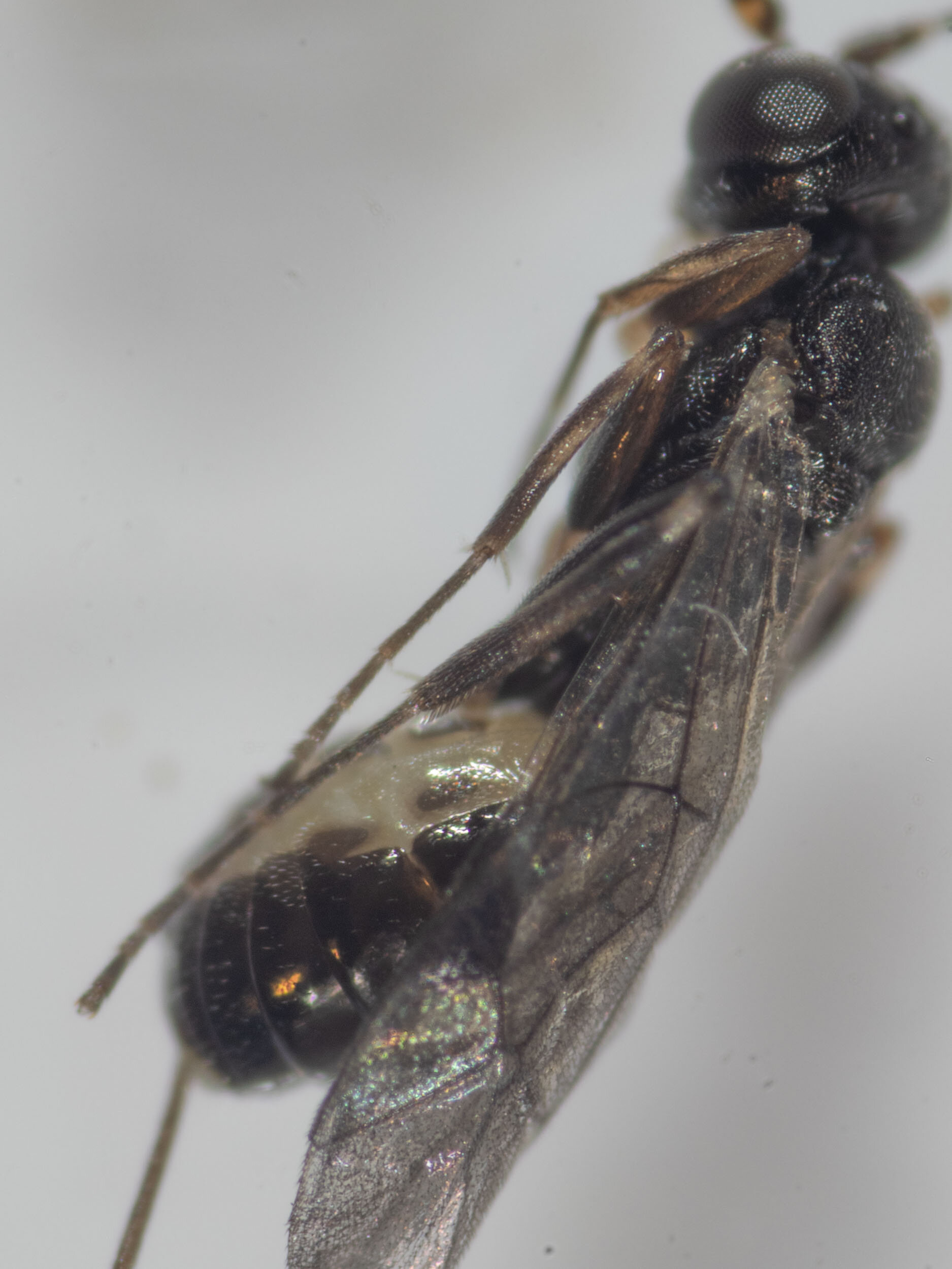
Midday - I noticed that 4 wasps had eclosed from the cocoons since last night. Based on their terminalia morphology, all appear to be males. Wing venation shows them to be Ichneumonidae.


I tore open one of the cocoons and found a female wasp inside in the process of emerging. She has a long ovipositor and similar body proportions to the 4 males that have just eclosed.


22:35 9/6/21 Another male has eclosed.


Morning 10/6/21
3 more males emerged overnight.


The cocoons from which these ichneumonid wasps emerged look identical to those of the microgastrine wasps. They are presumably hyperparasites of the latter’s larvae, developing within or on its body as the microgastrine itself continued to develop through to the pupal stage. The ichneumonid must have ingested the microgastrine completely some time after the latter had spun its cocoon as there was no trace of its body after the ichneumonid had eclosed as an adult wasp.
This means that the same caterpillar had been injected with eggs by two different species of female wasps - one, the primary parasite, a microgastrine; the other, the secondary parasite, an ichneumonid. After the eggs of both species hatched into larvae they continued to develop independently, feeding off the internal body tissues of the caterpillar host. (Although Kerri has read that sometimes the hyperparasite delays its development until its parasitic host has advanced to a late larval/prepupal stage, avoiding competition for the resources provided by the host caterpillar). The ichneumonid larva then attached itself onto (or inside?) the body of the microgastrine as it built its cocoon, then consumed the latter as it developed into a pupa.
An alternative scenario is that the ichneumonid wasp injected its eggs, not into the caterpillar, but into the microgastrine cocoons after they emerged from the caterpillar.
These images show the cocoon after the hyperparasitic ichneumonid has emerged. The neat circular escape hatch made by the microgastrine is replaced by a messier opening. I’ve dissected the cocoon open to show what’s left inside the pupal case - presumably the exoskeleton of the ichneumonid prepupa.


This is a workbook page … a part of our website where we record the observations and references used in making species identifications. The notes will not necessarily be complete. They are a record for our own use.