Under construction - the frog

Yesterday we spied a new clutch of frog eggs in our pond. A check under the microscope revealed that the embryos were a good deal younger than we had previously seen. They were at the neurula stage, which is when the brain and spinal cord first starts to form.
I removed an embryo from the jelly mass and took an image every 10-20 minutes to reveal the dynamics of this amazing process. In the sequence below, we are looking down on the future back of the embryo. The head end of the embryo is towards the bottom. The embryo is about 1 mm long.
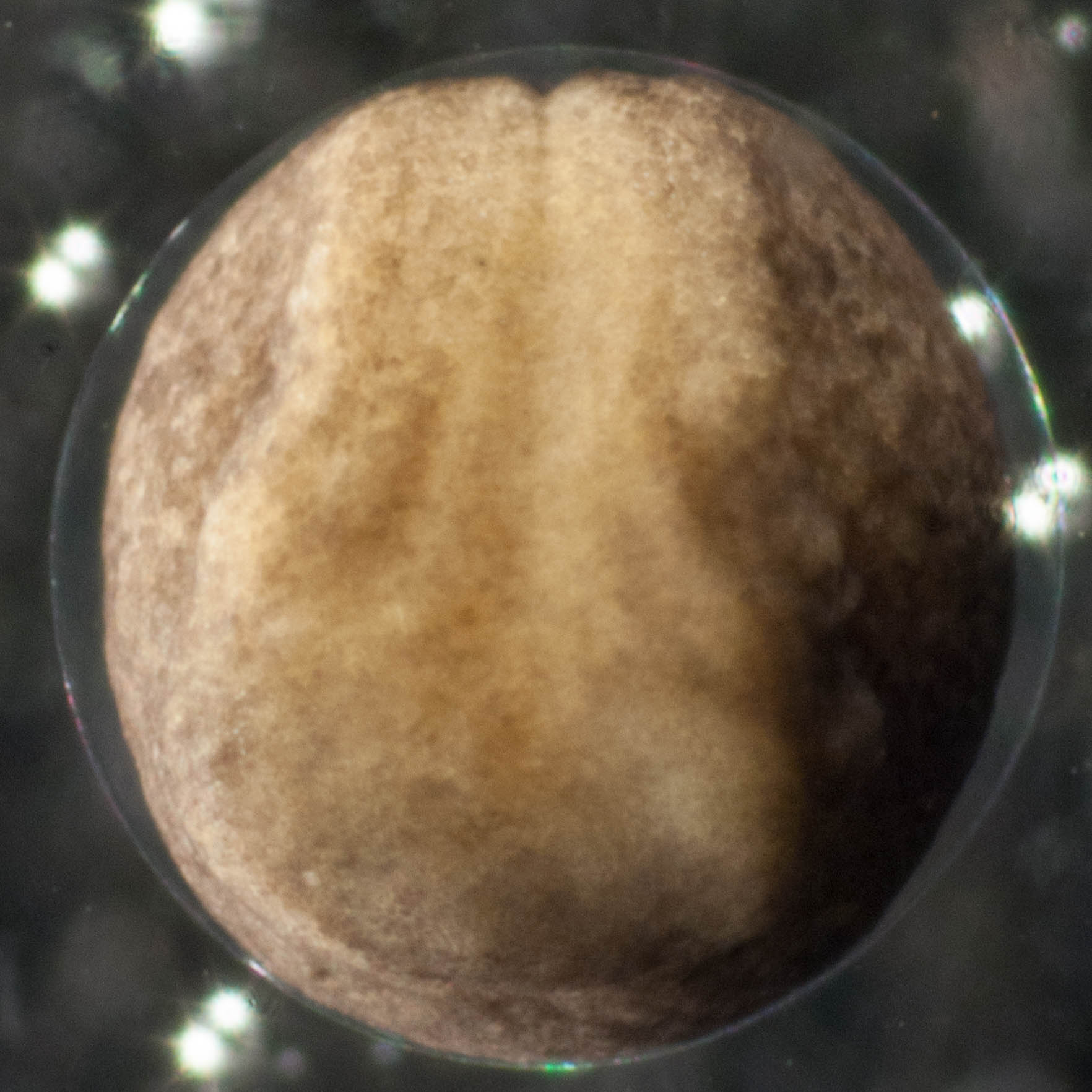
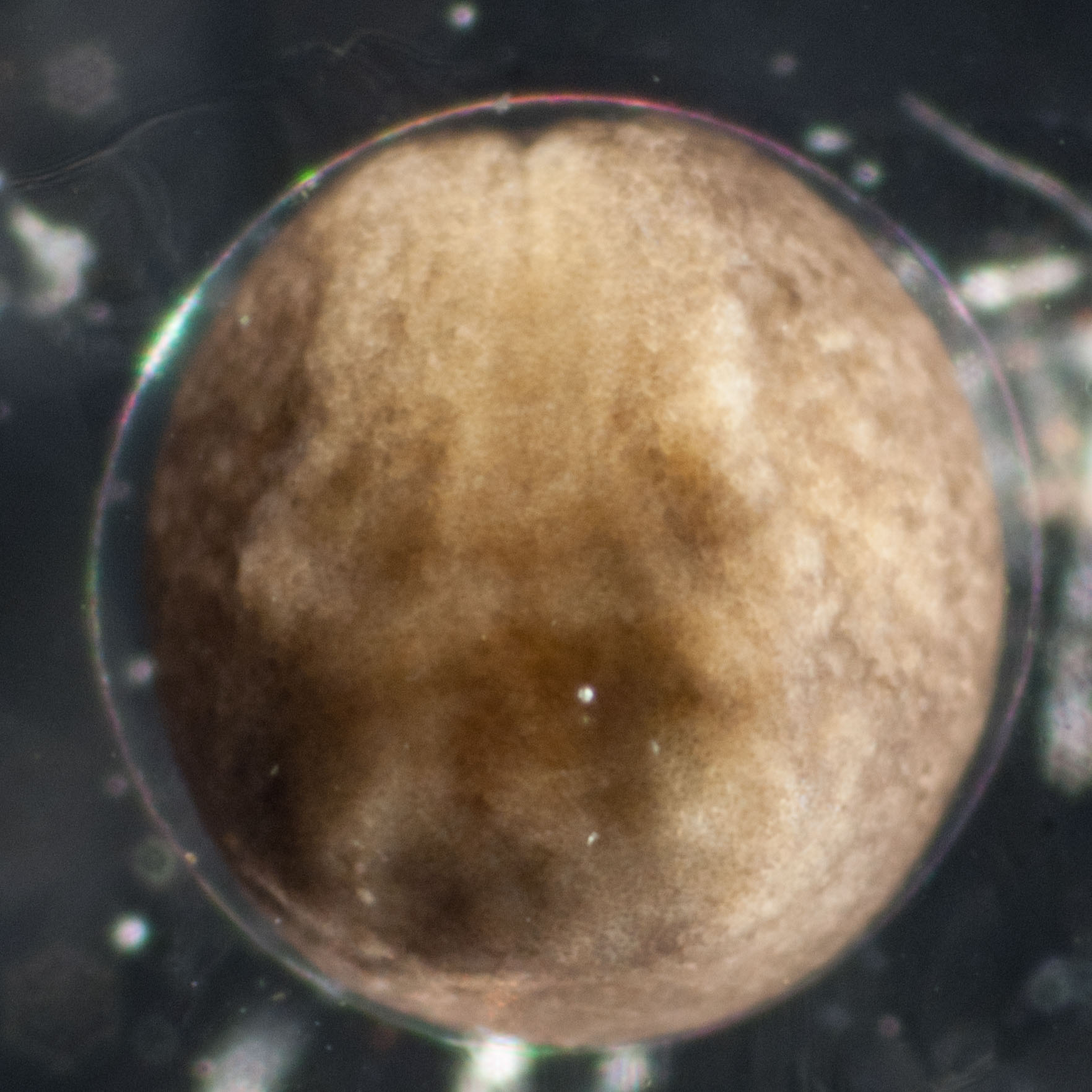
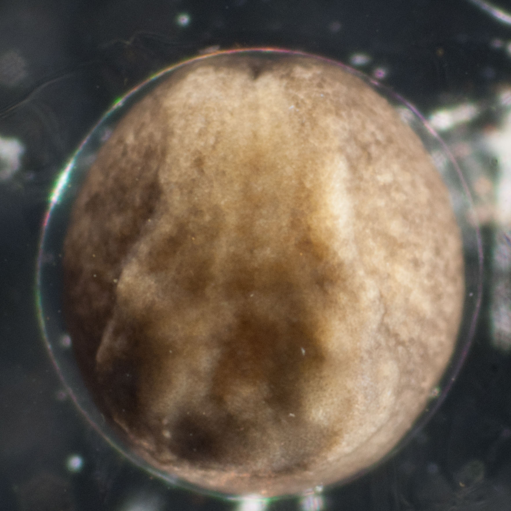
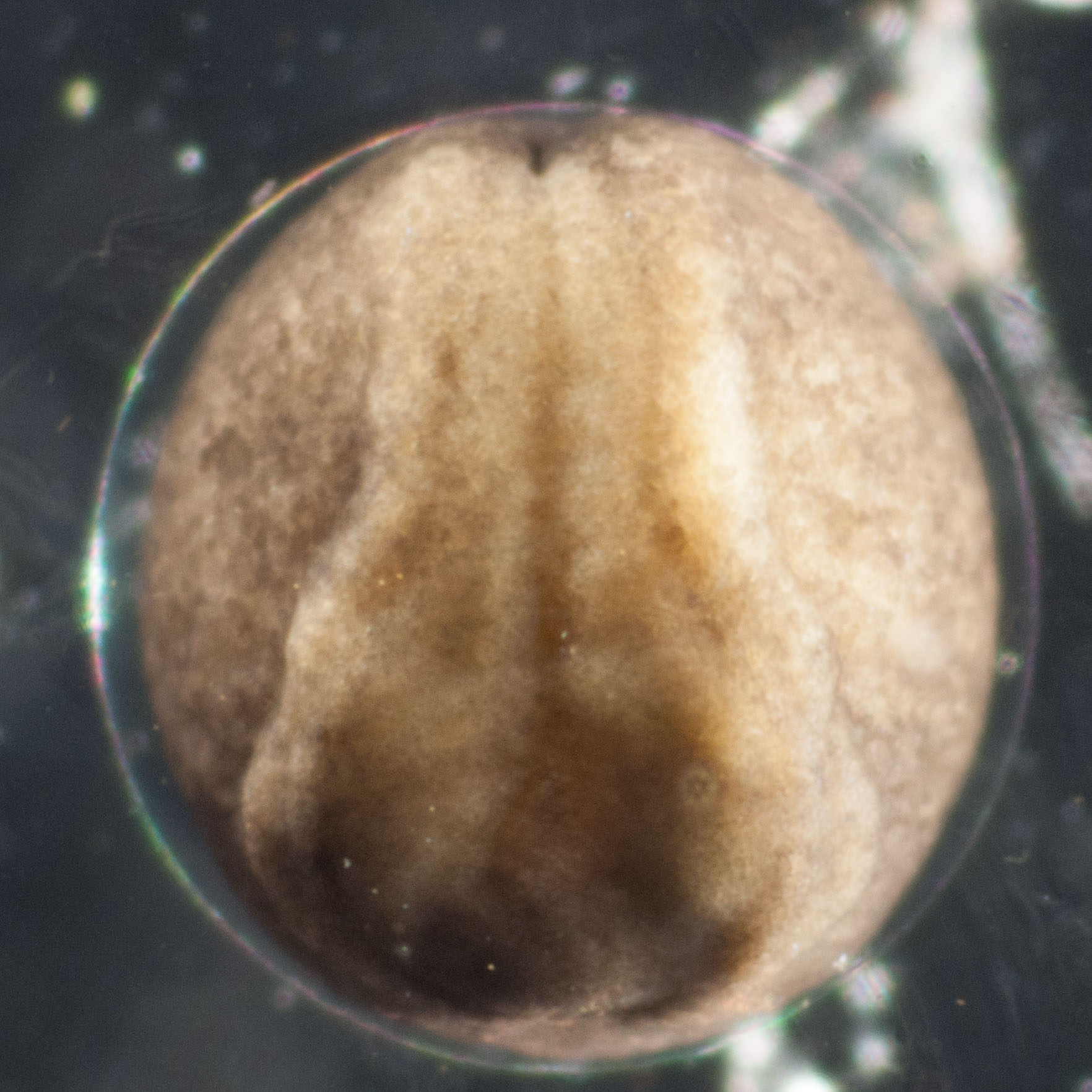
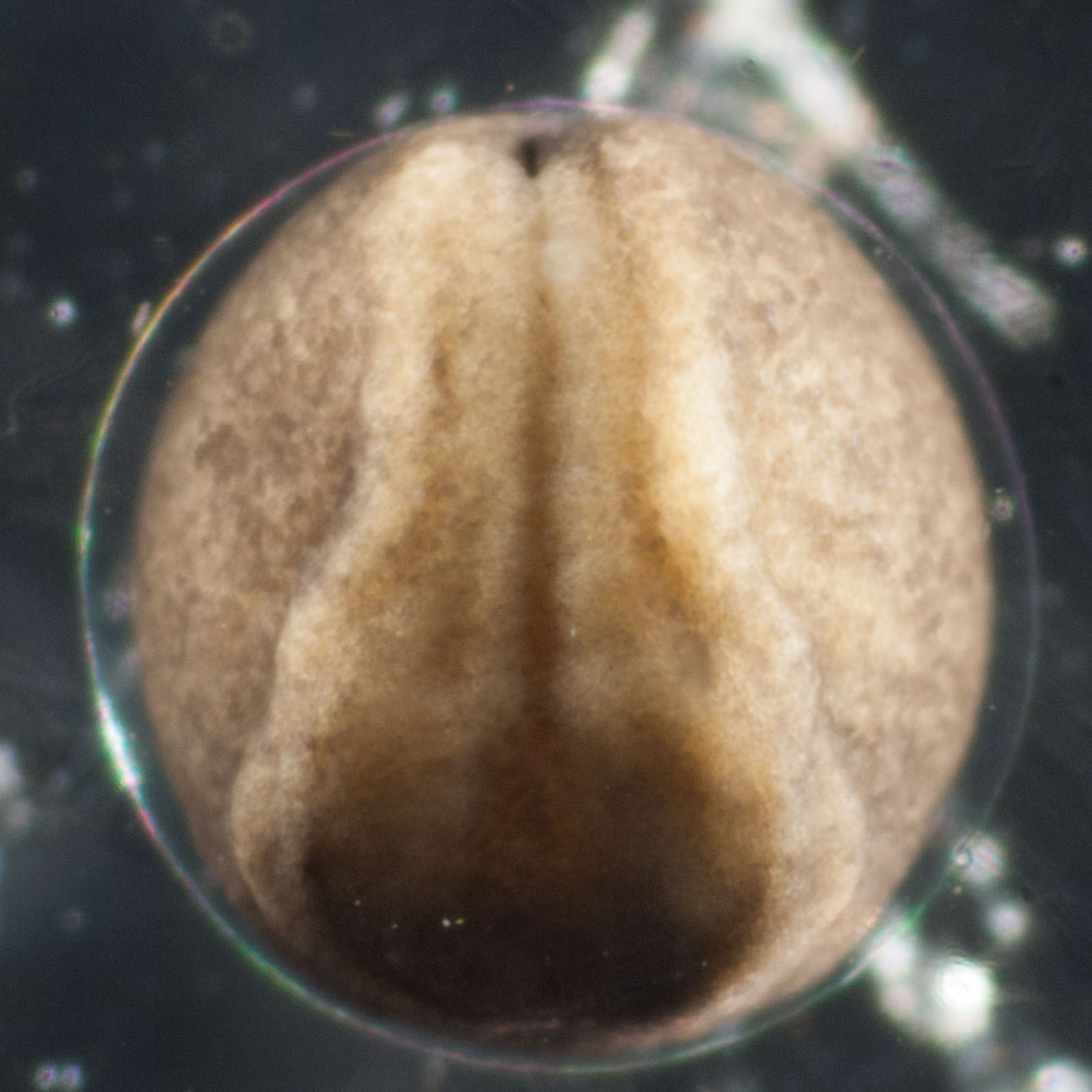
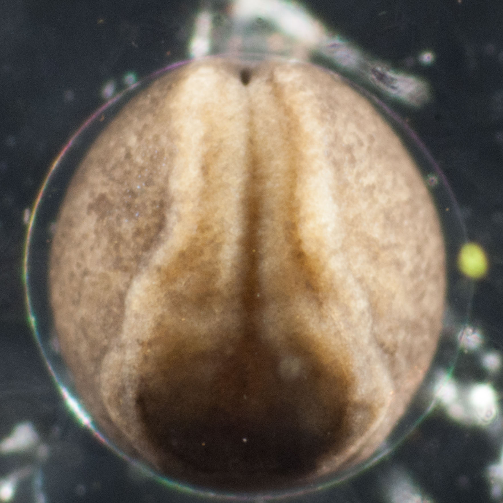
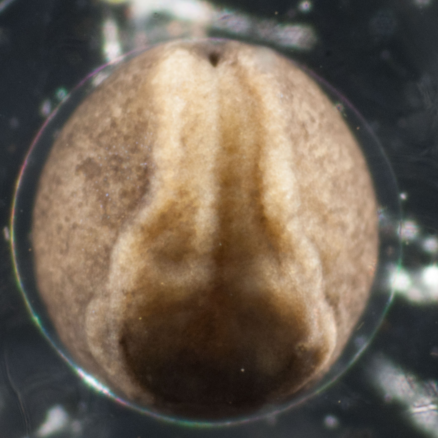
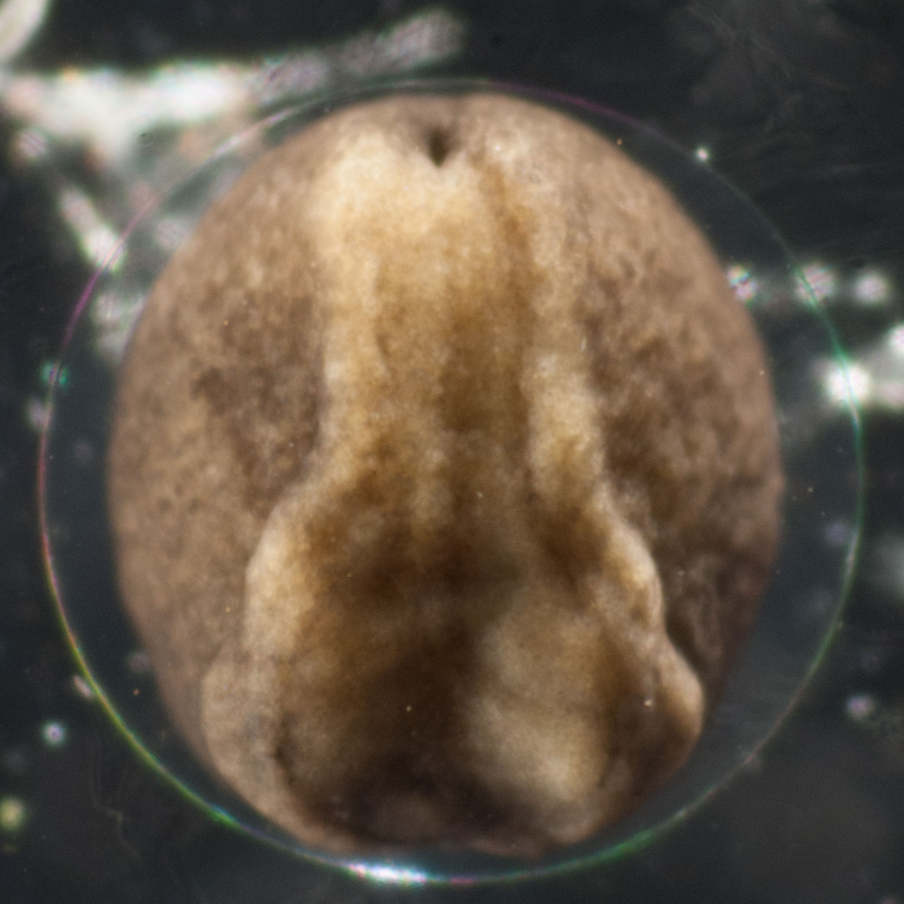

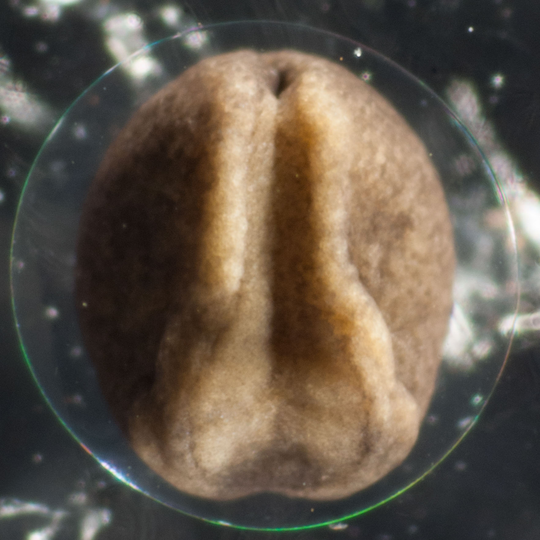


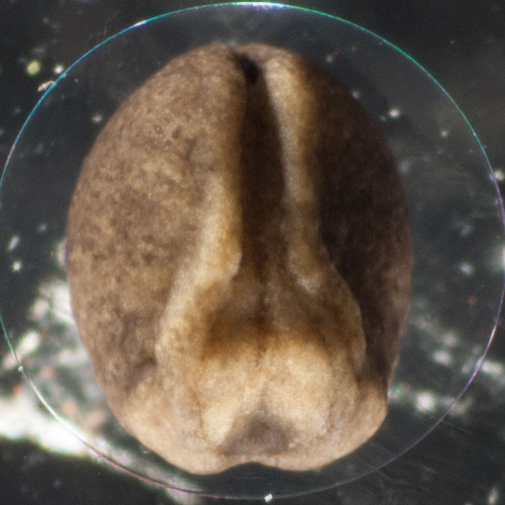
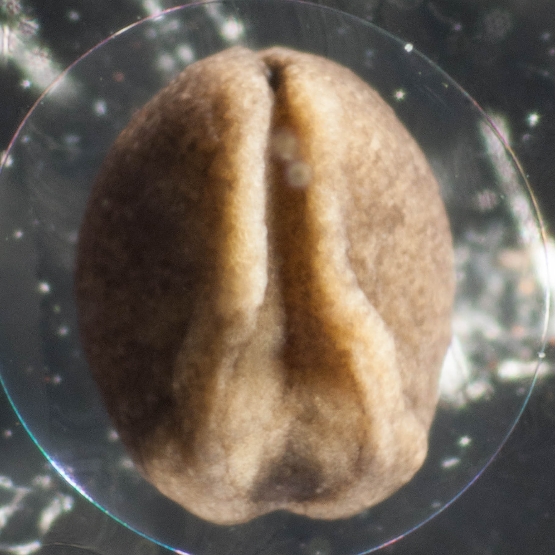
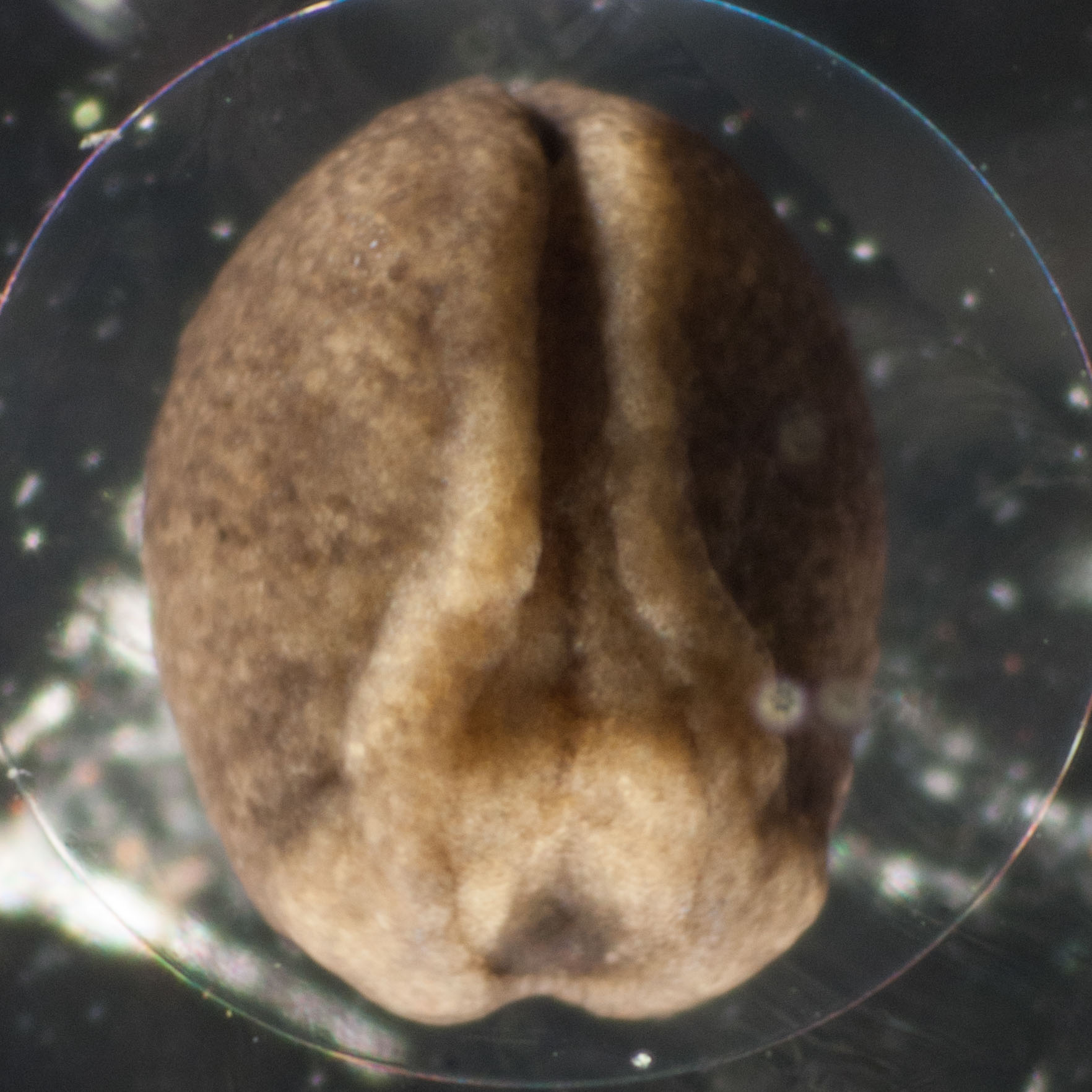
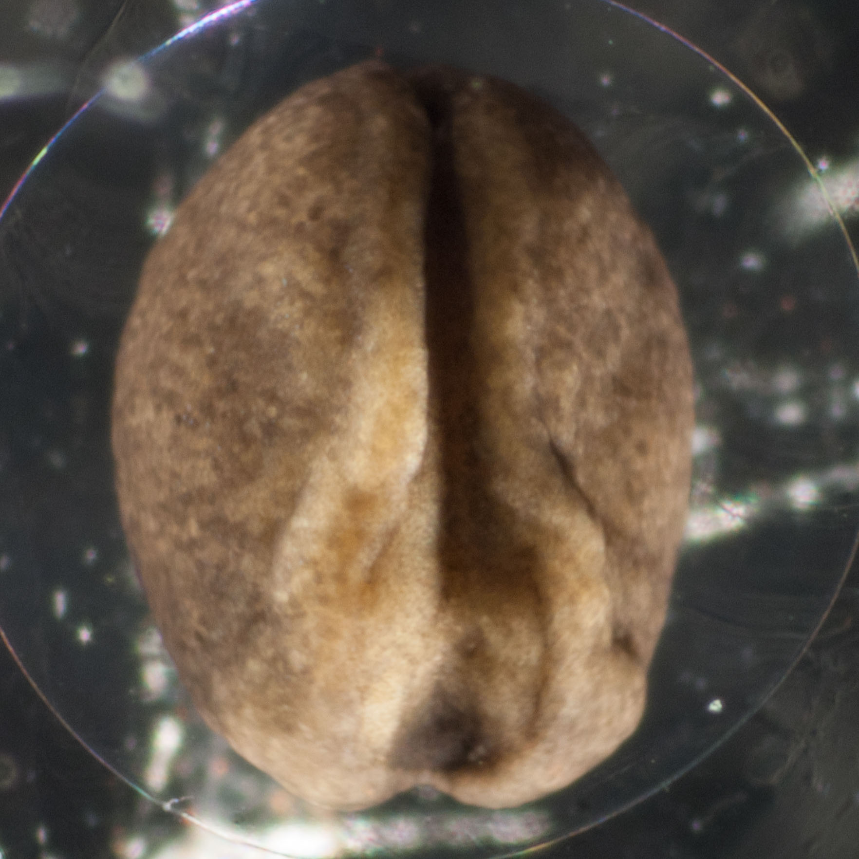
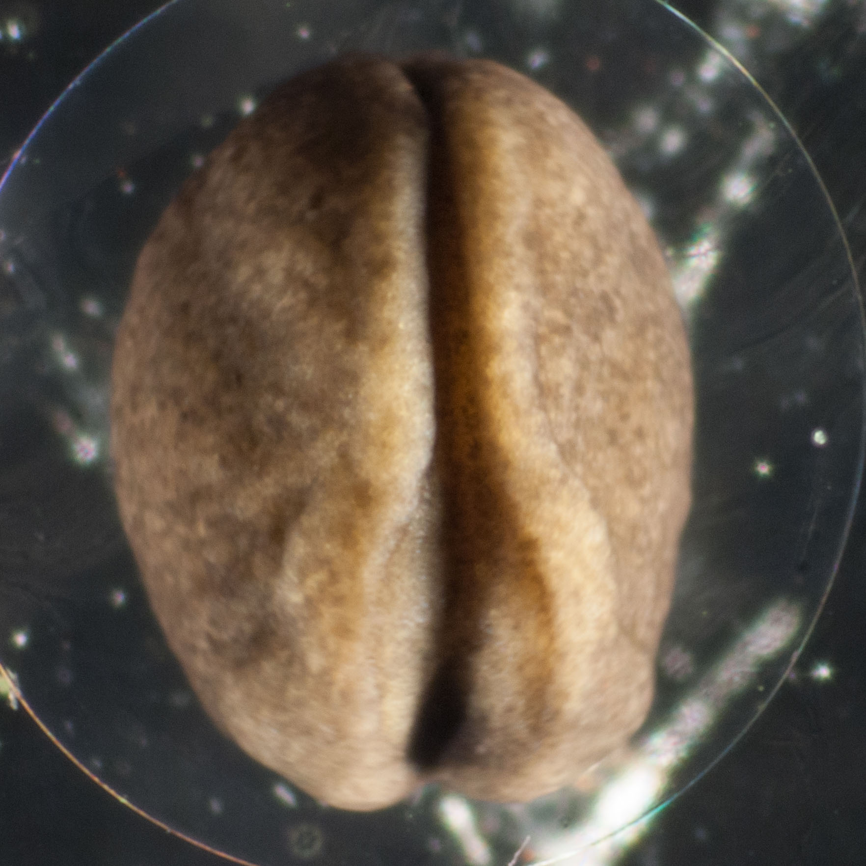
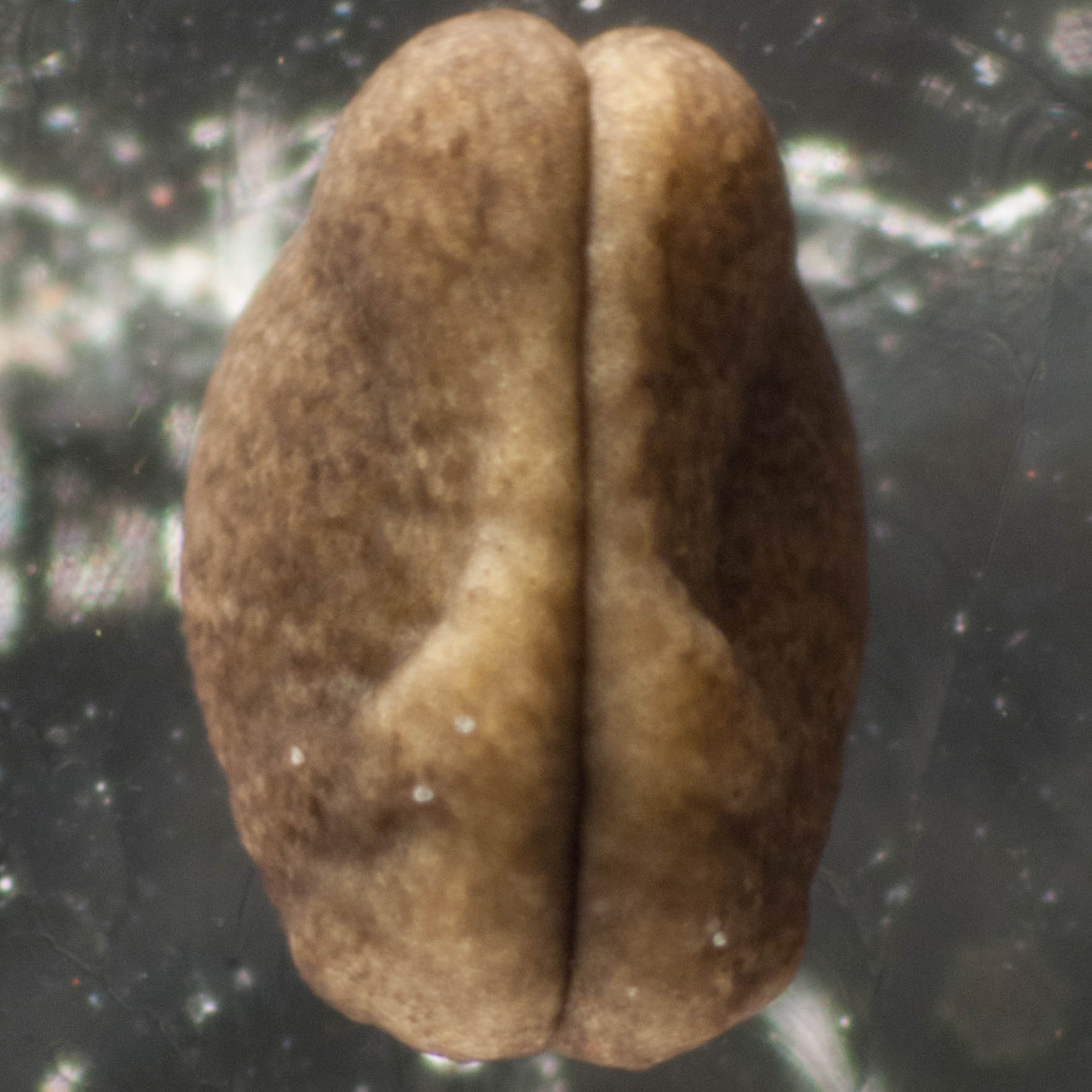
To help you understand what is happening, I've pulled out images 3, 10 and 18 and annotated them below.
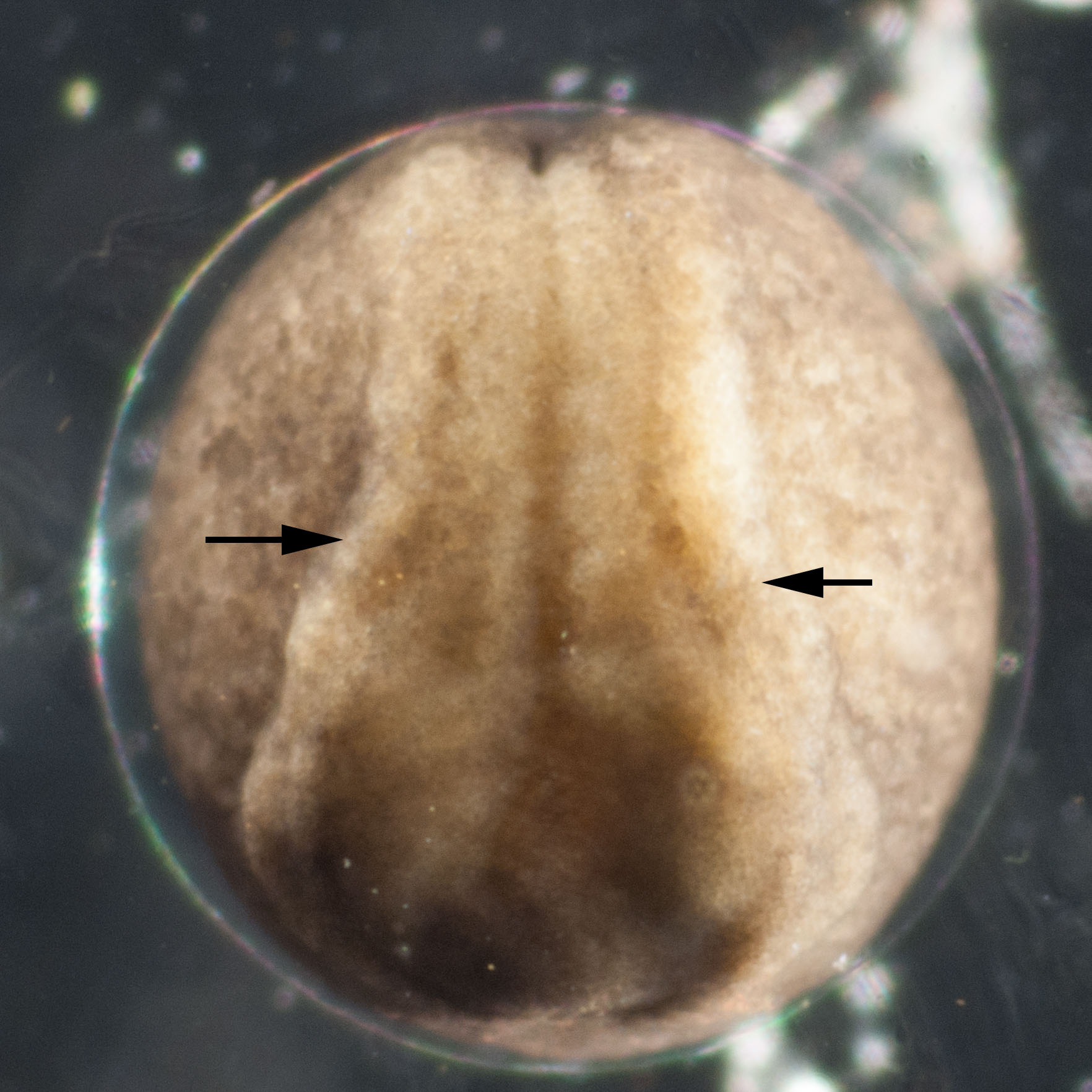
Image 3. A pair of folds (arrowed) rises up from the surface along the length of the embryo, on either side of the midline.
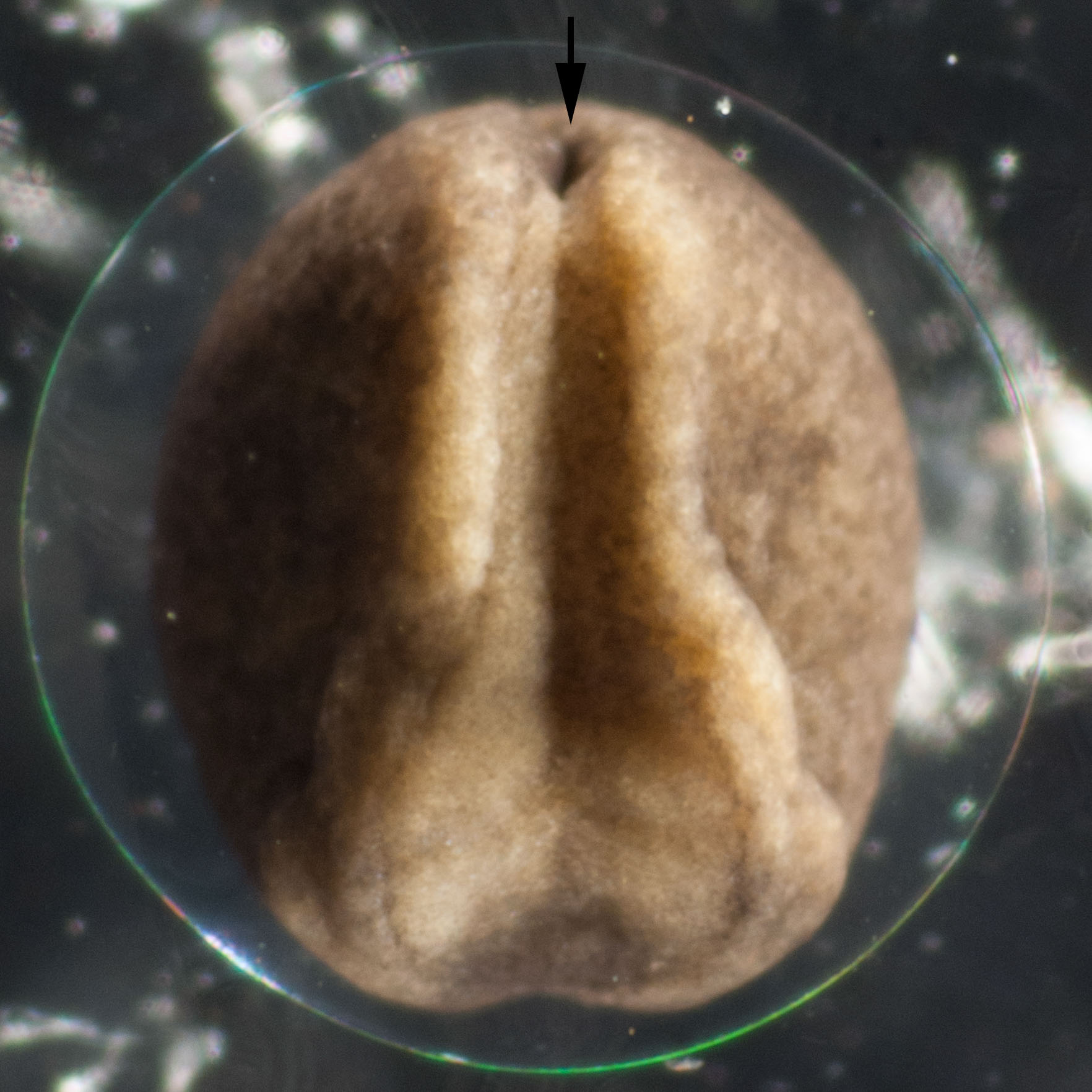
Image 10. The folds are more prominent and are approaching each other in the rear end of the embryo. The arrow points to a hole which becomes the anus.
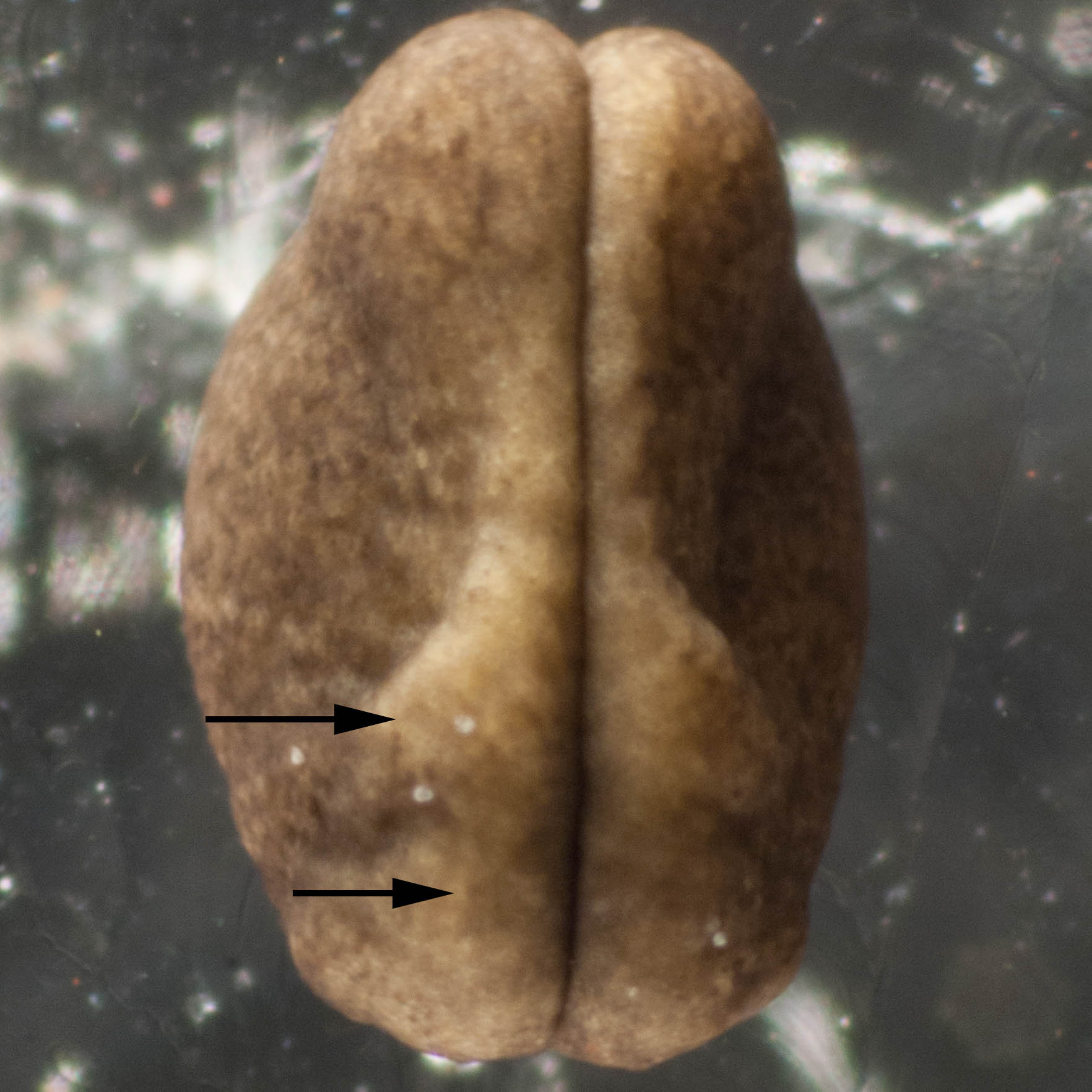
Image 18. The folds have come together at the midline along the whole length of the embryo. Soon they will fuse to form a tube just under the skin. This tube is the forerunner of the brain and spinal cord. The bulge shown by the lower arrow becomes the brain. The other bulge, indicated by the upper arrow, becomes the external gills.
The early stages in the formation of the spinal cord and brain seen here in the frog are common to all animals with backbones (vertebrates) - including humans.
Update: we can now confirm that the species is Litoria ewingii
