Pronotum (viewed dorsally, left wings removed)
Pronotum is long mid-dorsally (double arrow), the posterior edge weakly concave (single arrow heads)
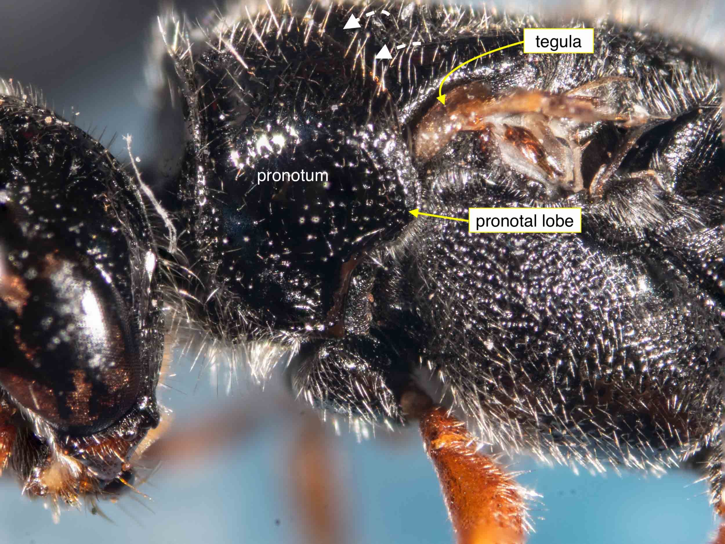
Pronotum (lateral view, wings removed)
The pronotum is free (i.e. distinct) from scutum (dotted arrows) and reaches the tegula, the posterolateral apex rounded (‘pronotal lobe’).
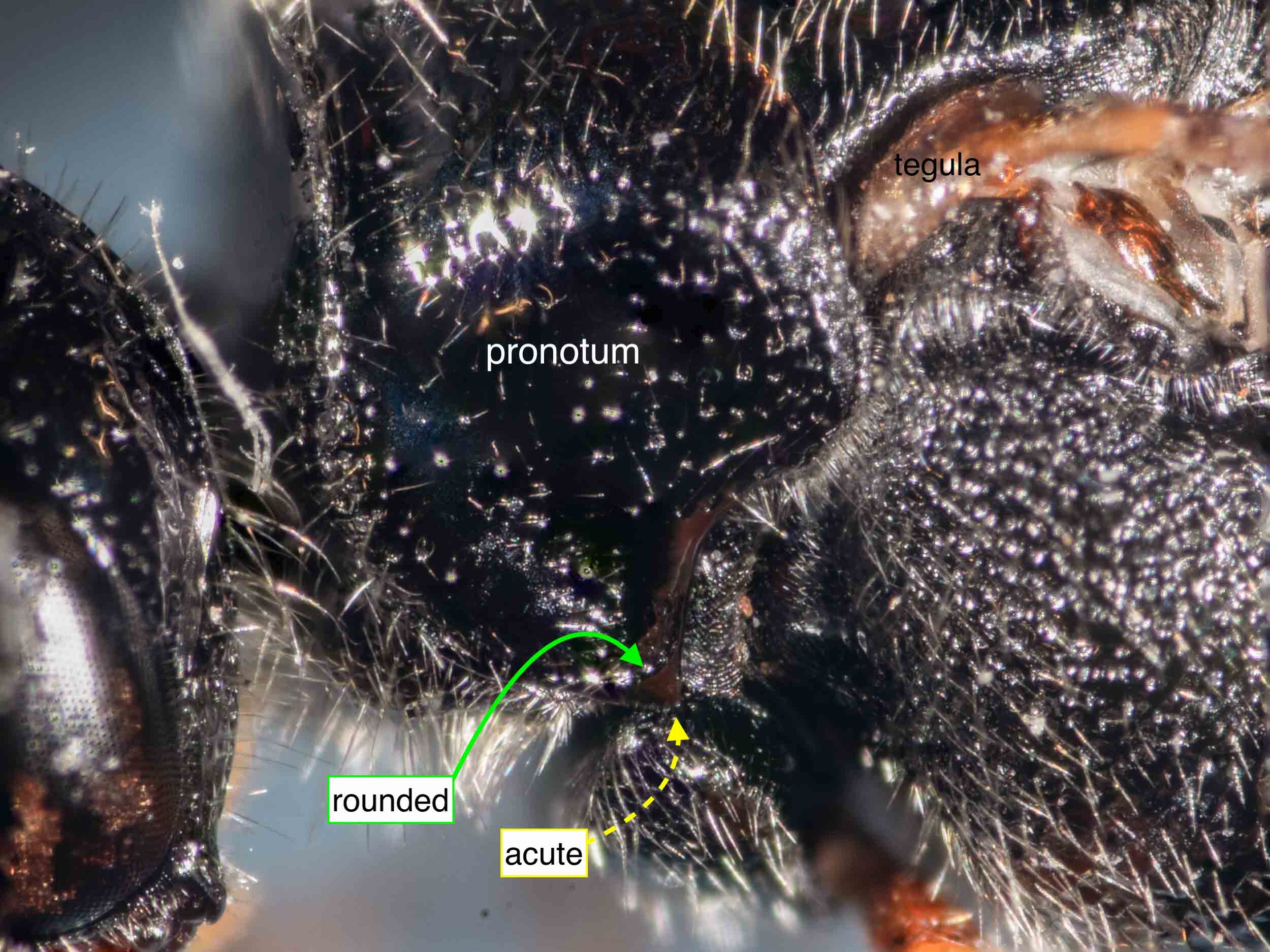
Pronotum (viewed laterally)
Ventrally the apex appears double-edged, both rounded and acute.

Mesosternum (viewed ventrally)
Pair of lightly pigmented (i.e. thin?) plates either side of midline (arrows) project posteriorly from mesosternum and partially conceal the mesocoxae.

Mesosoma, ventral
Bases of mid coxae partially concealed by mesosternal lobes. Fig. 42.32L, Tiphiidae*, mesosoma ventral, male. (Naumann, 1991)
(* most Australian genera in Tiphiidae at the time of Naumann’s work were subsequently elevated to the family Thynnidae.)

Frons (viewed antero-dorsally)
Antennal bases (broken arrows) very slightly obscured by small overhanging ridges (solid arrows).
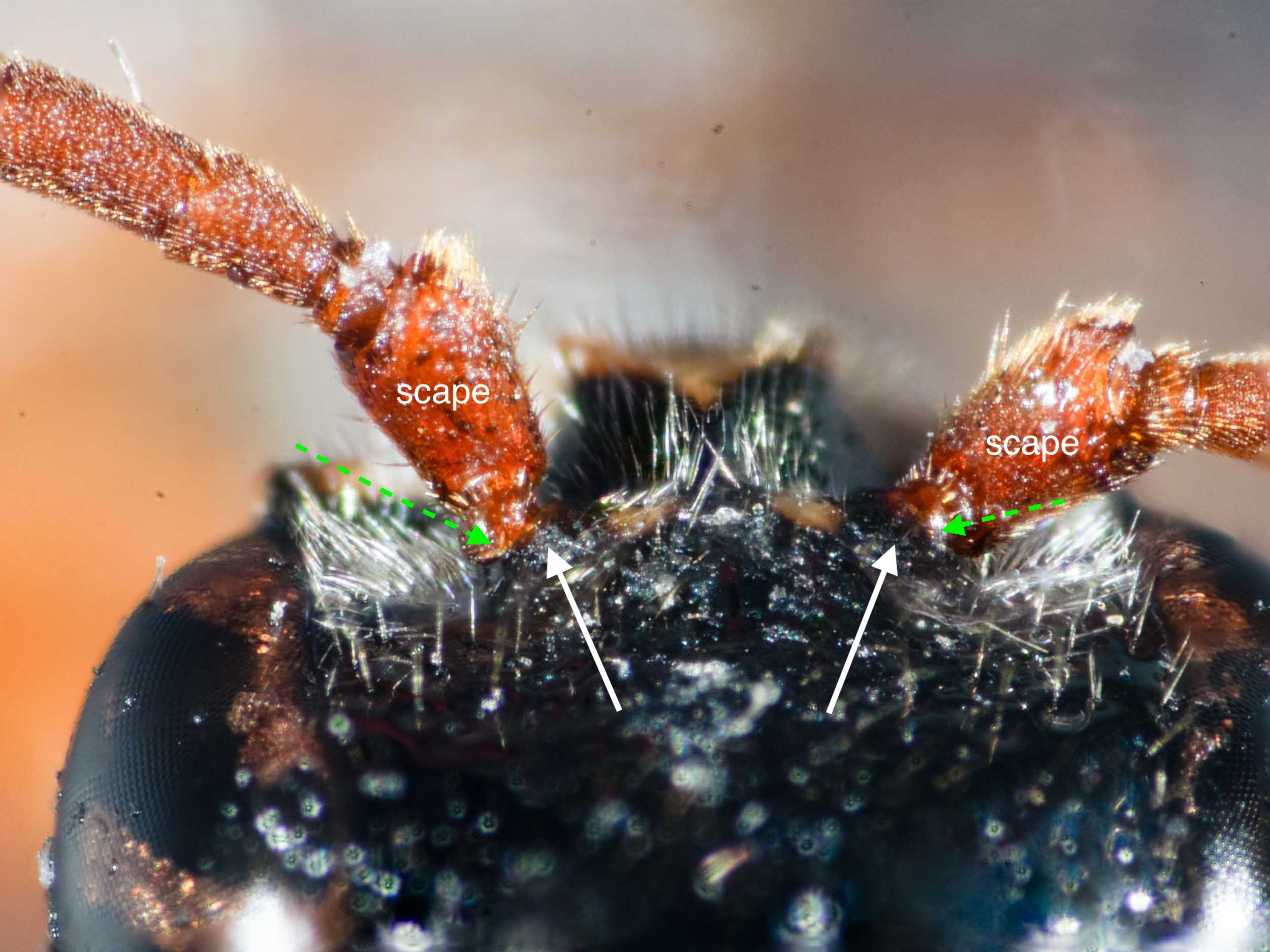
Frons (viewed dorsally)
Antennal bases (broken arrows) very slightly obscured by small overhanging ridges (solid arrows).

Frons of male Thynninae (a) and Anthoboscinae (aa)
Arrows indicate location of torulus (antennal socket).
Extract from Thynnidae key in Brown 1993, page 180. (See associated text in next image)

Contrasting frons structure of Thynninae and Anthoboscinae
Extract from Thynnidae key in Brown 1993, page 180. (See associated figure in next image)

Metasoma (viewed laterally)
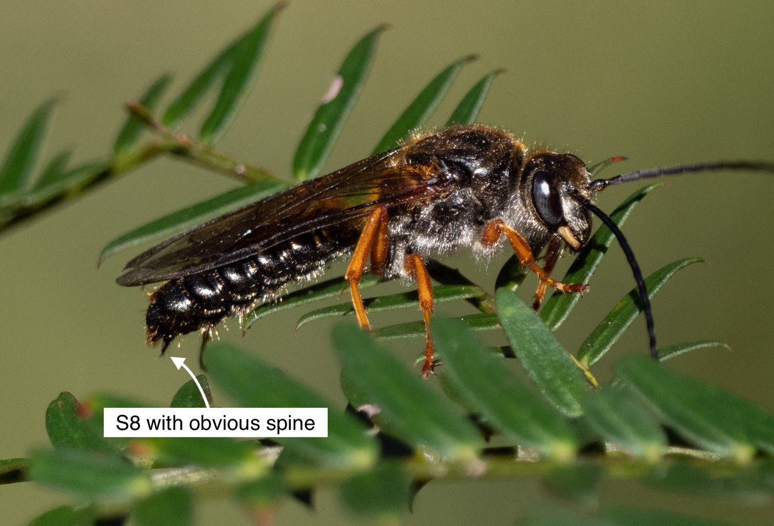
Seven exposed tergites, consistent with male Thynnidae (females have just six). At the apex of the metasoma is a simple cup-like structure (arrow) without visible spines, hooks or other projections.

Apex of metasoma (viewed laterally)
Arrow showing apex, which appears to be the epipygium

Apex of metasoma (viewed dorsally)
Arrows showing epipygium/T7

Apex of metasoma (viewed lateroventrally)
Arrows showing epipygium/T7

Forewing (left, viewed dorsally, flattened)
The first submarginal cell shows a trace of a spur vein, although it is not cuticularised and therefore less obvious than in many Thynninae.

Entire (before removal of wings)

Head and mesosoma (viewed laterally)

Body size
Overall length ~10mm

Wing size
Forewing length ~7mm

Venation (left, removed, flattened, viewed dorsally)
Forewing vein 2m-cu is received distally to 2r-m (which contrasts with the arrangement in Diamminae … see this iNaturalist sighting for illustration in Diamma bicolor male)

Head (frontal view)

Antennal segments
Flagellum with 11 segments (‘flagellomeres’) = male

Ocelli & vertex

Head (viewed anterodorsally)

Head (viewed dorsally)

Propodeum (left wings removed)

Mesosoma (viewed laterally, left wings removed)

Metasoma (viewed ventrolaterally)

Mid and hind leg bases (viewed ventrally)


