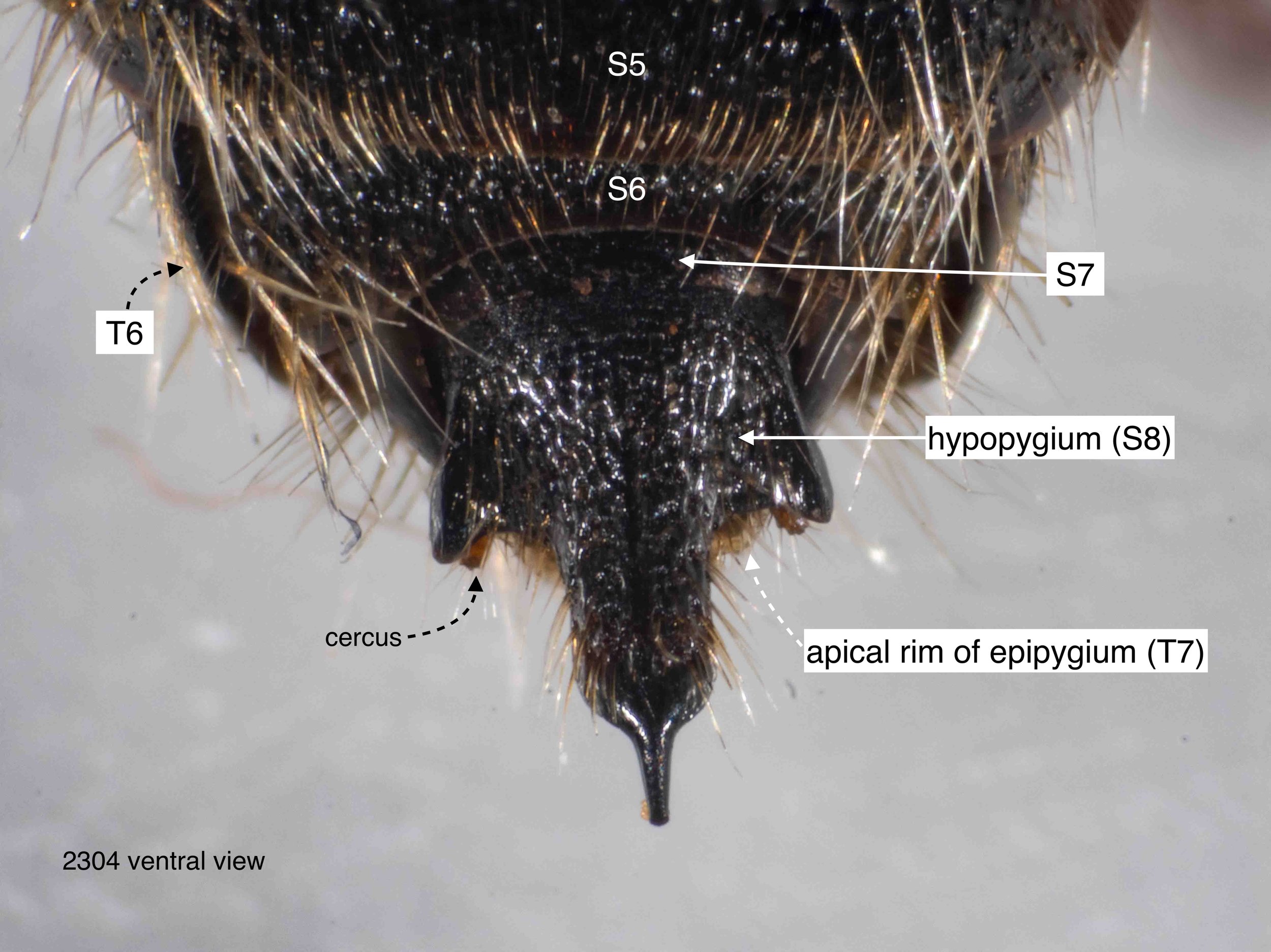
hypopygium in situ
The shape of the hypopygium is apparent when viewed ventrally, at least the apical part. Note that the basal section is concealed beneath the more anterior sternites (S7, S6, S5).
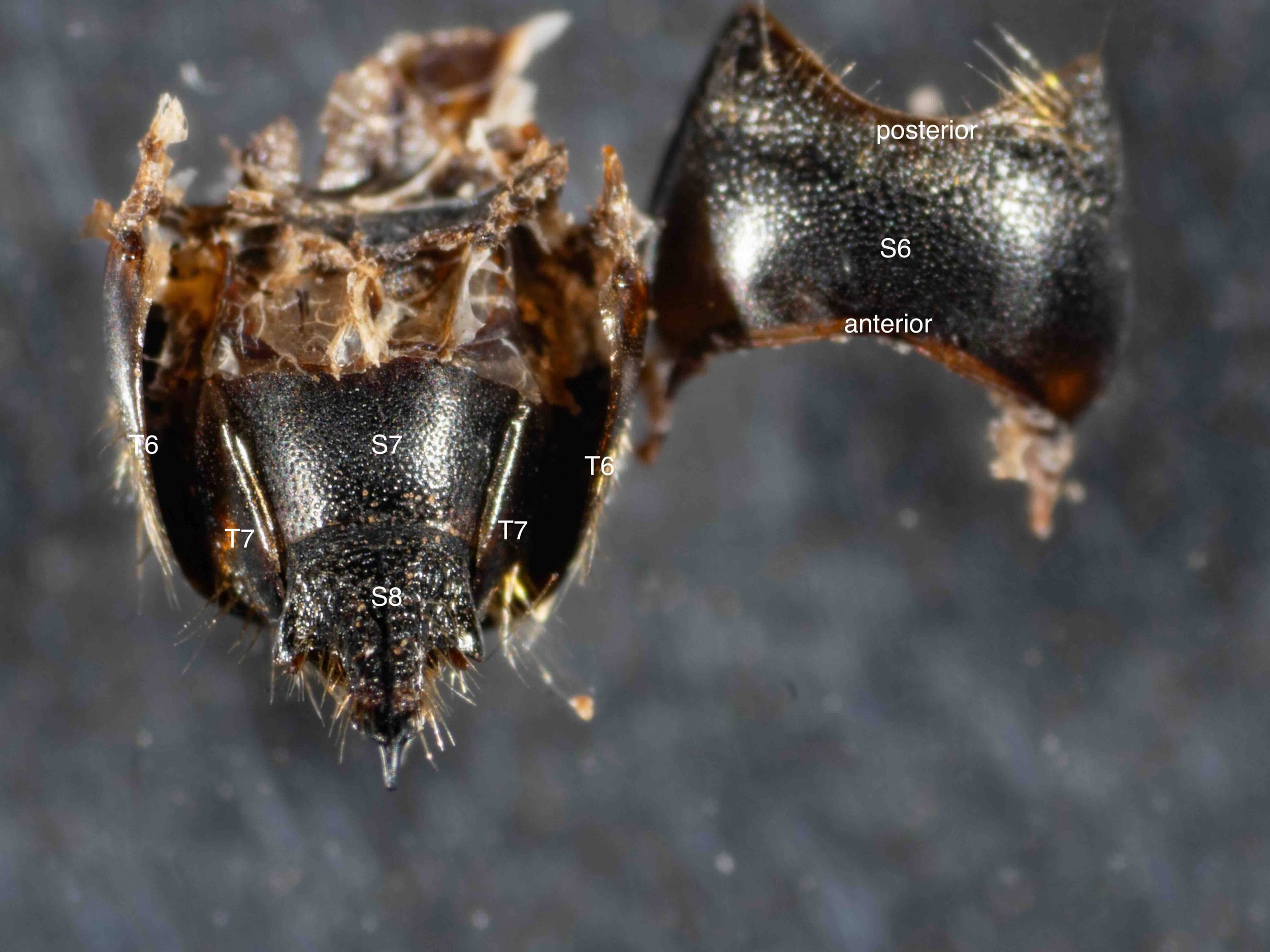
removal of S6
With S6 removed, the shape and extent of S7 is apparent. Note the close fit between the apical rim of S7 and S8.
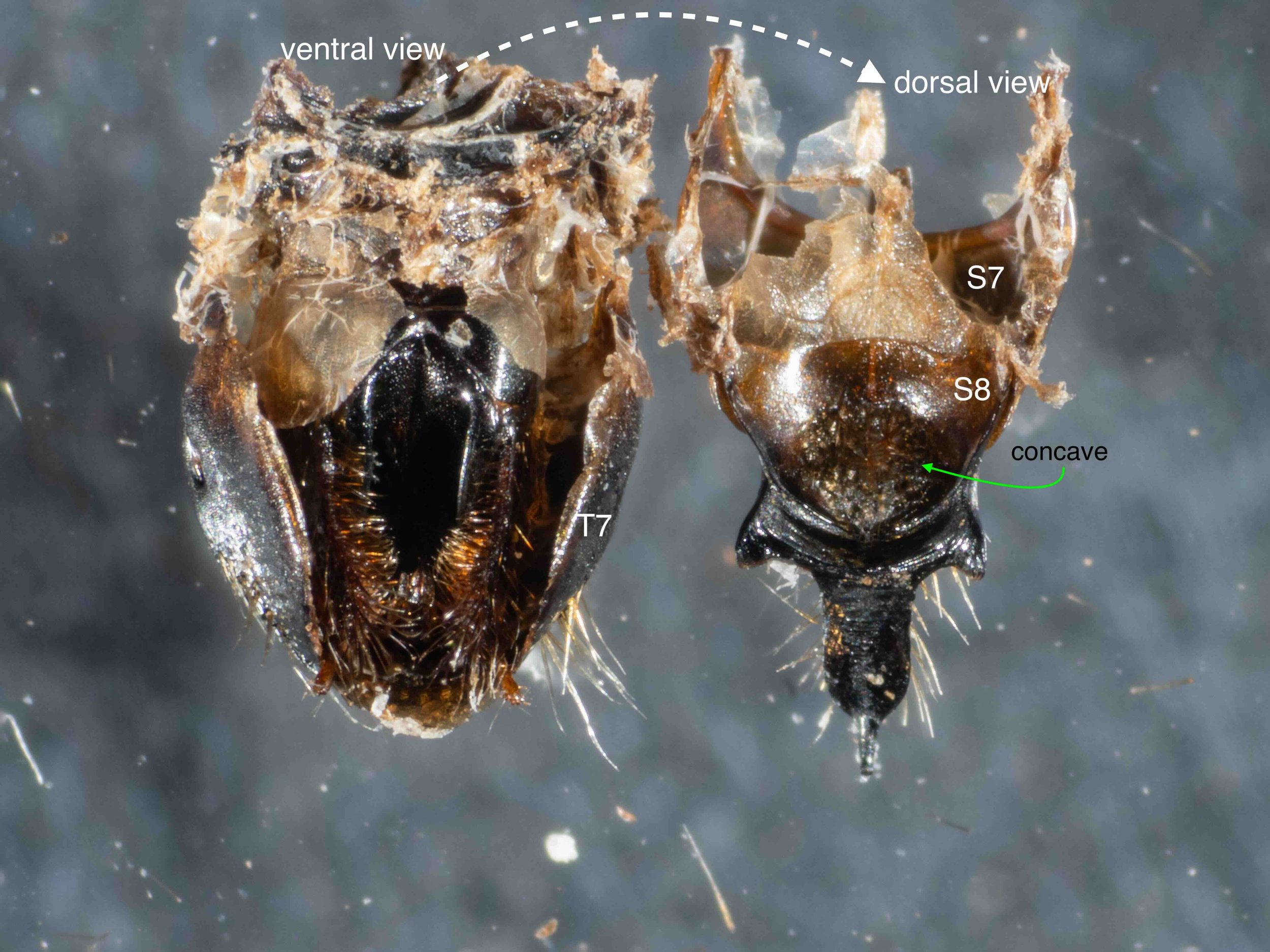
S7+S8 removed and flipped
The two terminal sternites (S7 & S8) are closely adherent and lifted off as a single unit. This revealed the genitalia (phallus), cradled by T7. Note that I have also removed T6 at this stage.
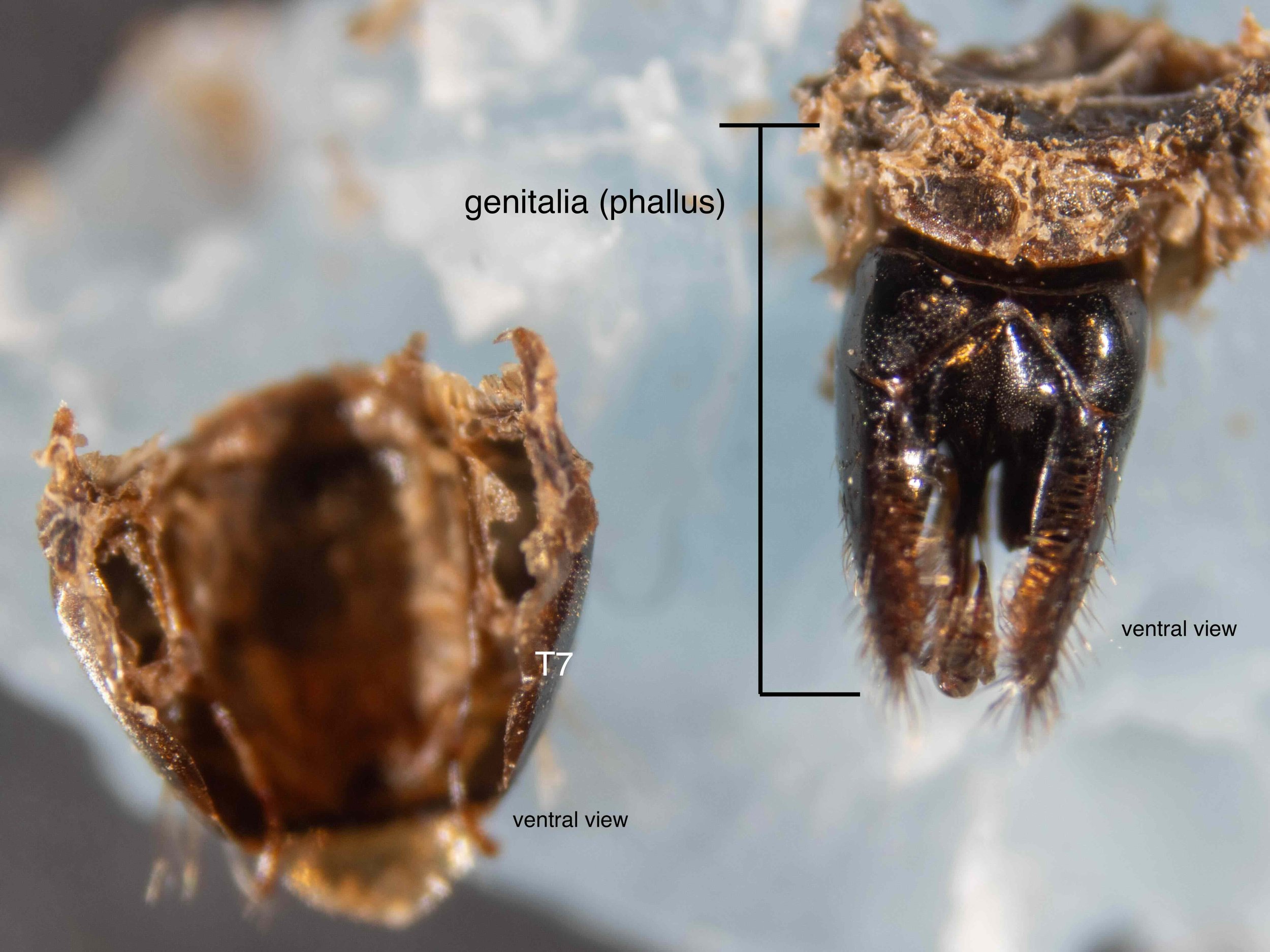
Genitalia (phallus) removed from T7
With just a little persuasion, the phallus was lifted out of the cup formed by T7. The basal part of the phallus bears evidence of its former connection to the genital cavity wall, but the more apical elements are ‘free’.
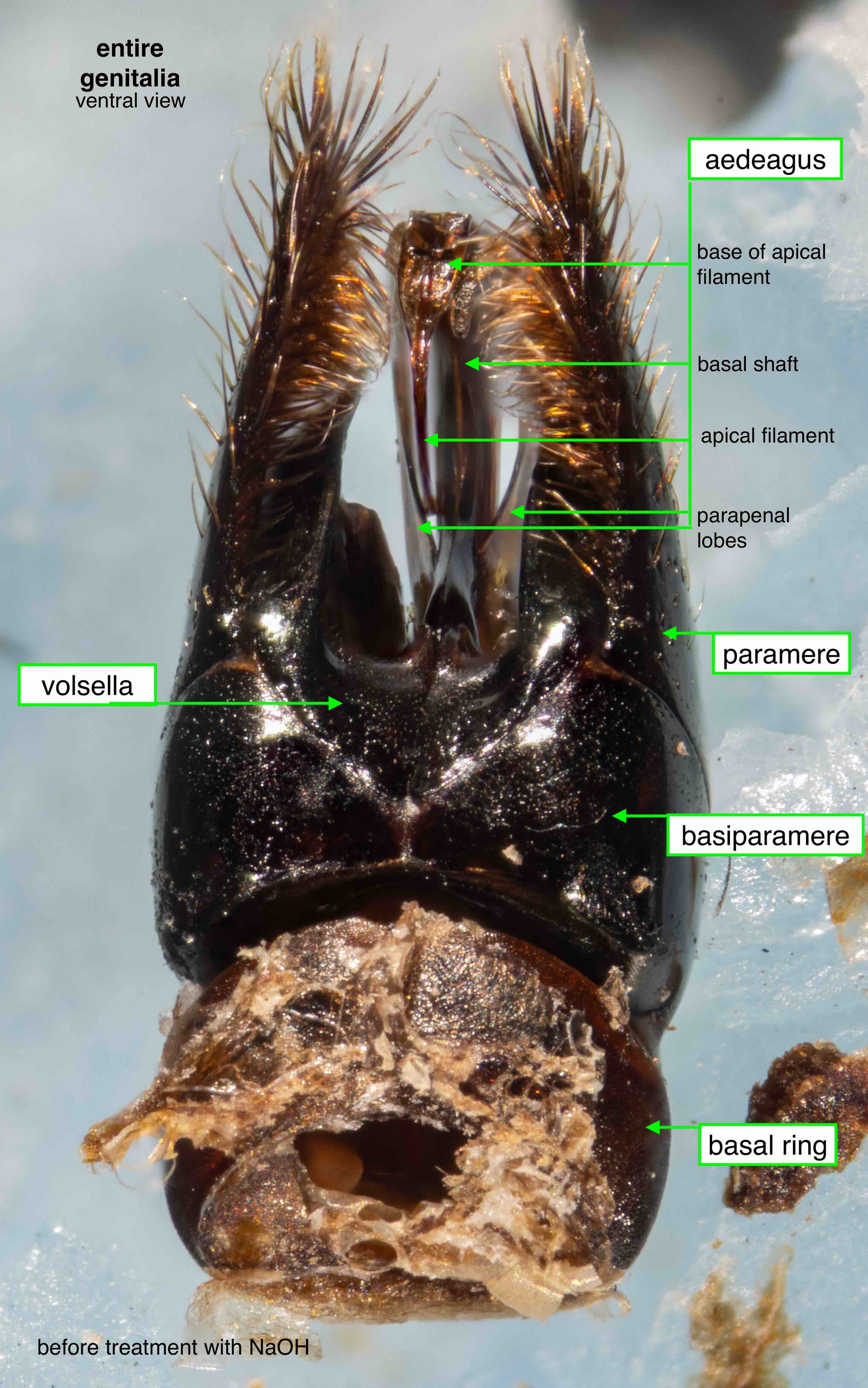
entire phallus
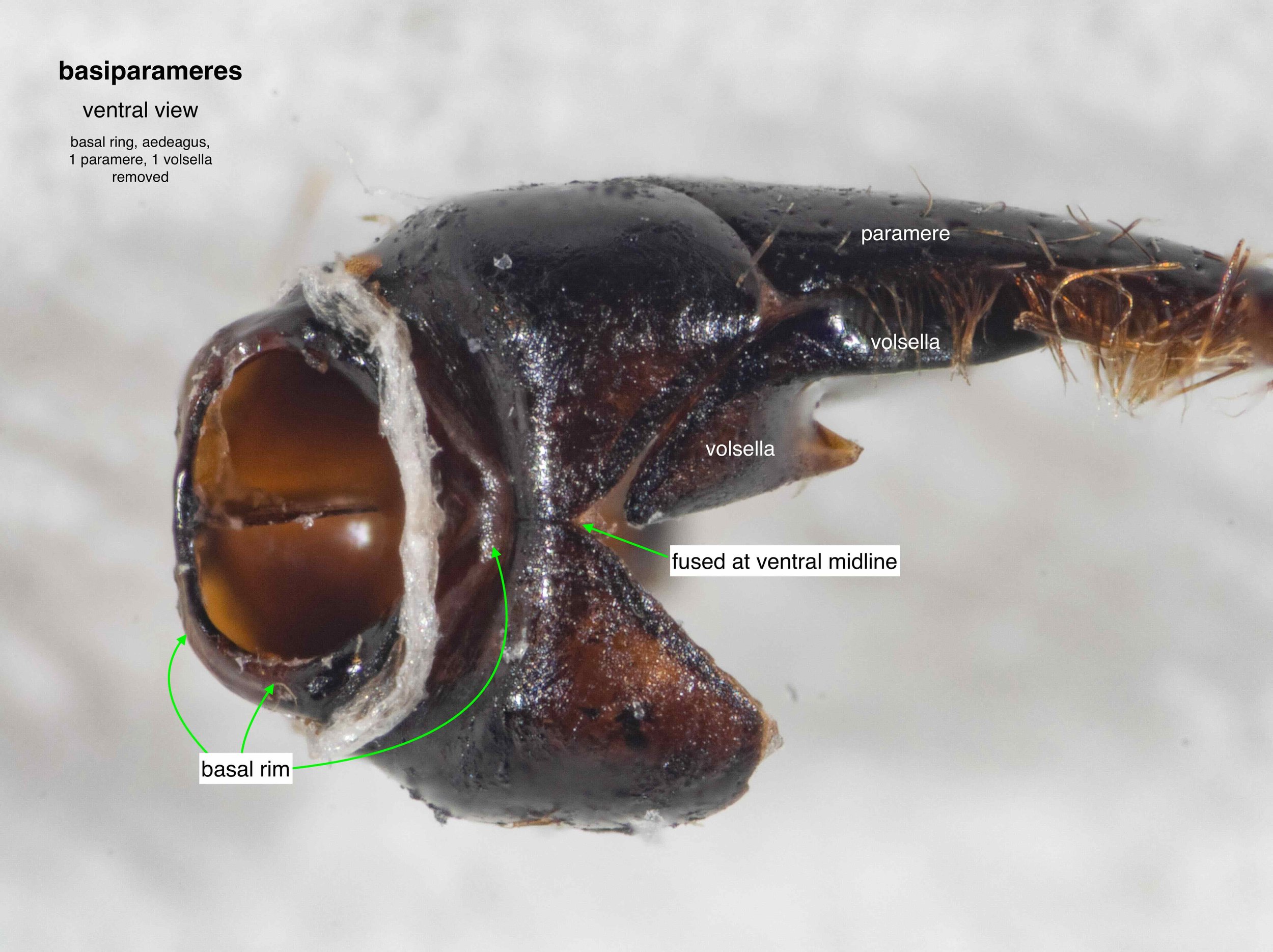
basiparameres
The right and left sides are also fused ventrally, but to a lesser extent.
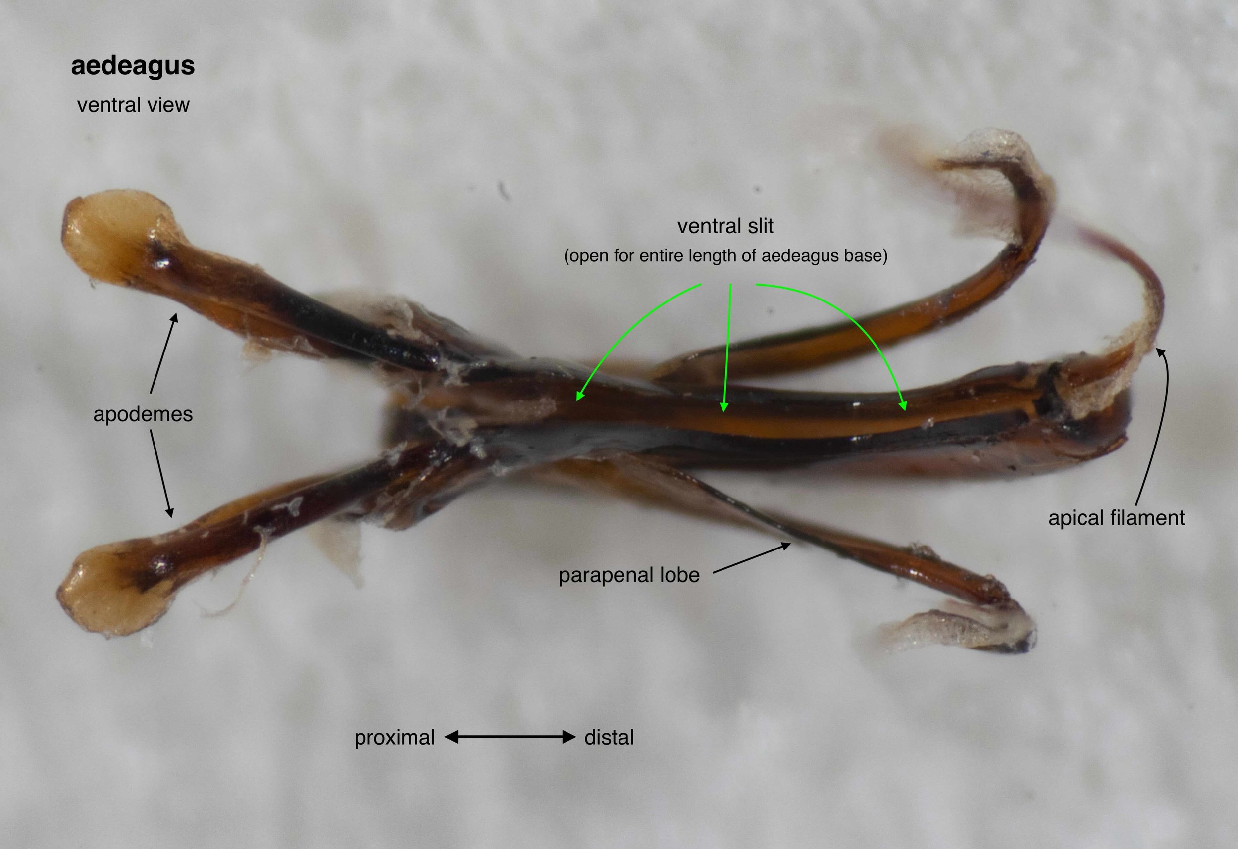
aedeagus
In this species the central shaft of the aedeagus bears a long, ventral slit. This is likely to vary between taxa.
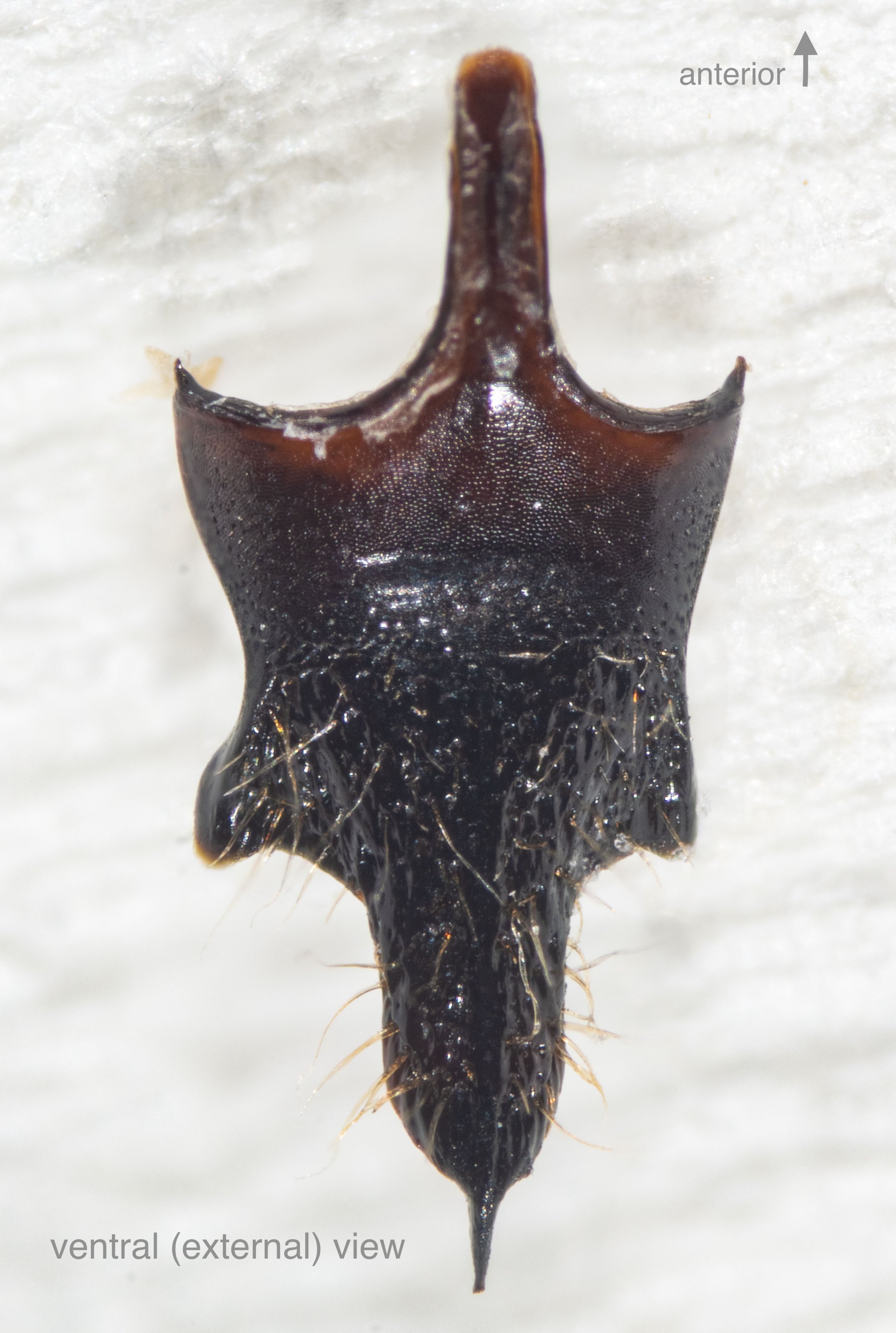
S8 (hypopygium)


