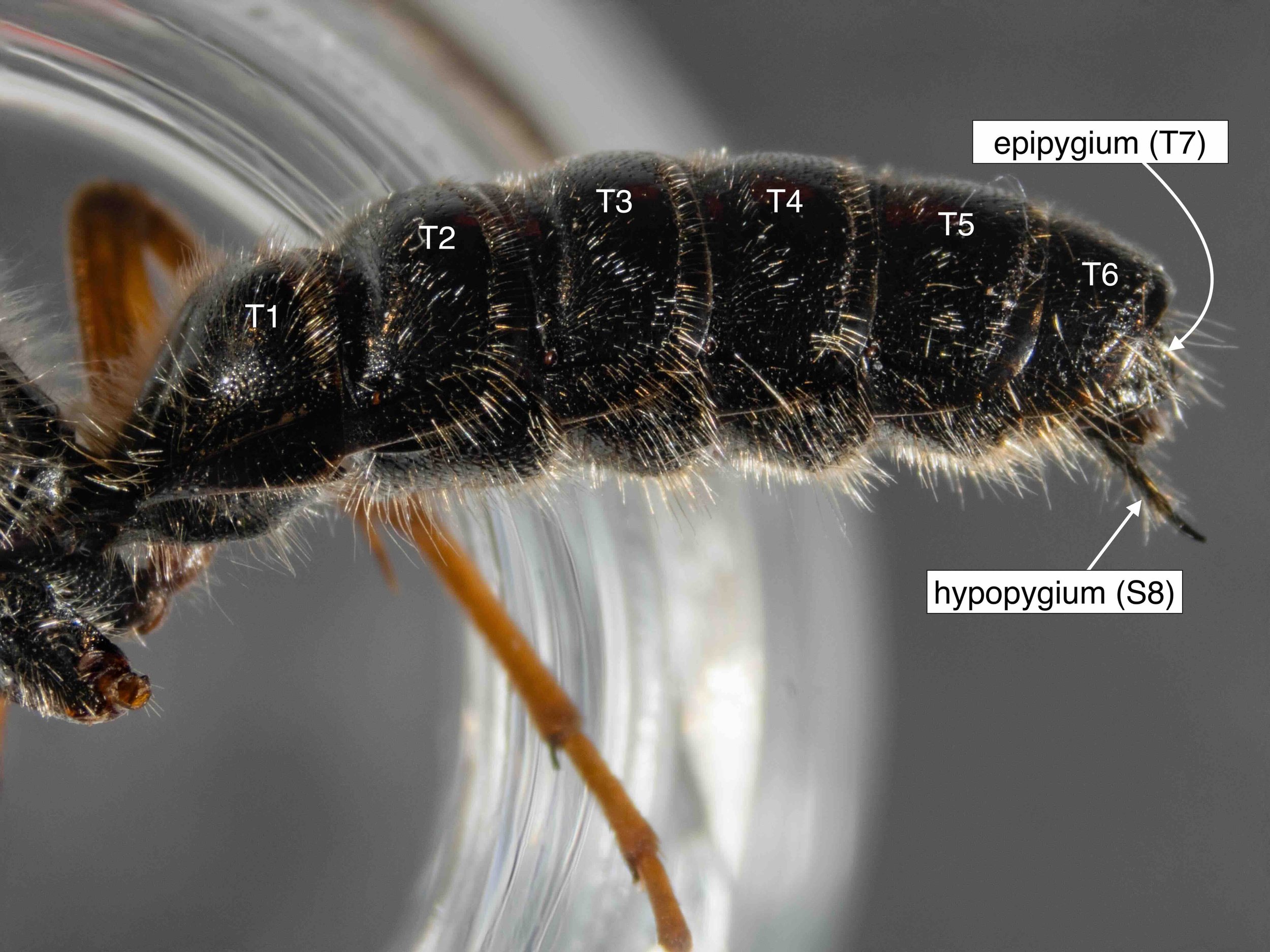
entire metasoma
The coupling apparatus is located at the very end of the body. In many species, including this one, the hypopygium has an apical spine that extends beyond the tip of the metasoma.
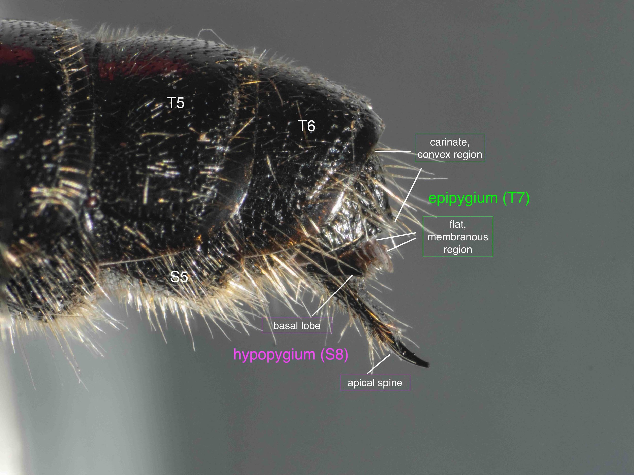
terminal segments of metasoma
Up close, parts of T7 and S8 are visible. Note that S6 and S7 are less apparent, especially when viewed laterally.
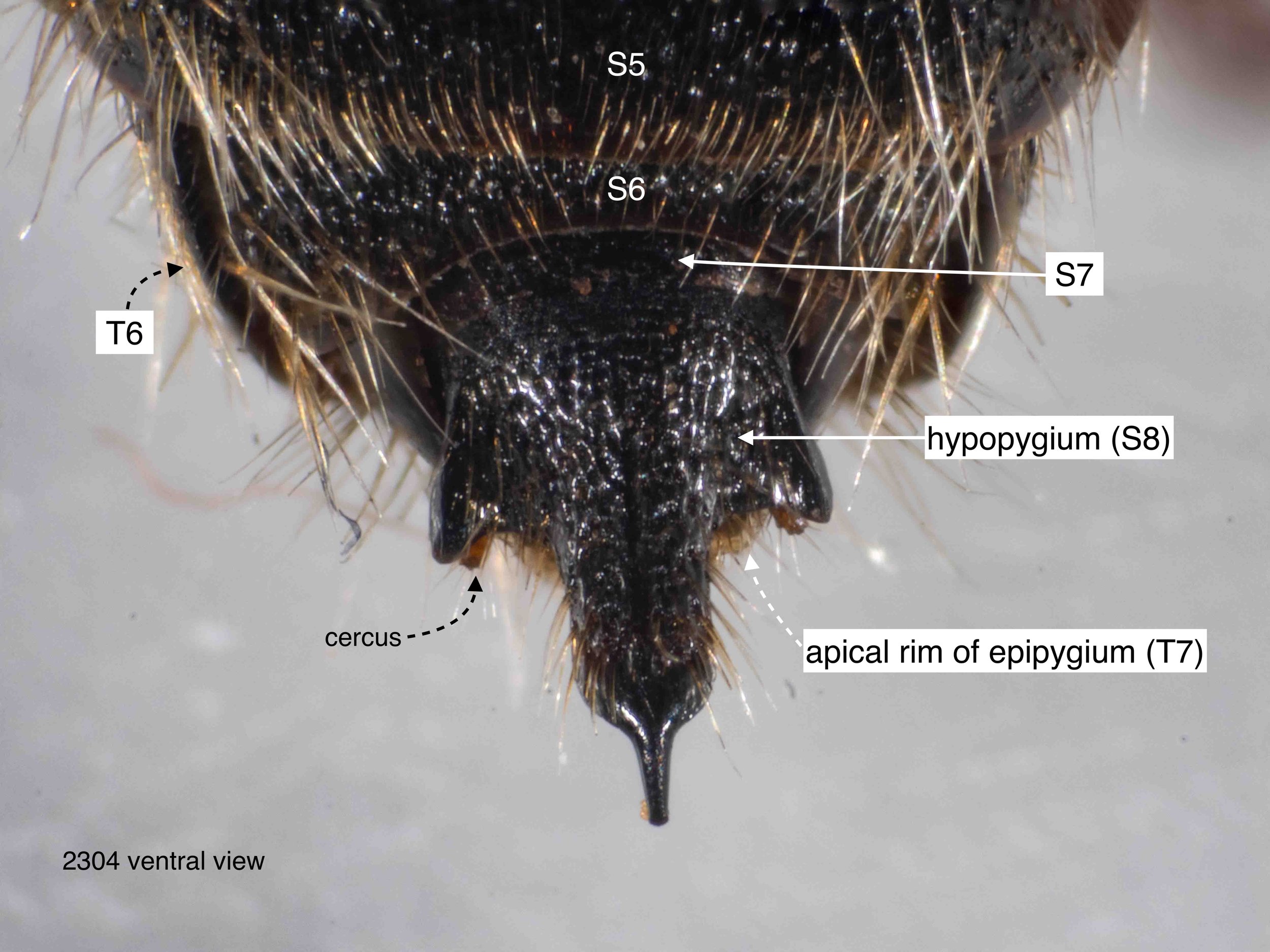
hypopygium in situ
The shape of the hypopygium is apparent when viewed ventrally, at least the apical part. Note that the basal section is concealed beneath the more anterior sternites (S7, S6, S5).
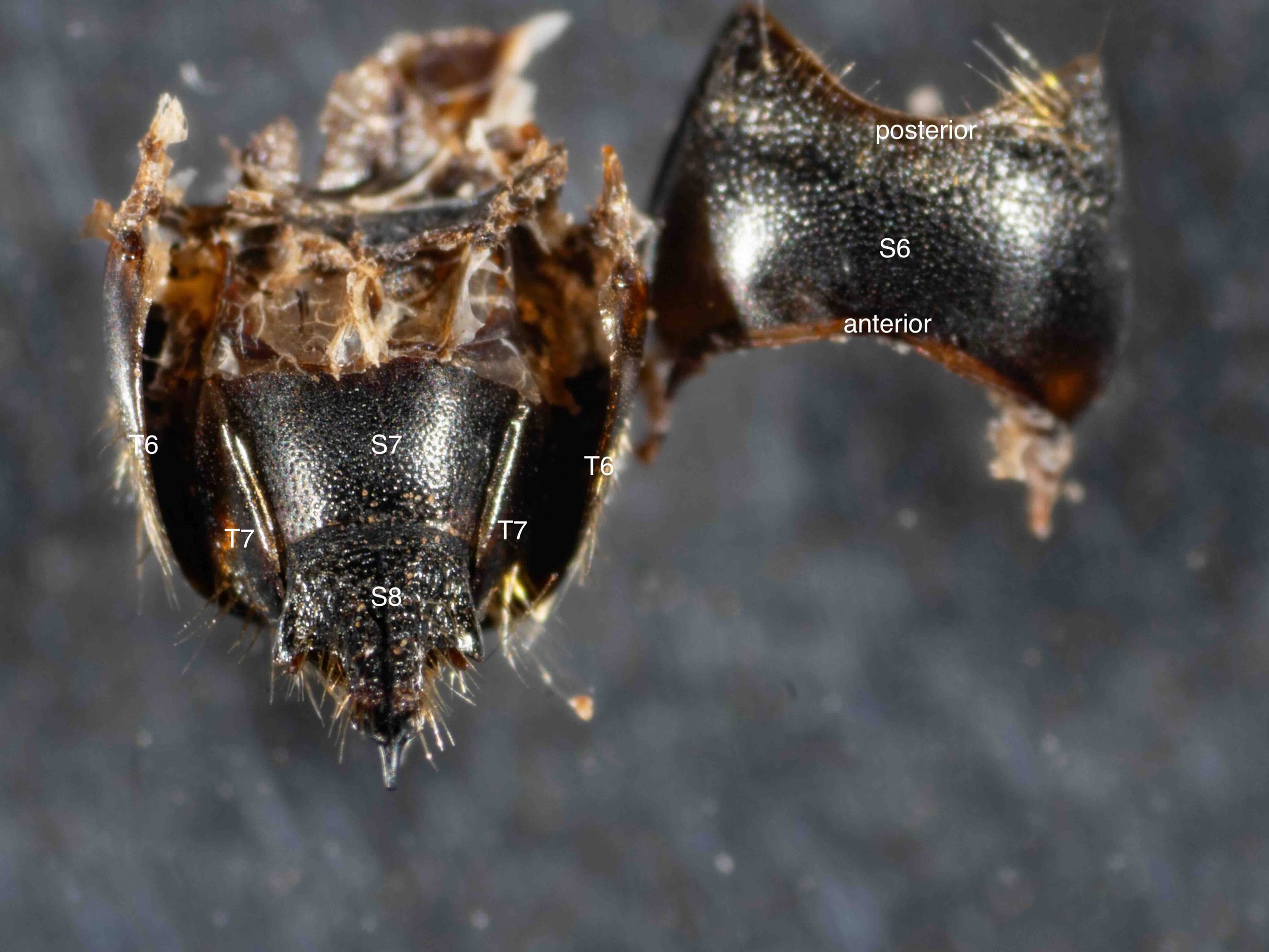
removal of S6
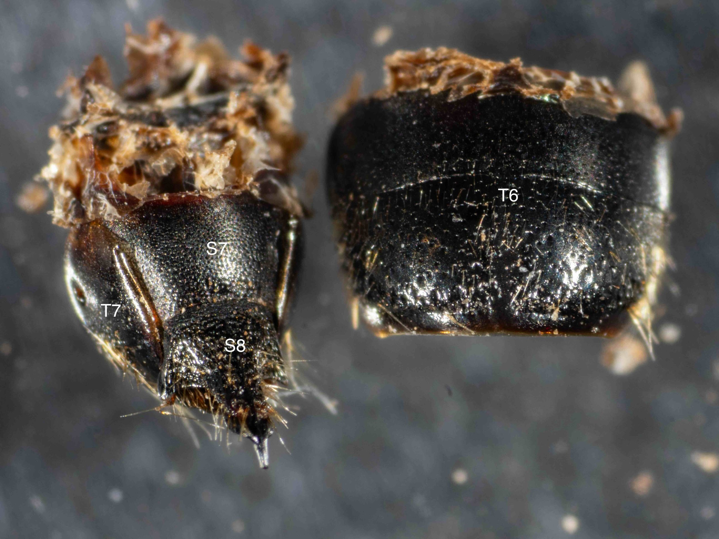
With S6 removed, the shape and extent of S7 is apparent. Note the close fit between the apical rim of S7 and S8.
S7+S8 removed and flipped
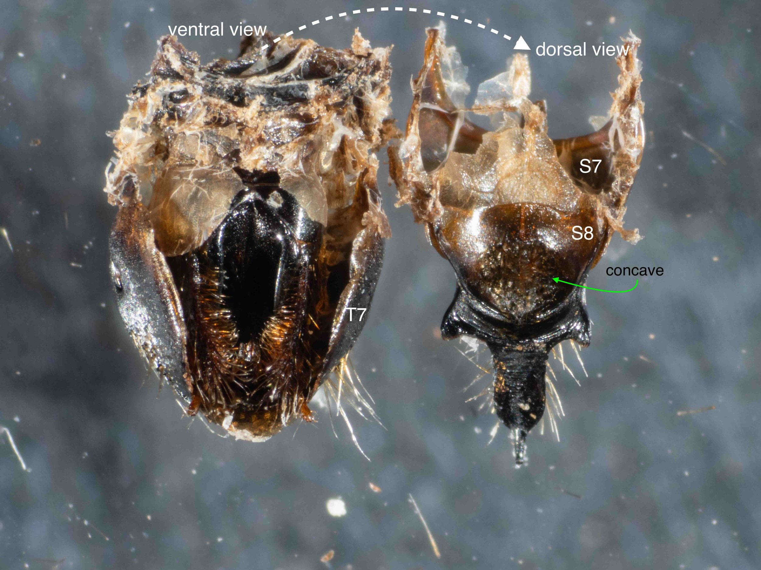
The two terminal sternites (S7 & S8) are closely adherent and lifted off as a single unit. This revealed the genitalia (phallus), cradled by T7. Note that I have also removed T6 at this stage.
S8 separated from S7
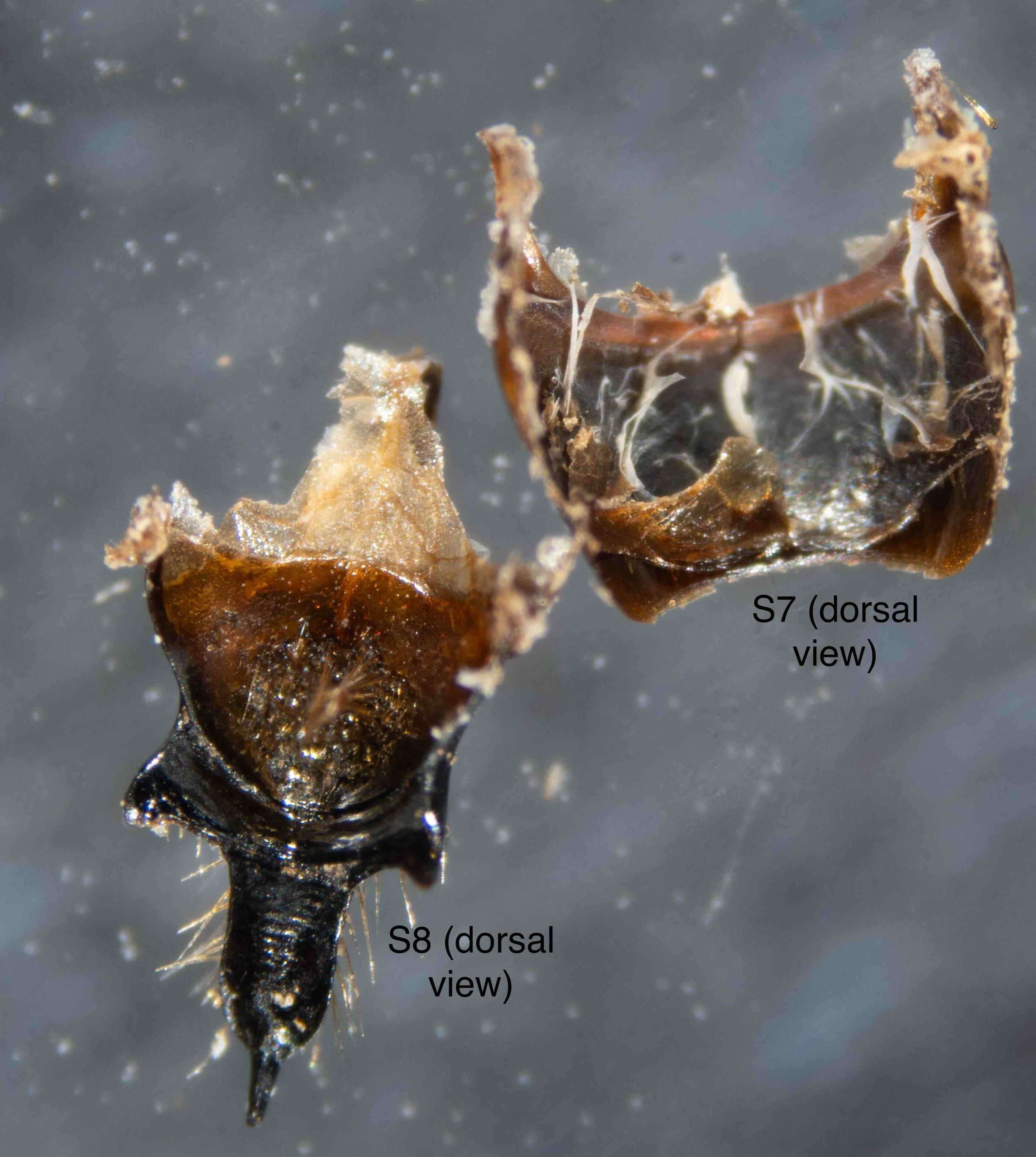
Despite their close attachment, S7 and S8 are distinct sclerites. The basal part of S8, normally concealed, is quite extensive.
Genitalia (phallus) removed from T7
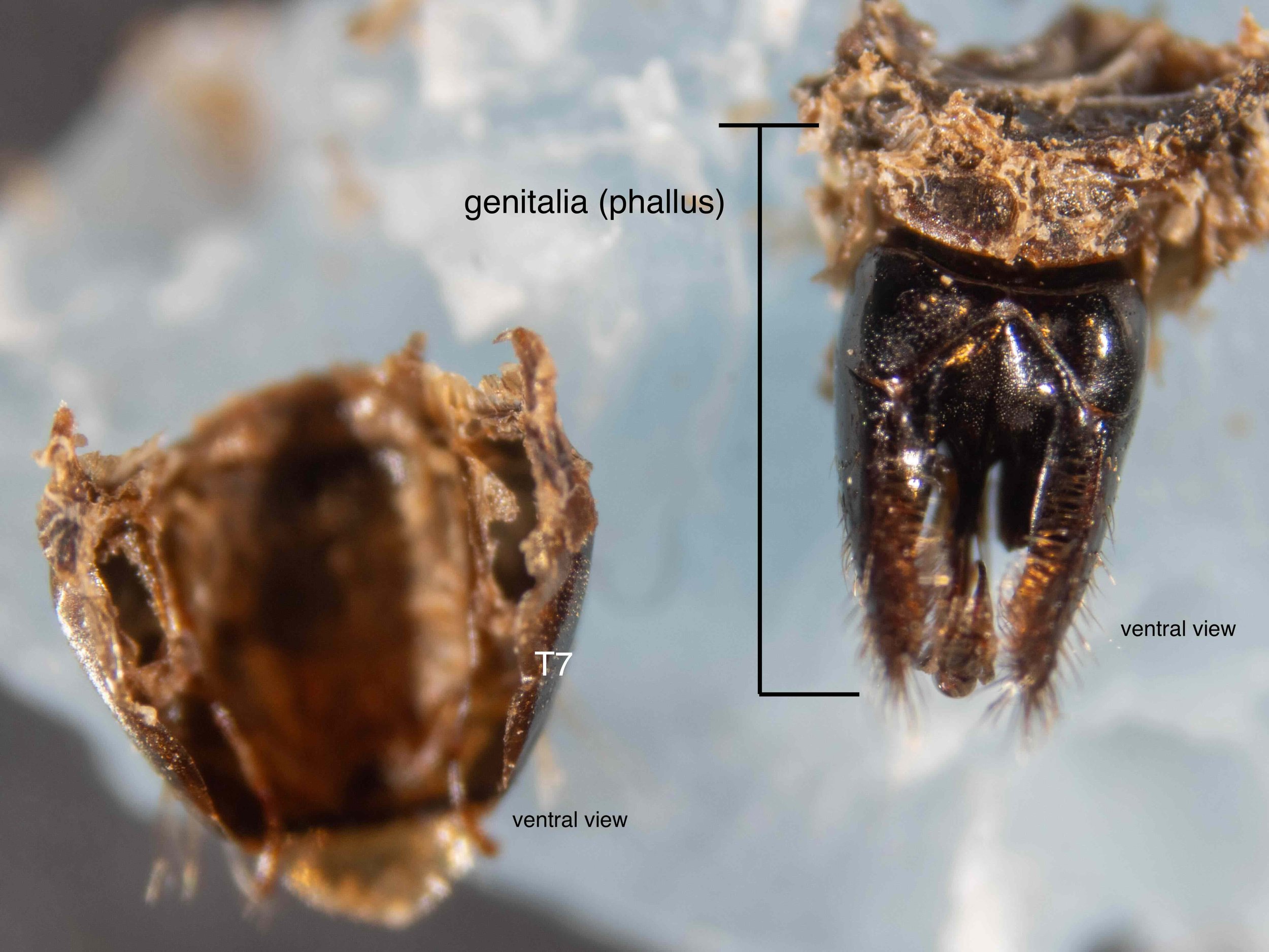
With just a little persuasion, the phallus was lifted out of the cup formed by T7. The basal part of the phallus bears evidence of its former connection to the genital cavity wall, but the more apical elements are ‘free’.
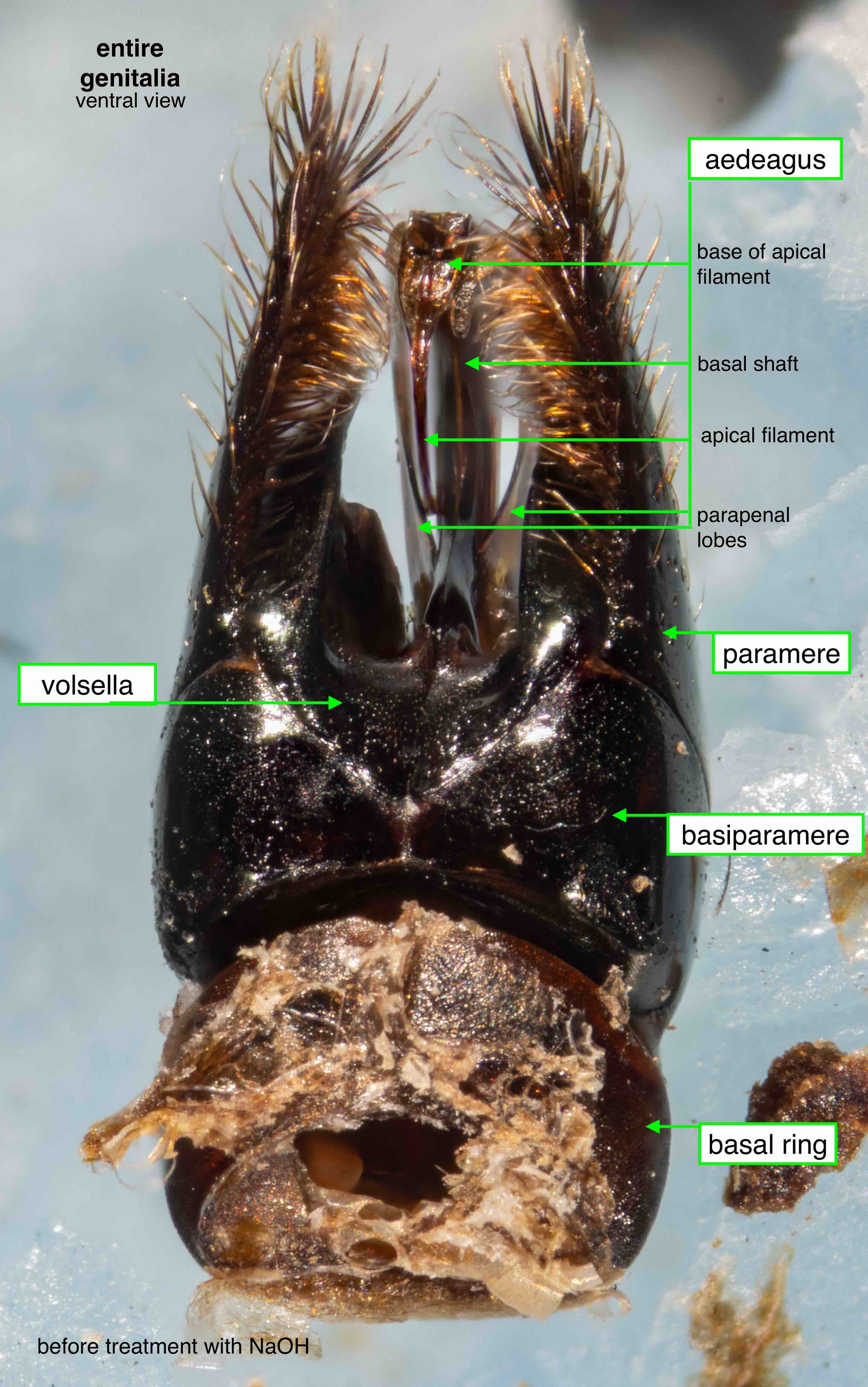
entire phallus
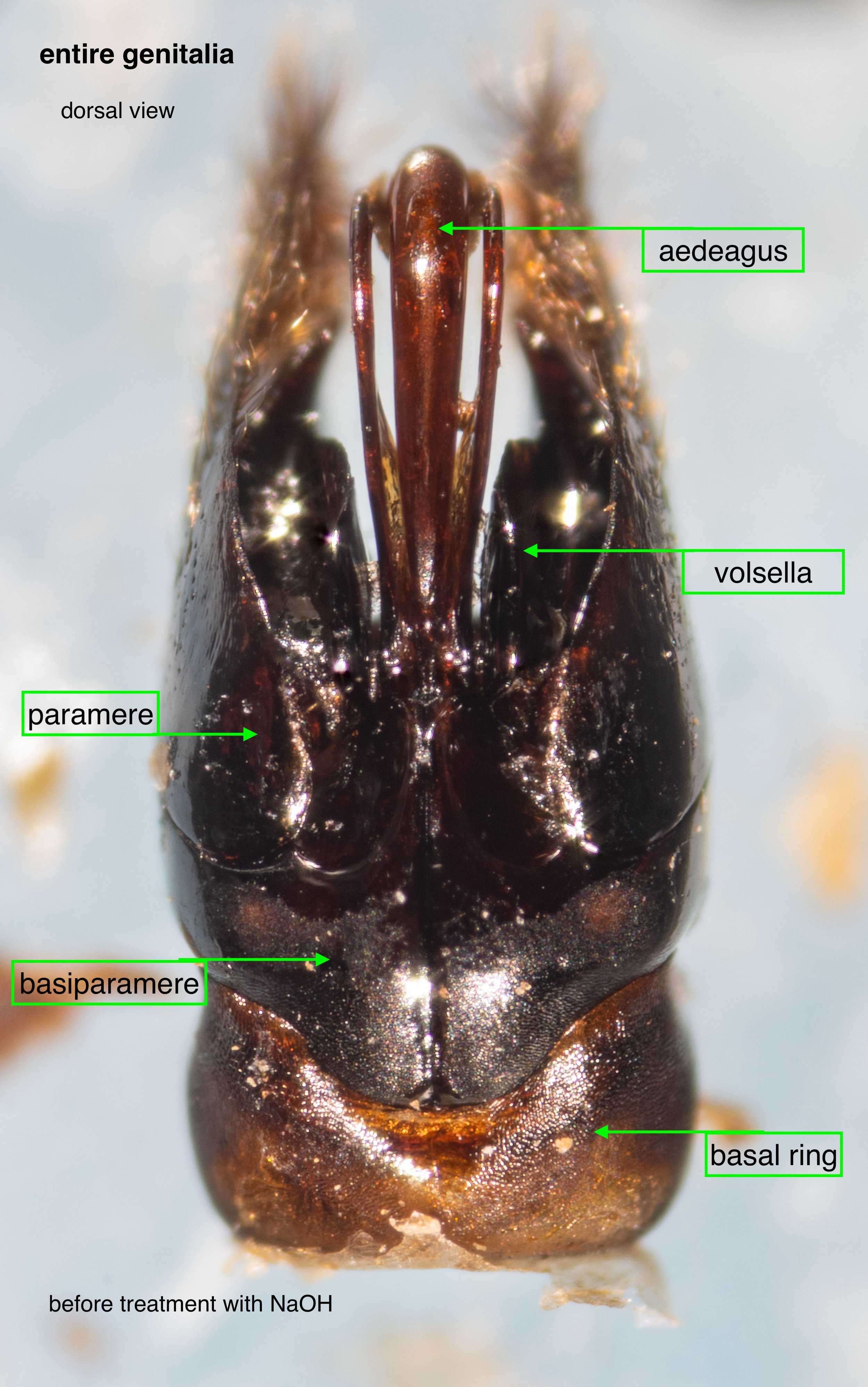
entire phallus
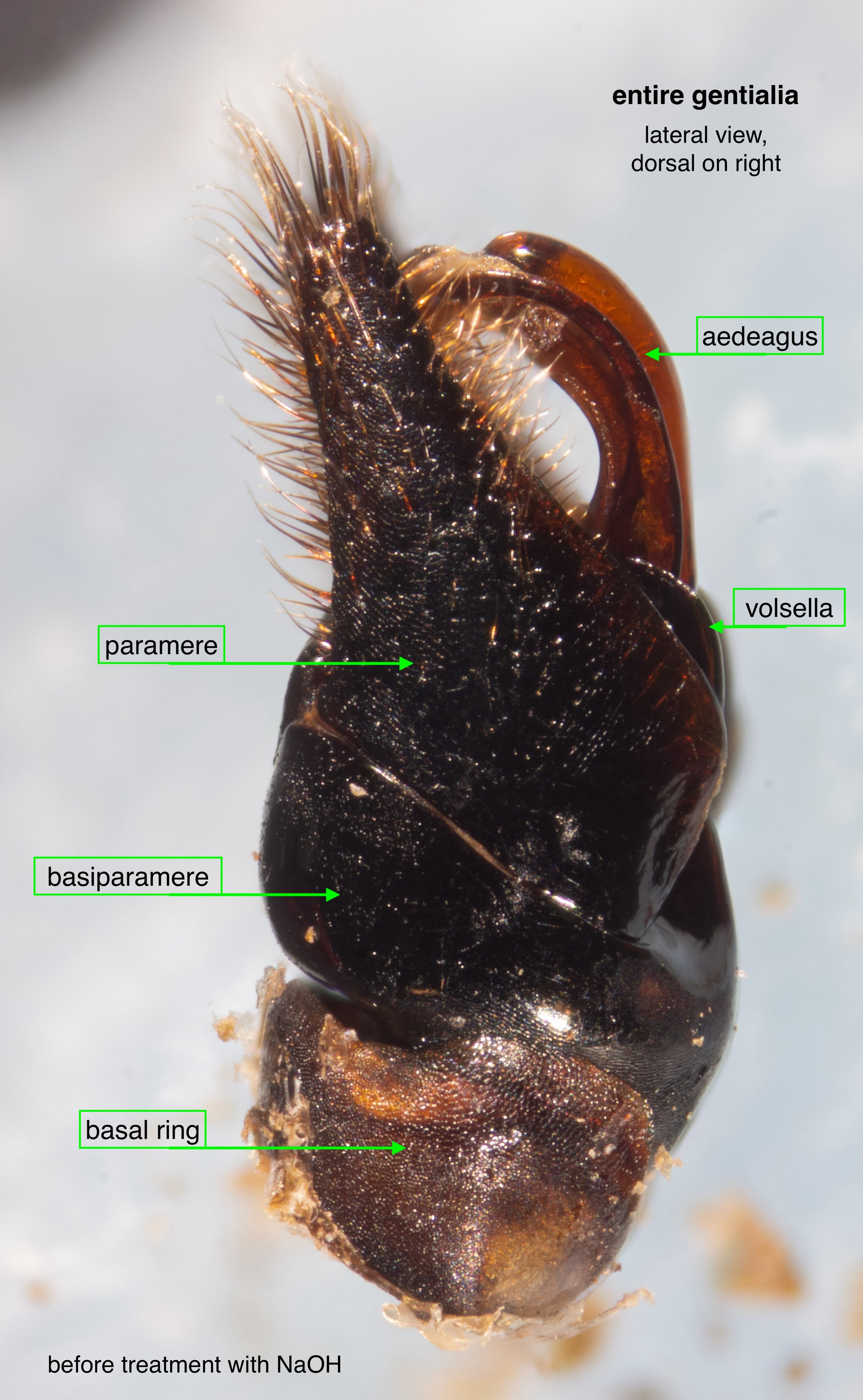
entire phallus
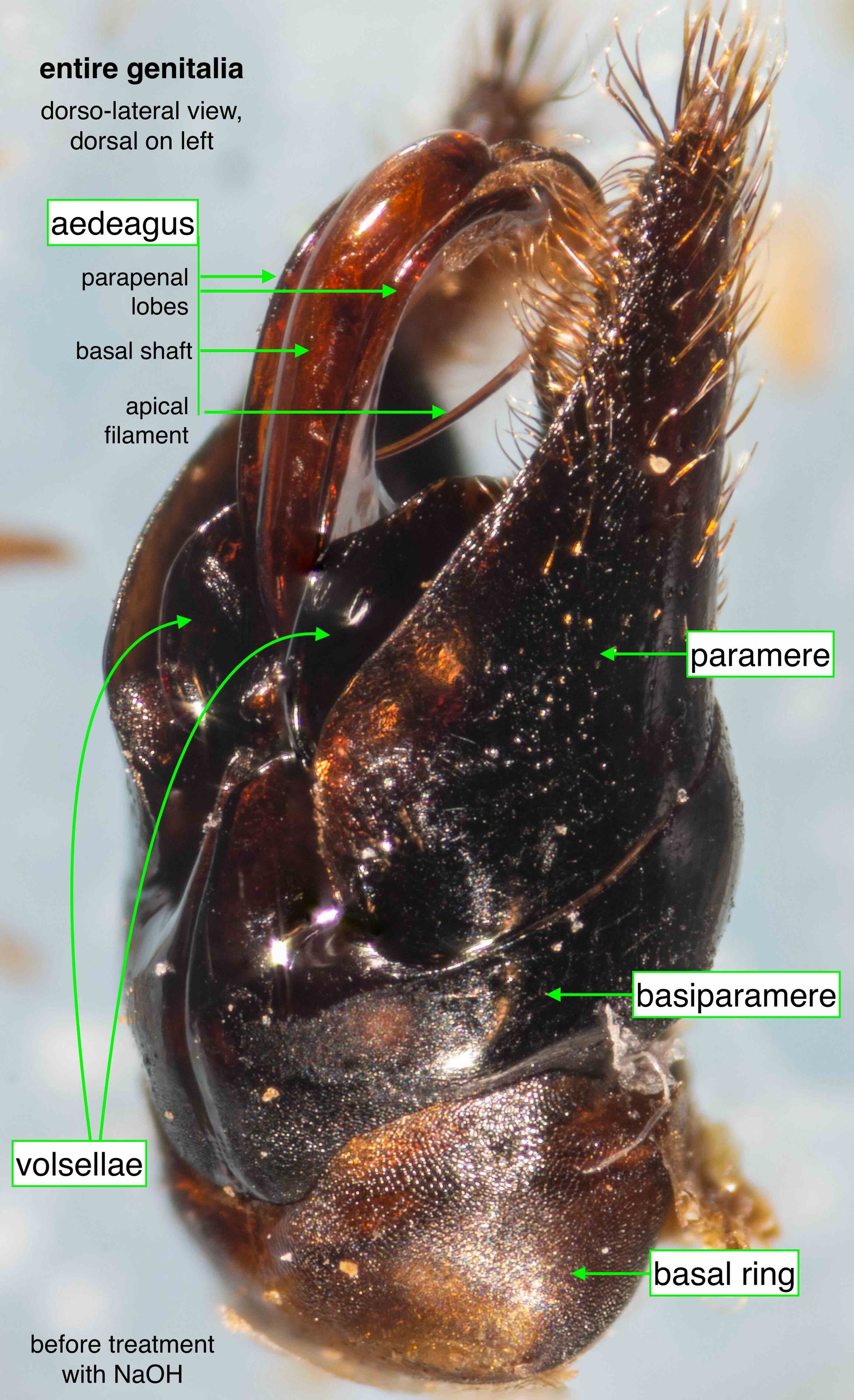
entire phallus
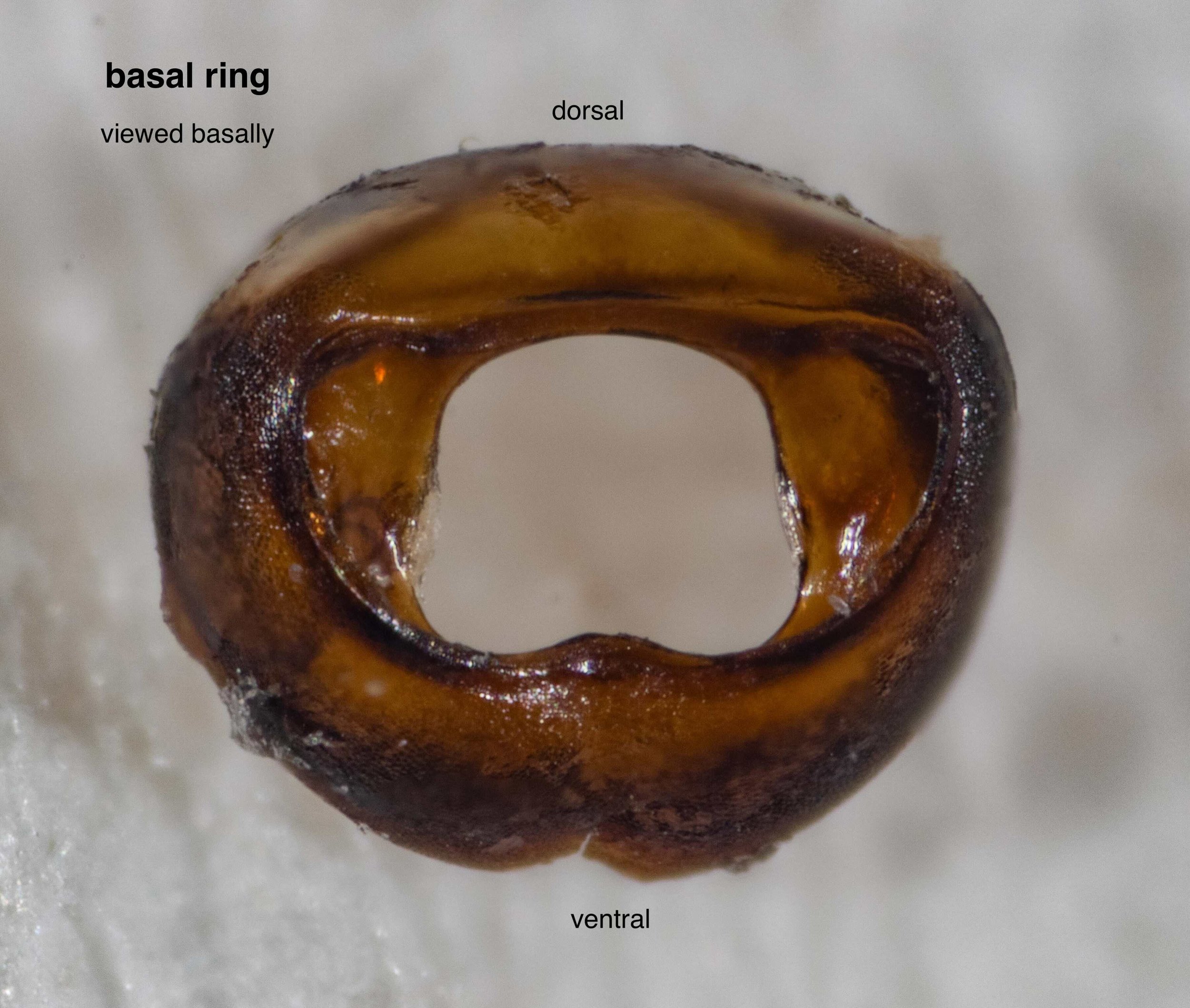
basal ring
Strongly sclerotized (thick & hard), the basal ring is elaborated internally. These projections are sites of muscle attachment. The internal structures of the body cavity connect with those of the phallus via the central hole.
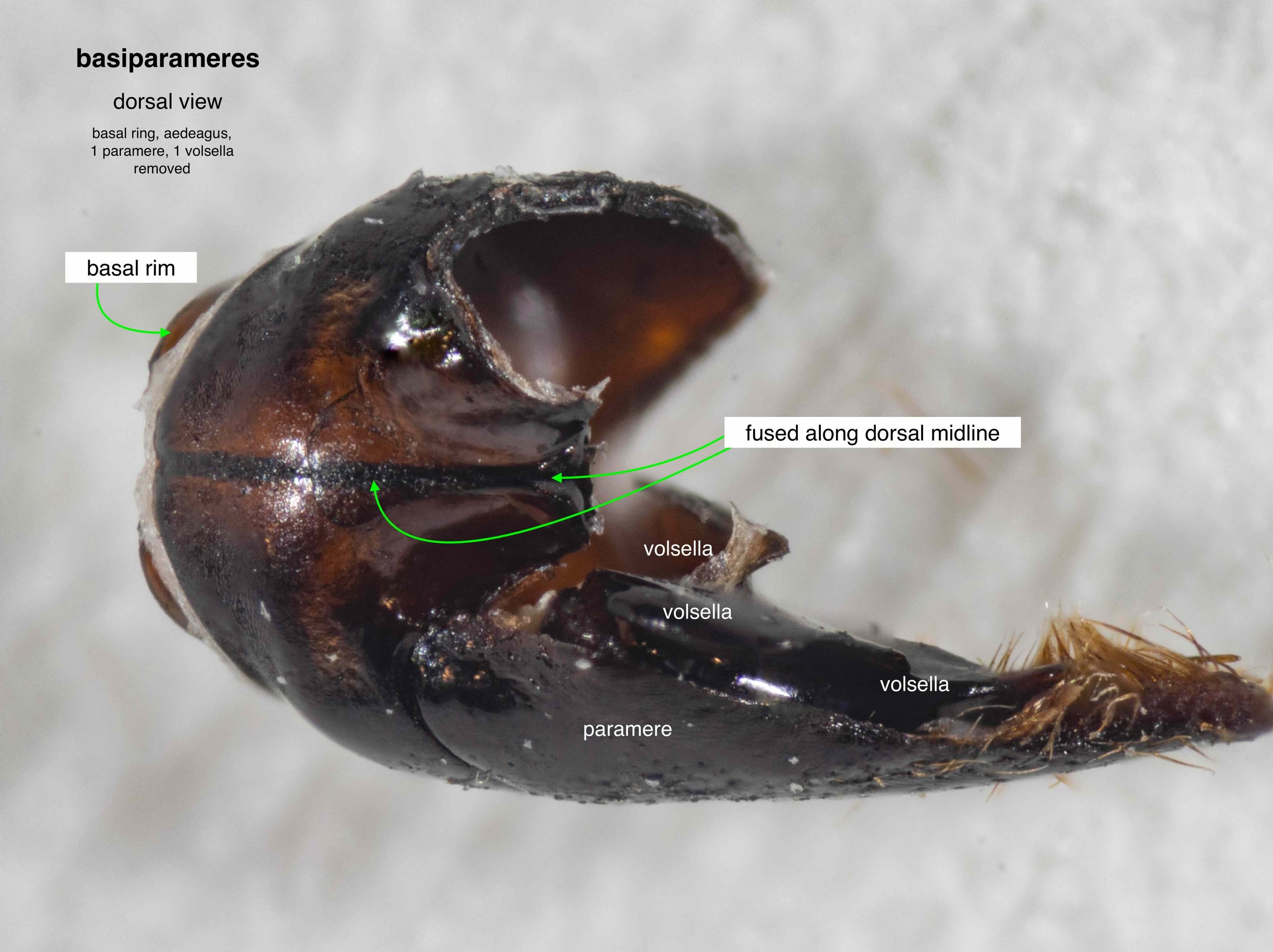
basiparameres
The dorsal suture between the left and right basiparameres is a useful feature in genus-level identification. Note that the ‘right-hand’ volsella and paramere have been removed, along with the aedeagus and basal ring.
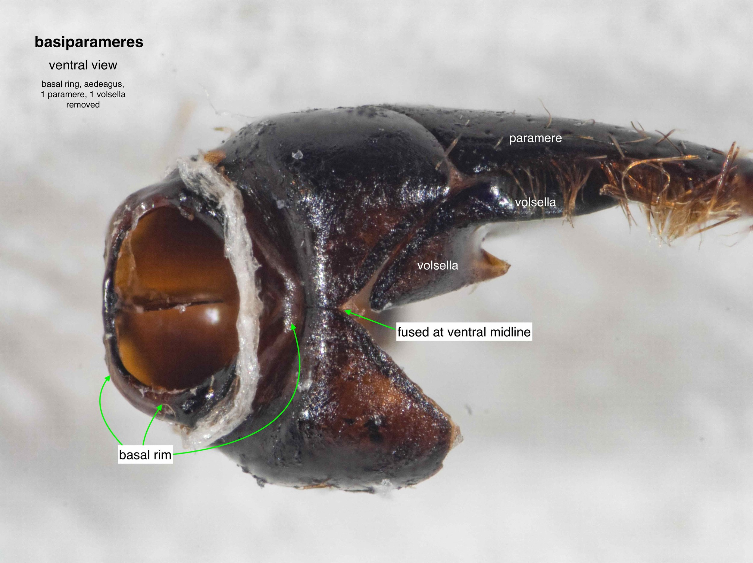
basiparameres
The right and left sides are also fused ventrally, but to a lesser extent.
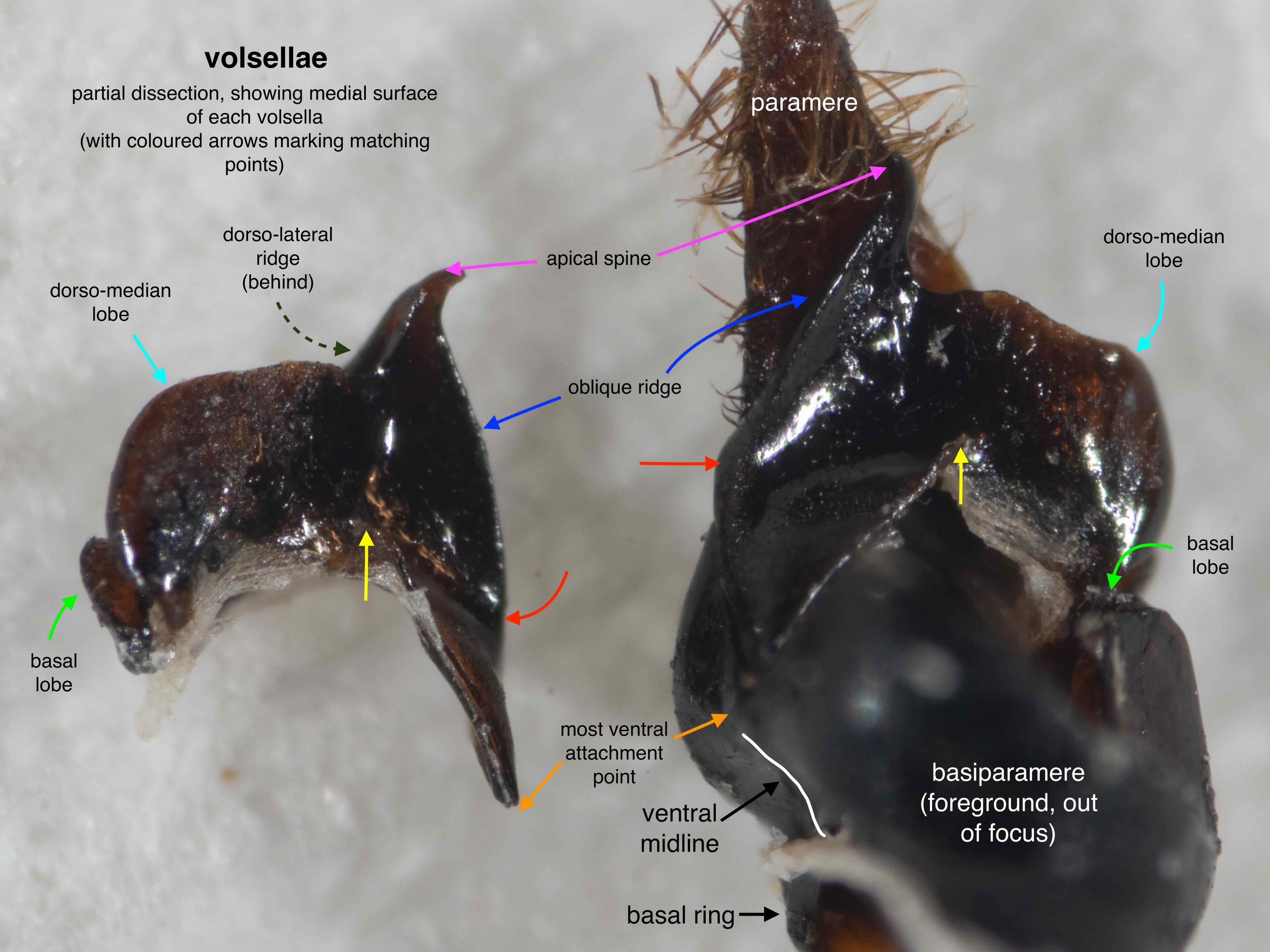
volsellae
The shape is highly complex and normally concealed by the parameres. Here the isolated volsella shows its lateral face, while the partner shows the shape as seen medially.
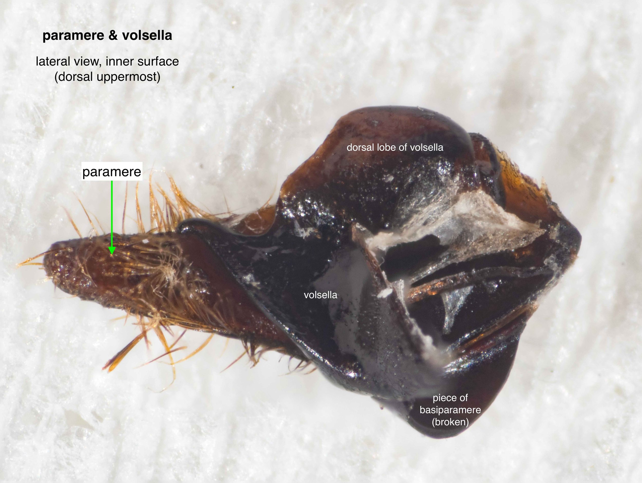
volsella & paramere
Another medial view of the volsella, this time with the basiparamere removed. Note that the hairs on the paramere have been damaged during the cleaning process.
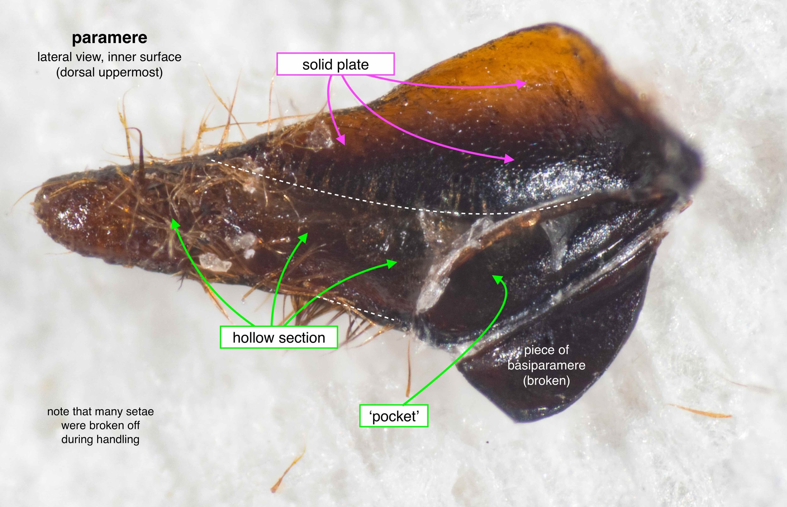
paramere
With the volsella removed, the hollow nature of the paramere is revealed.
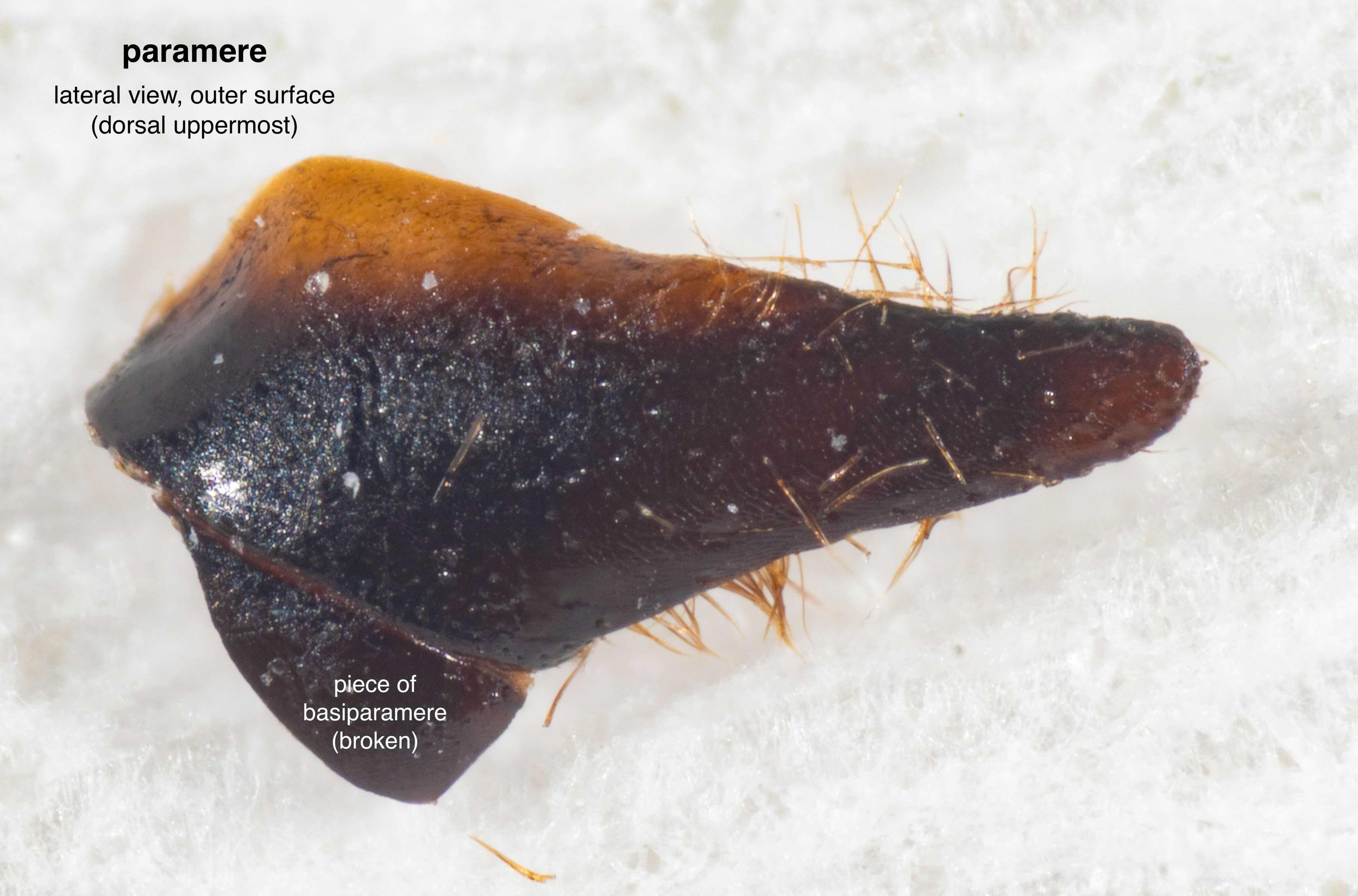
paramere
The shape of the paramere is useful for genus-level ID, including the shape of the apex and the extent of the suture between the paramere and basiparamere. The outer face of the paramere, as seen here, is typically visible during coupling … although it’s still tricky in a field photo, unless the insects are large and the image sharp.
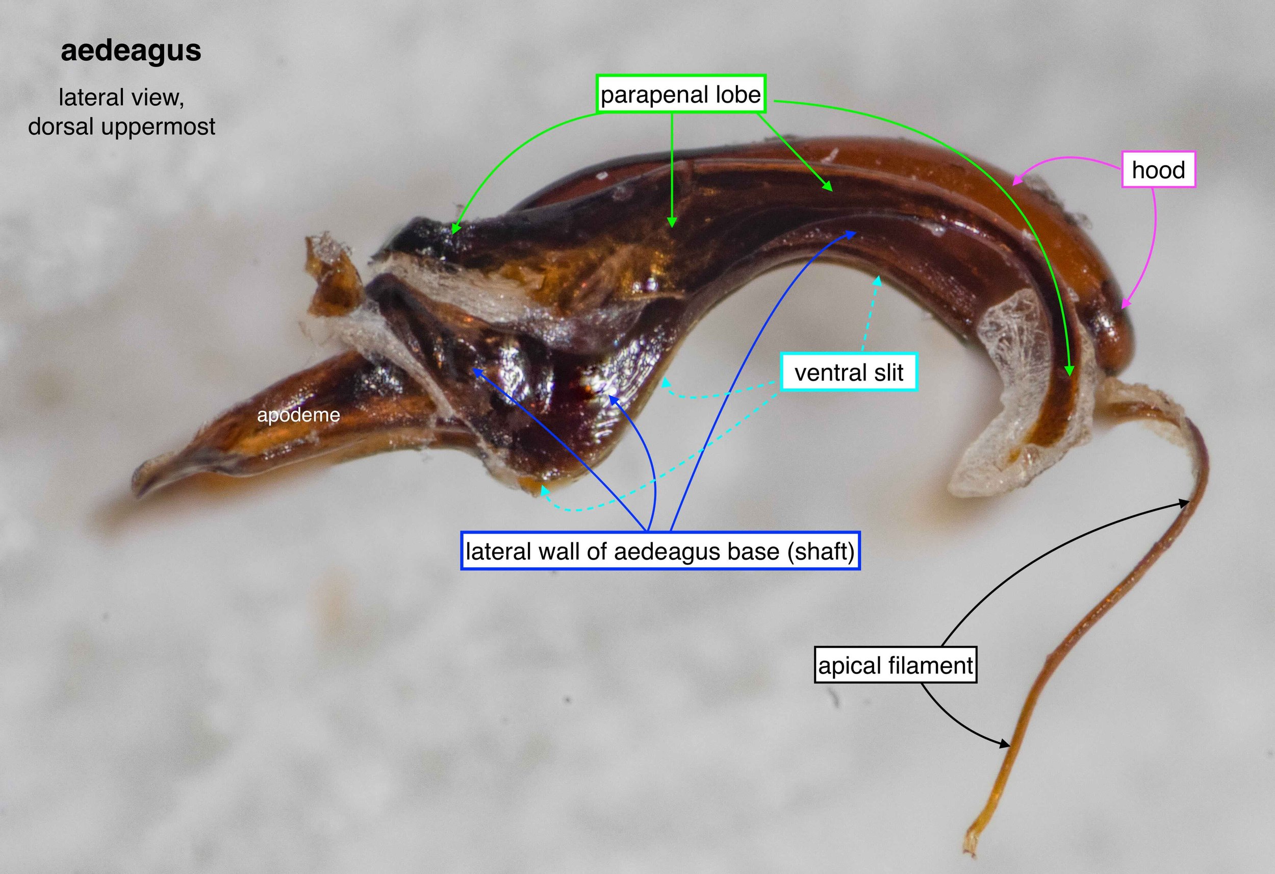
aedeagus
A single sclerite, but a complex and partially delicate one.
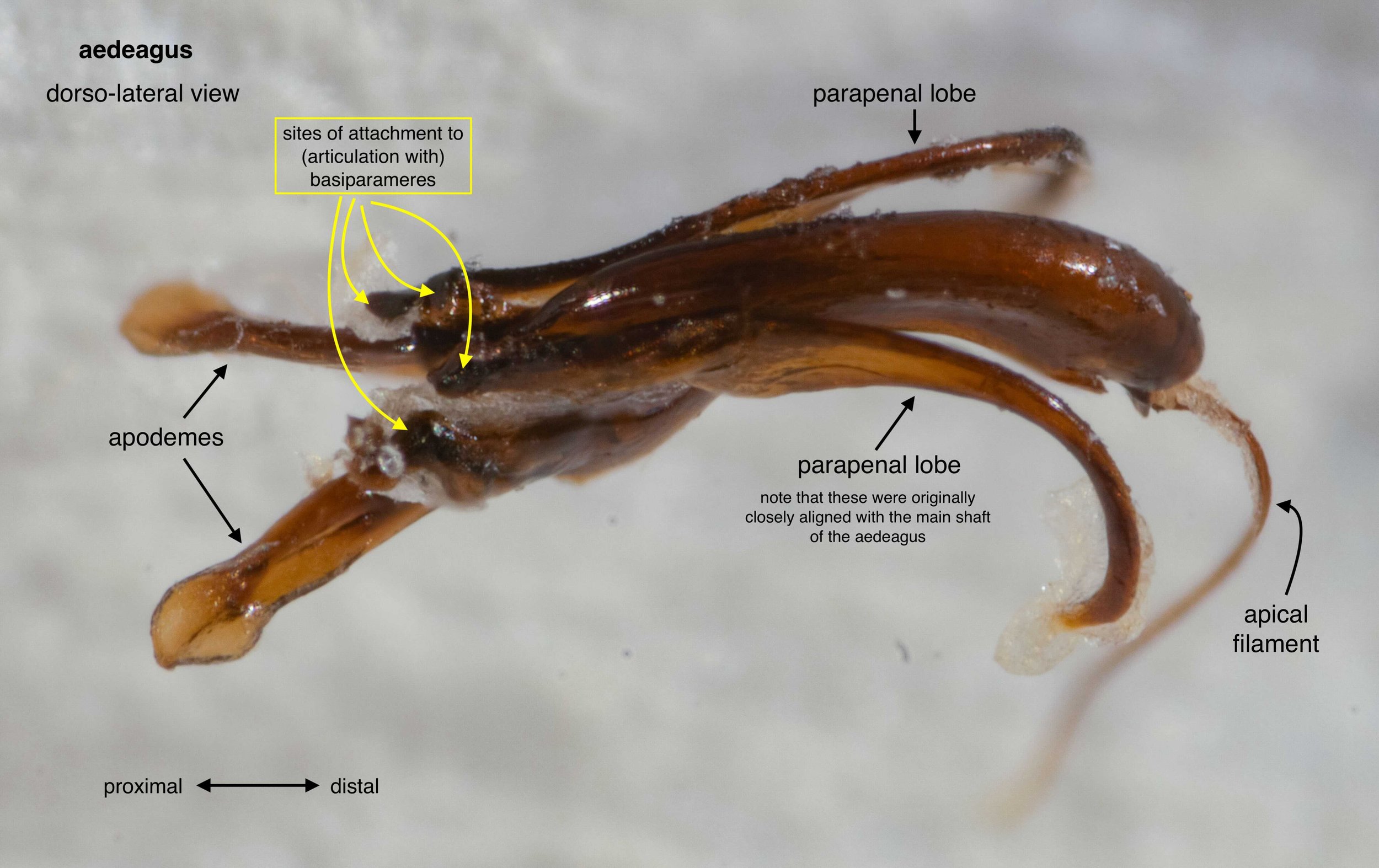
aedeagus
Basal projections are sites of muscle attachment. The apical filament present in this species is not found in all Thynninae. As to its exact function, I’m unsure.
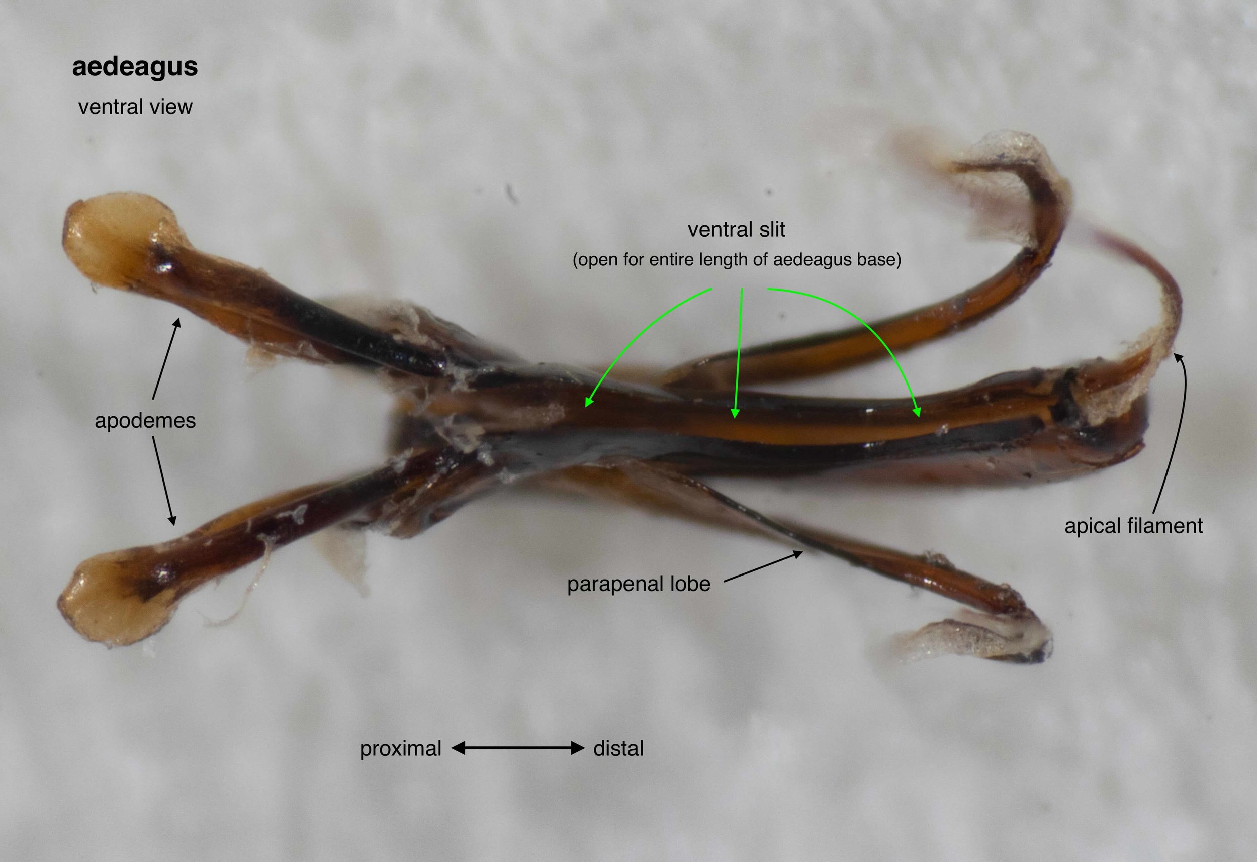
aedeagus
In this species the central shaft of the aedeagus bears a long, ventral slit. This is likely to vary between taxa.
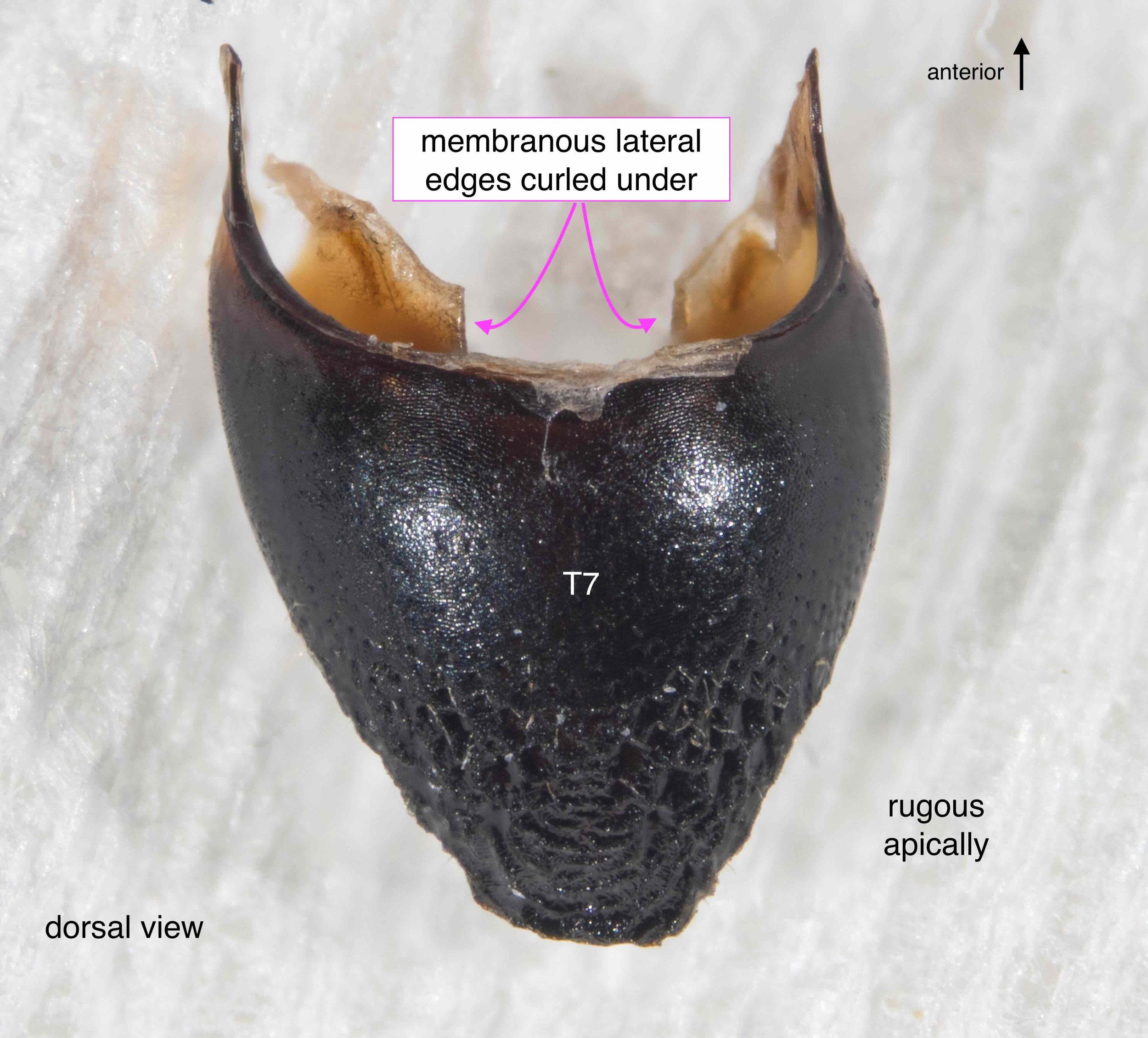
T7 (epipygium)
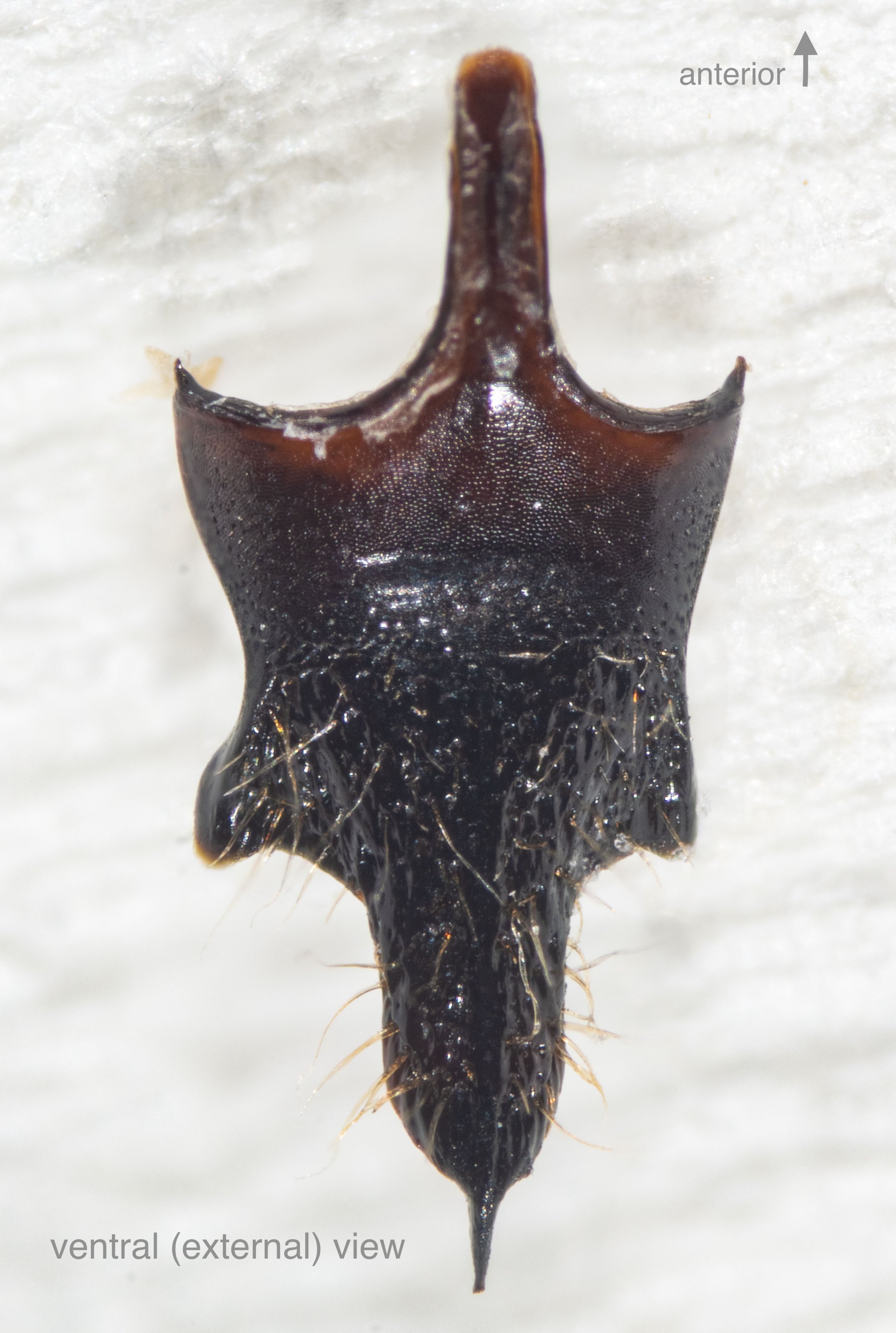
S8 (hypopygium)

S8 (hypopygium)


