
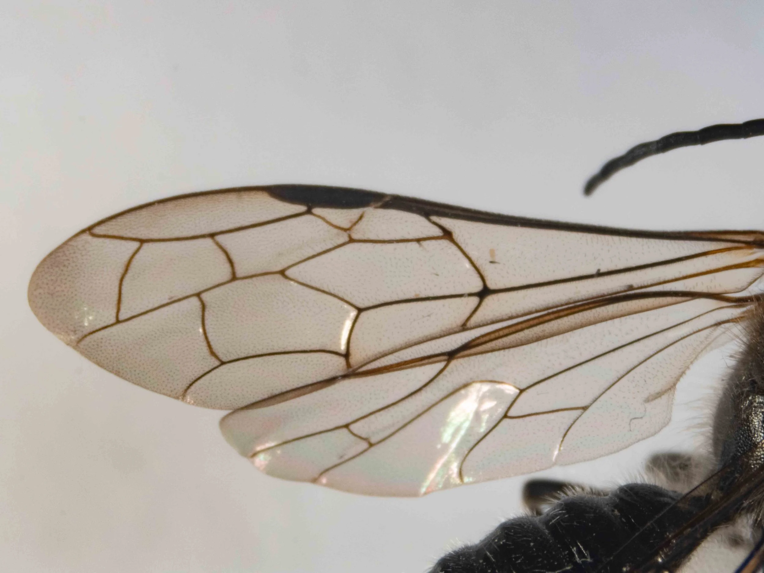
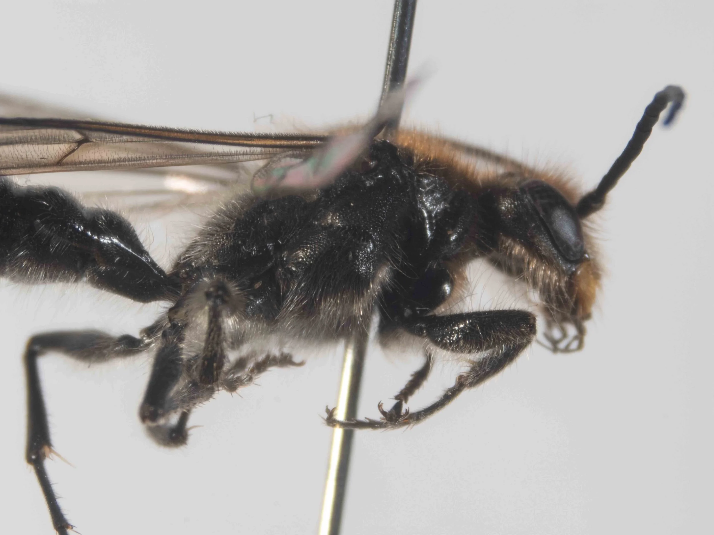
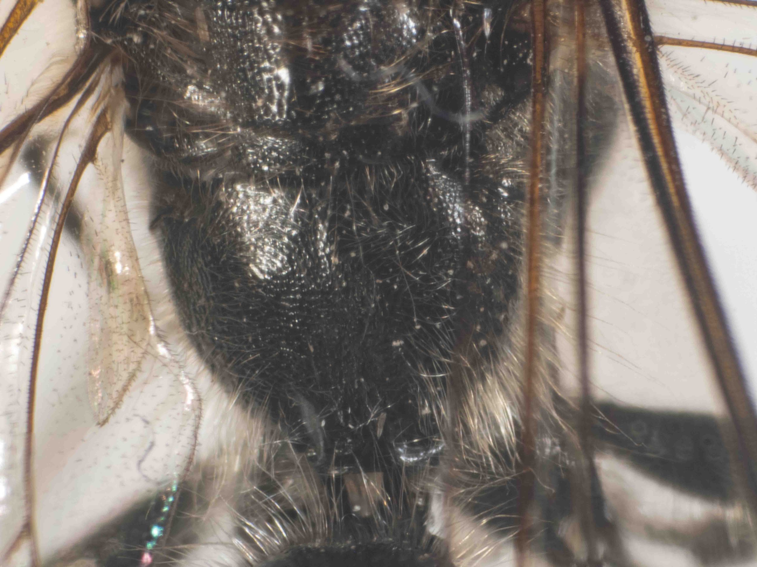
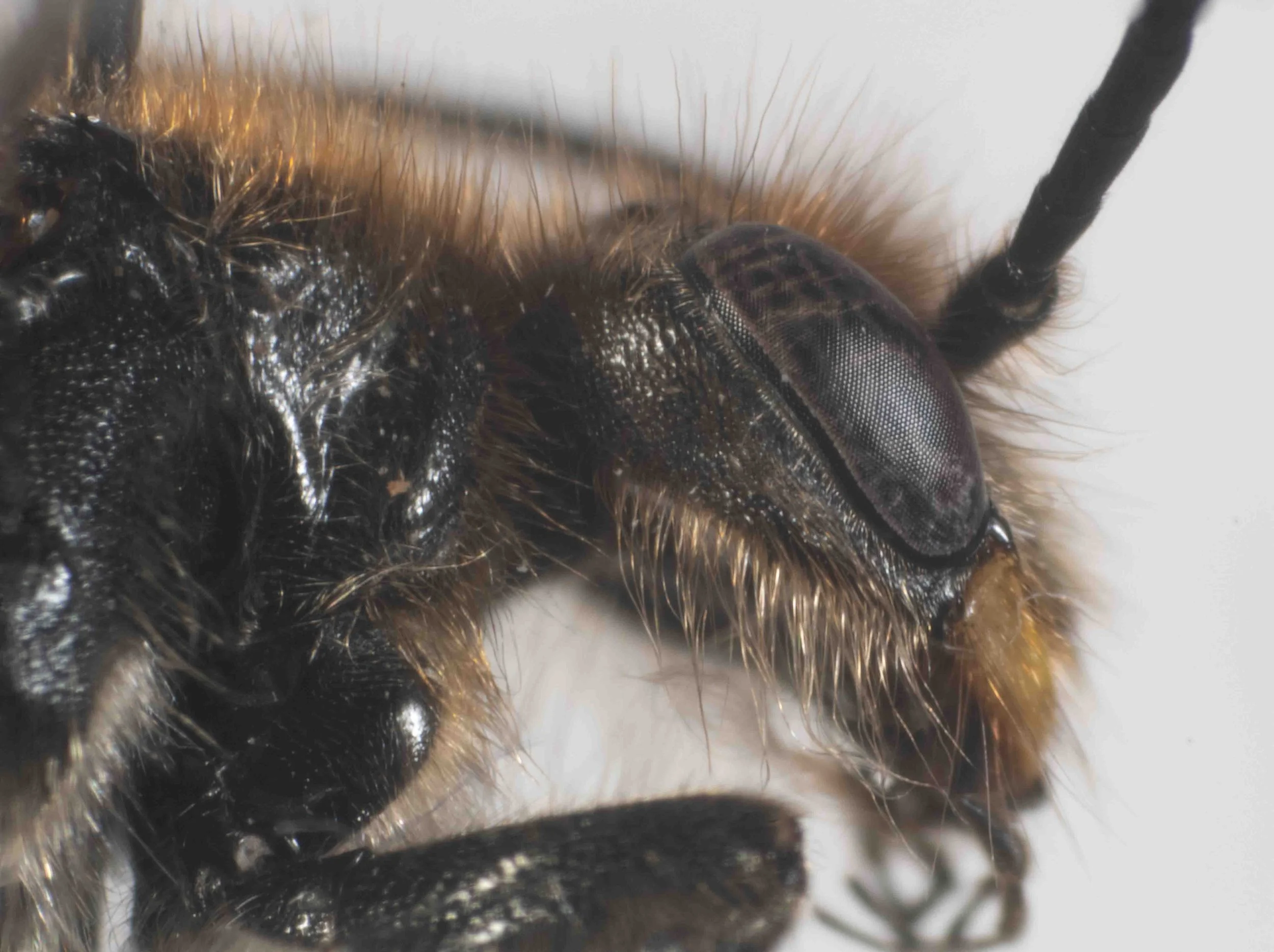
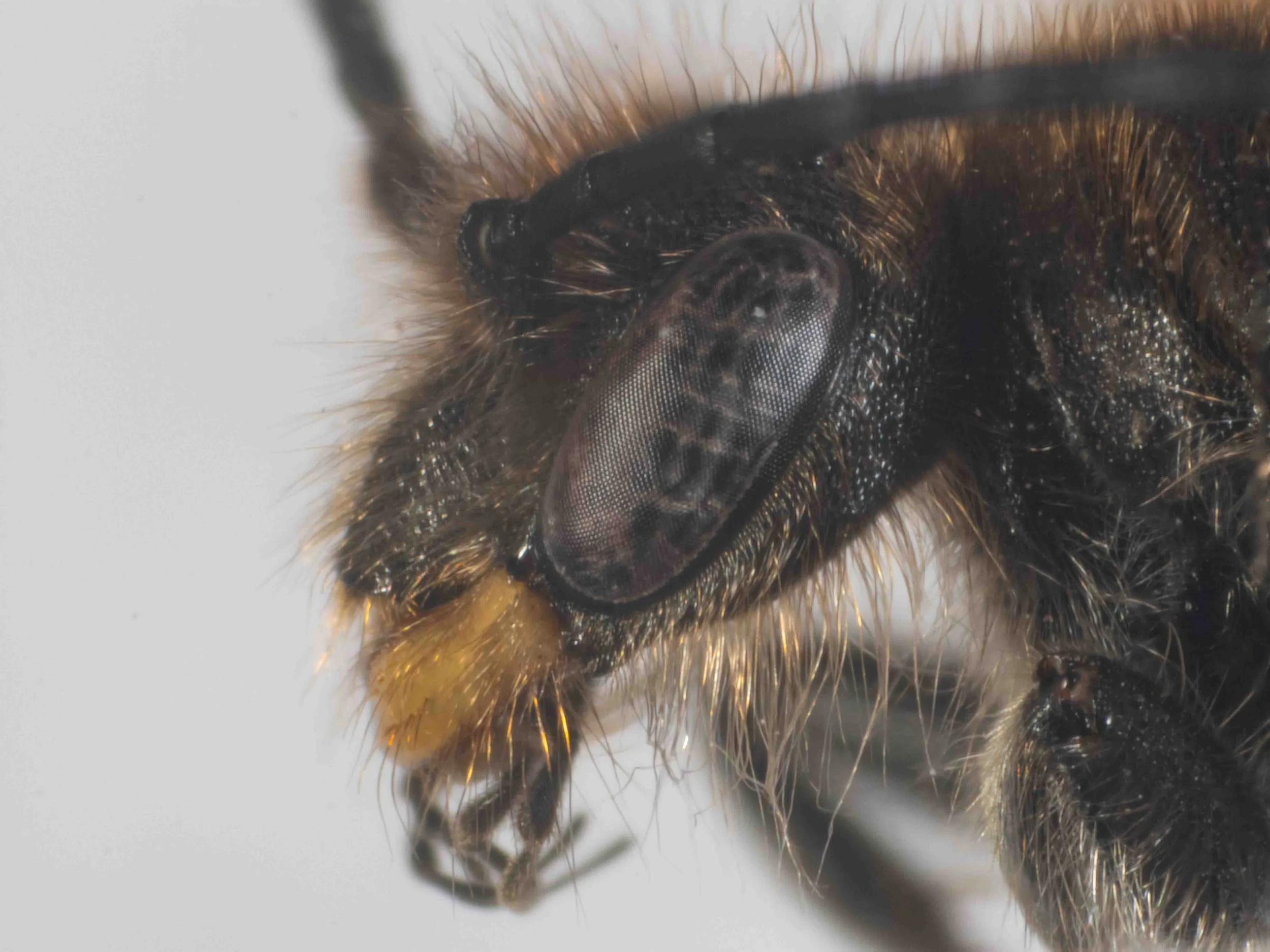
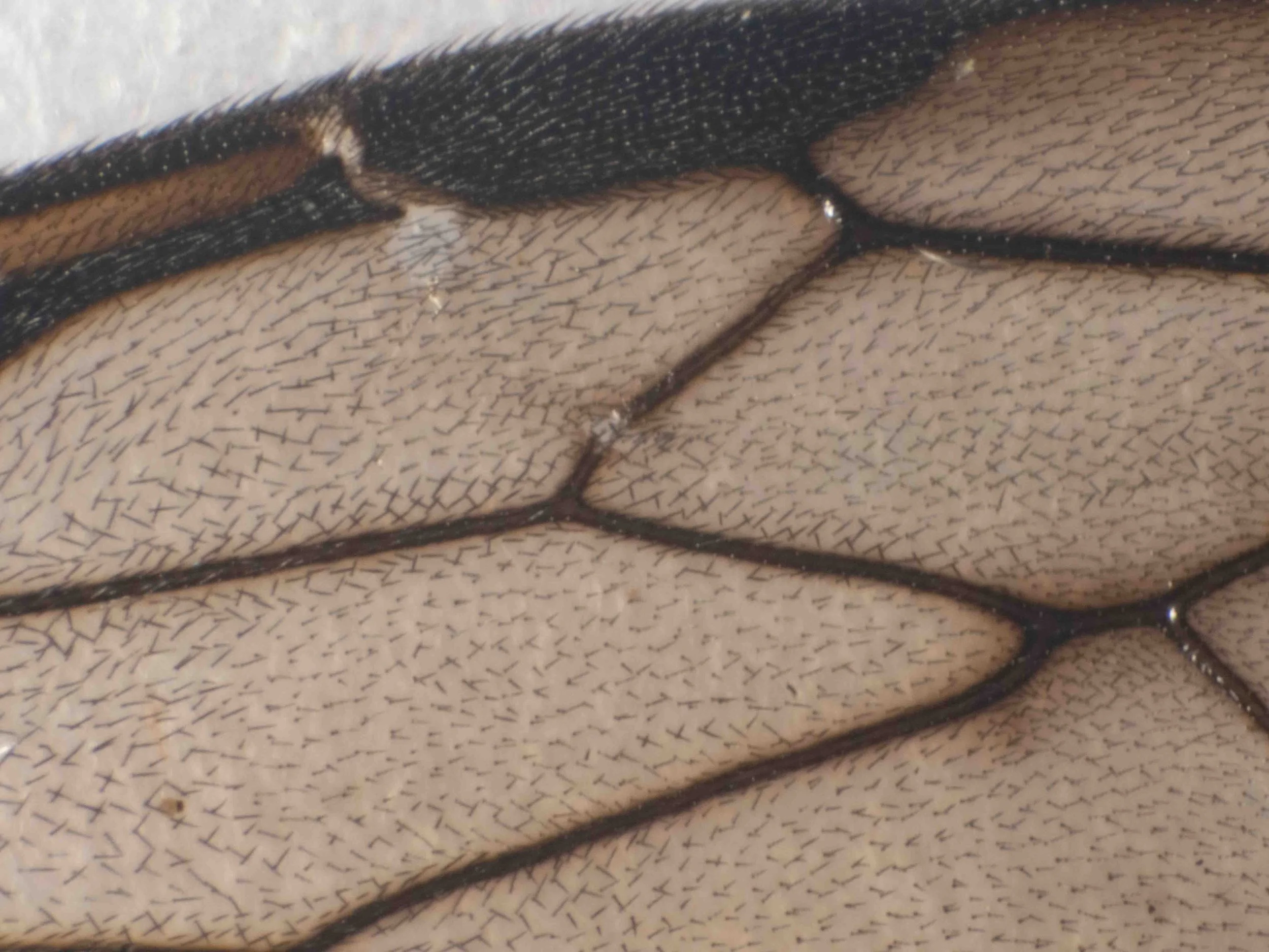
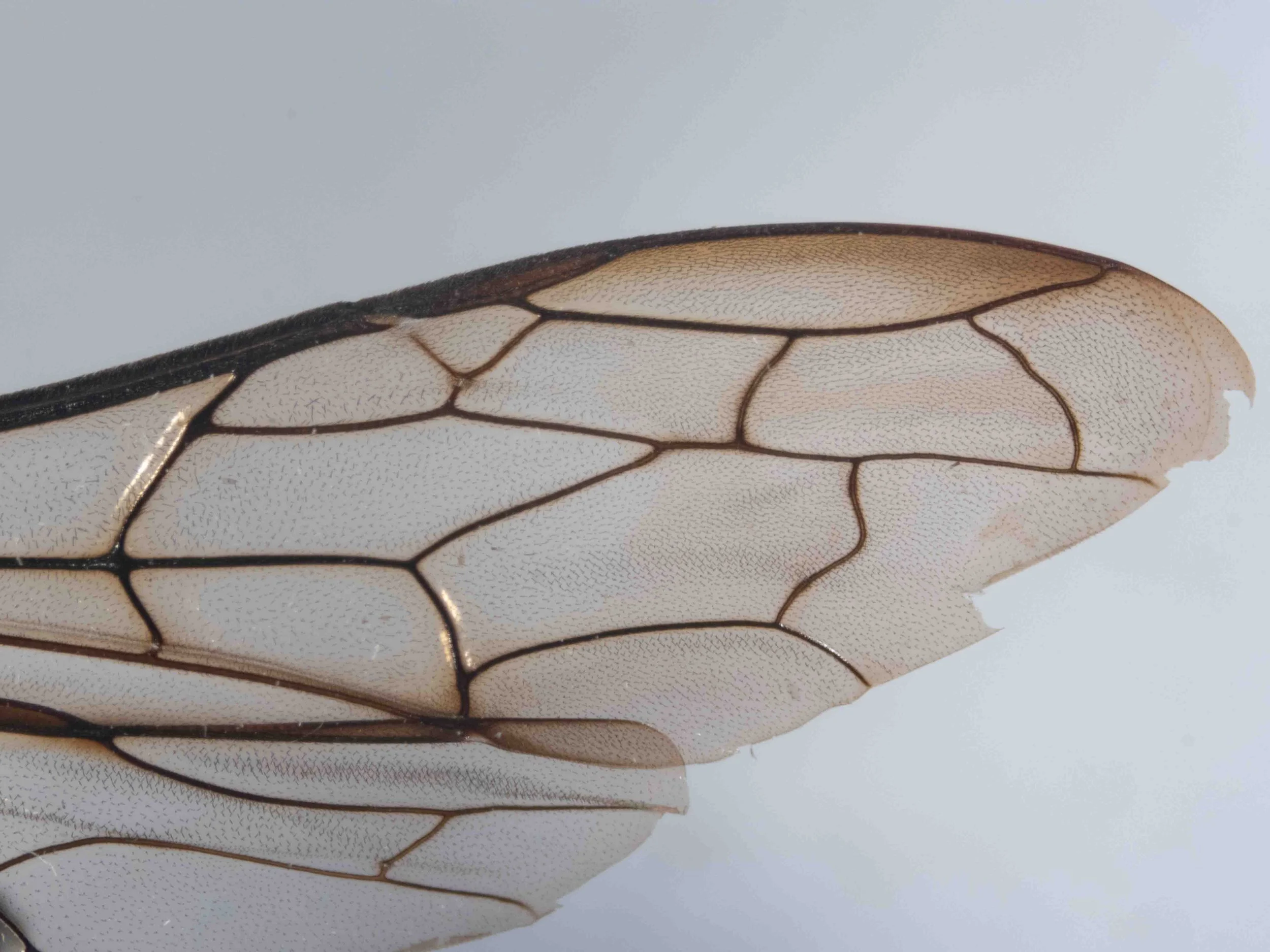
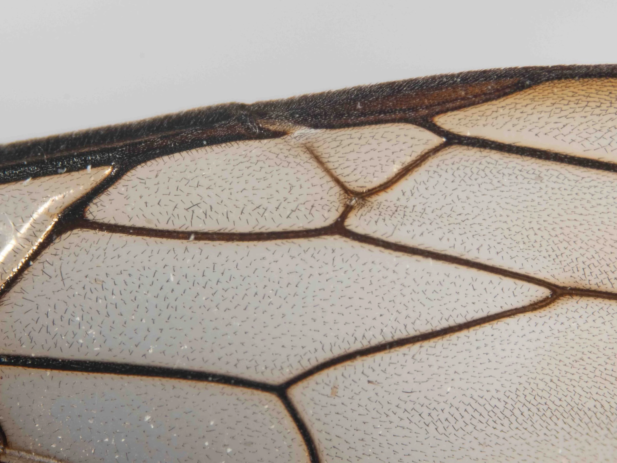
forewing venation
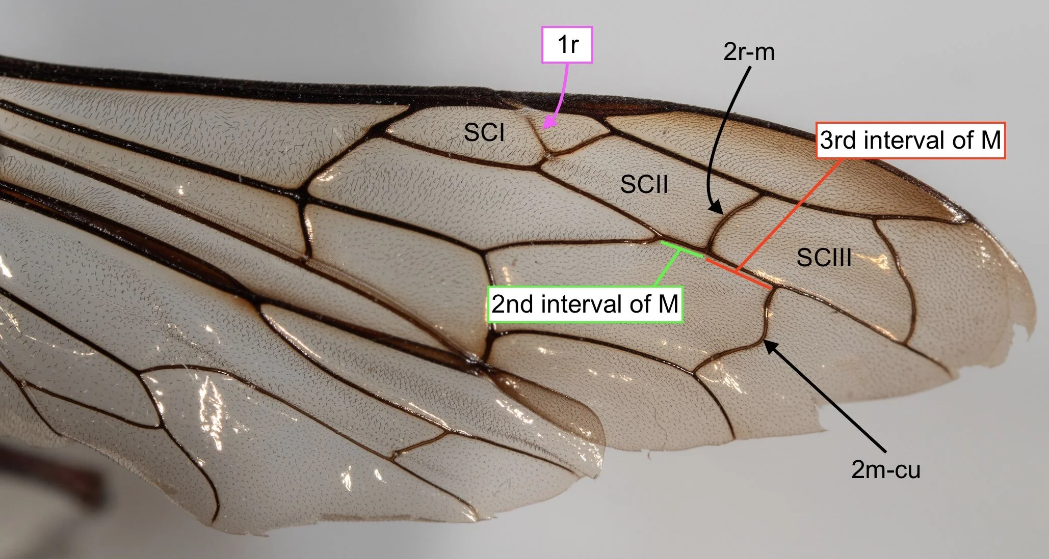
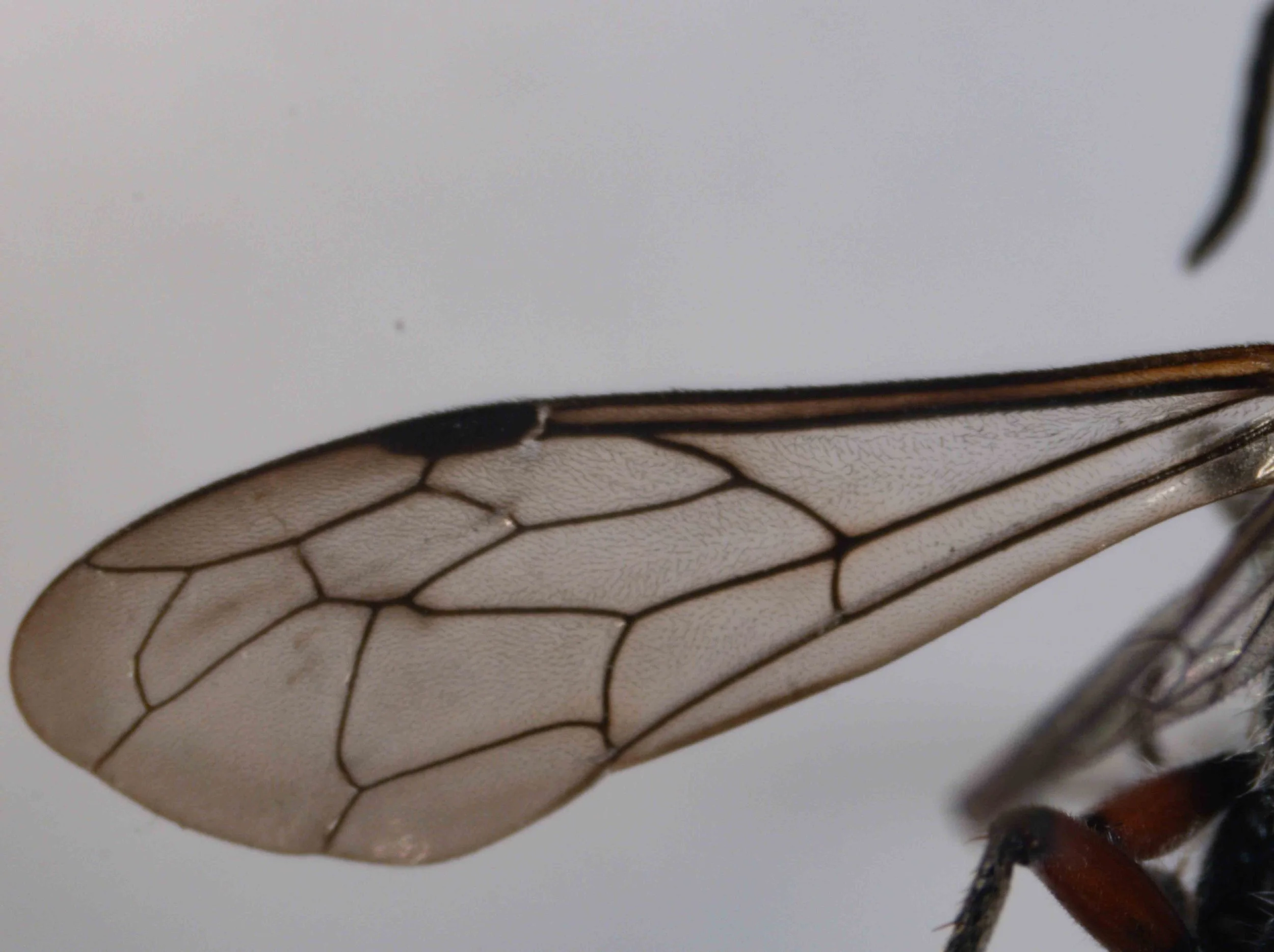
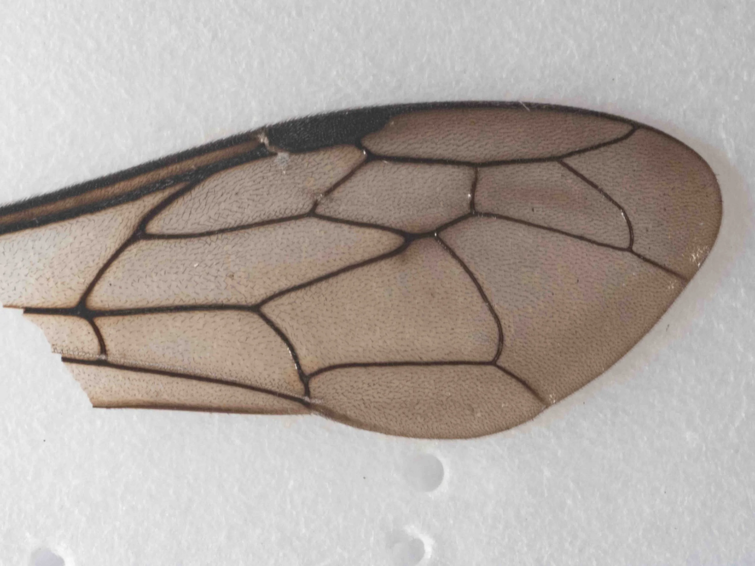
The short vein within the first submarginal cell (SCI) is variously described as vein 1r or the ‘spur vein’. This vein is obvious in many Thynninae, including virtually all Thynnini. However, it is absent or barely indicated in Iswaroidini, another of the Thynninae tribes.
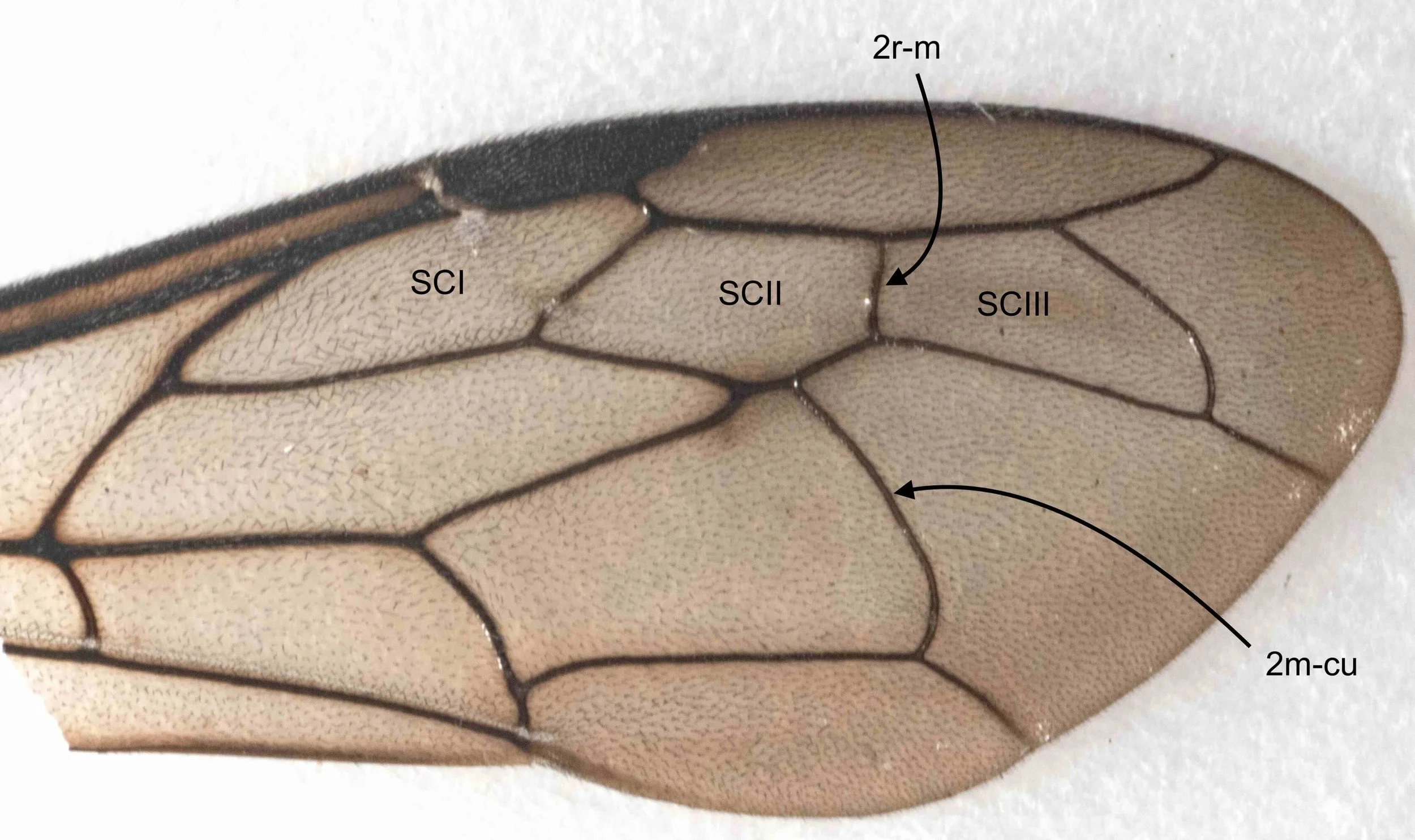
The relative positions of the ‘recurrent veins’, particularly 2m-cu, and the 2nd submarginal cell cross vein (2r-m) is particularly important in distinguishing the Thynninae tribes.
forewing venation
The subfamily Diamminae consists of a single species, Diamma bicolor. While the females are easily identified (large, metallic blue, often seen moving freely above ground), males are less distinctive. Their forewing venation, however, provides a very good clue. In Diamminae, 2m-cu meets SCII. In nearly all other Thynninae, 2m-cu meets SCIII.
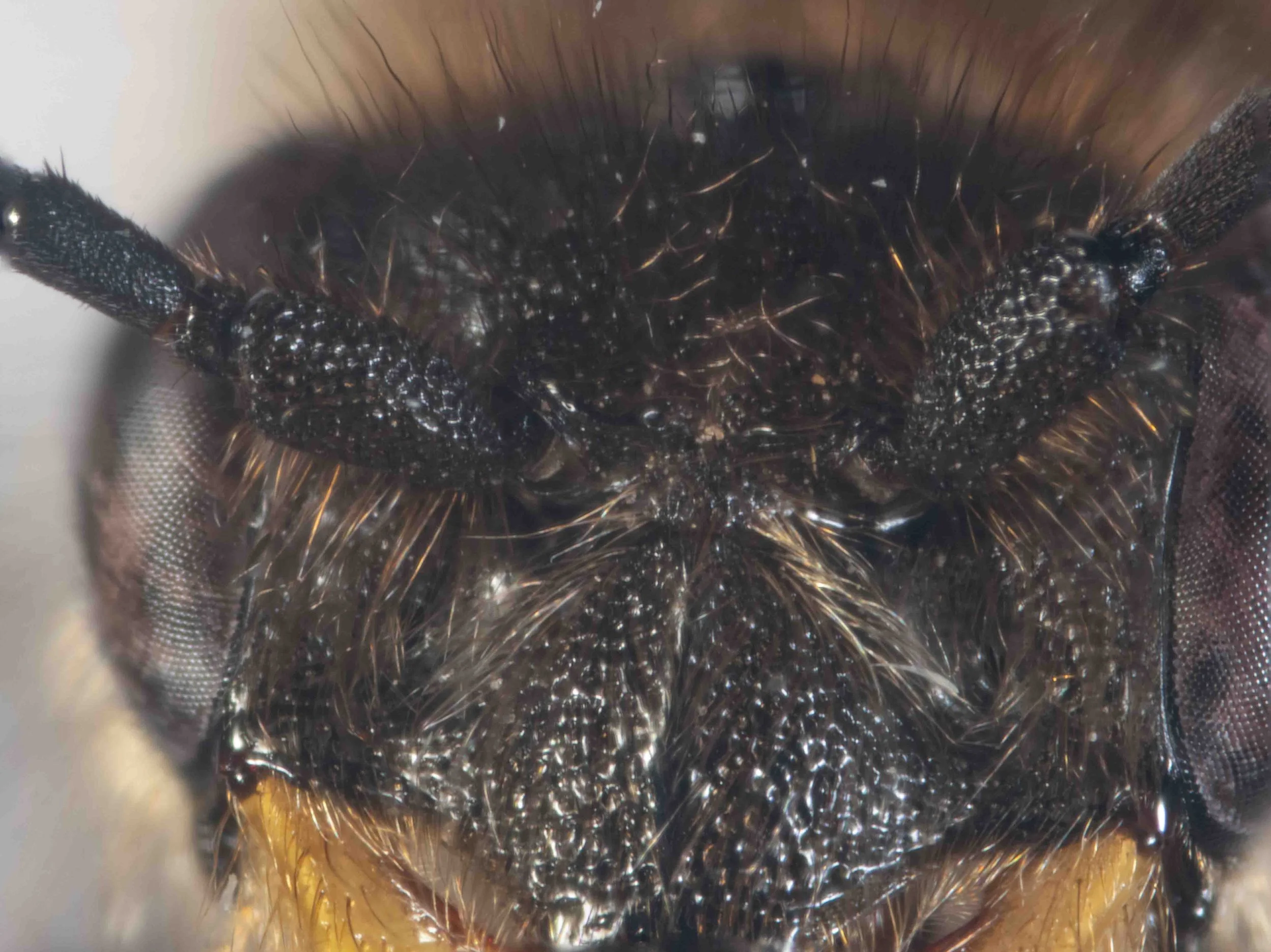
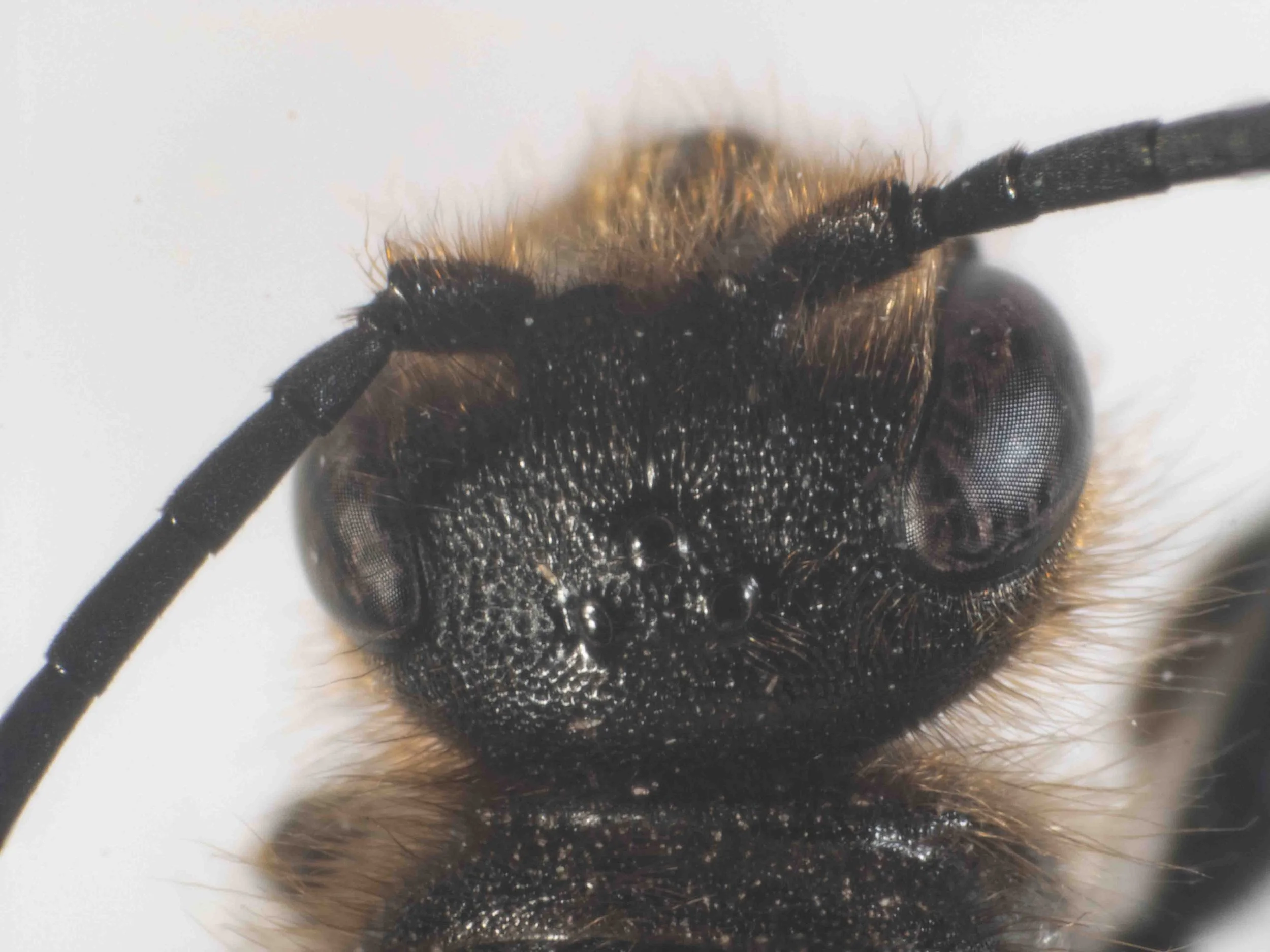
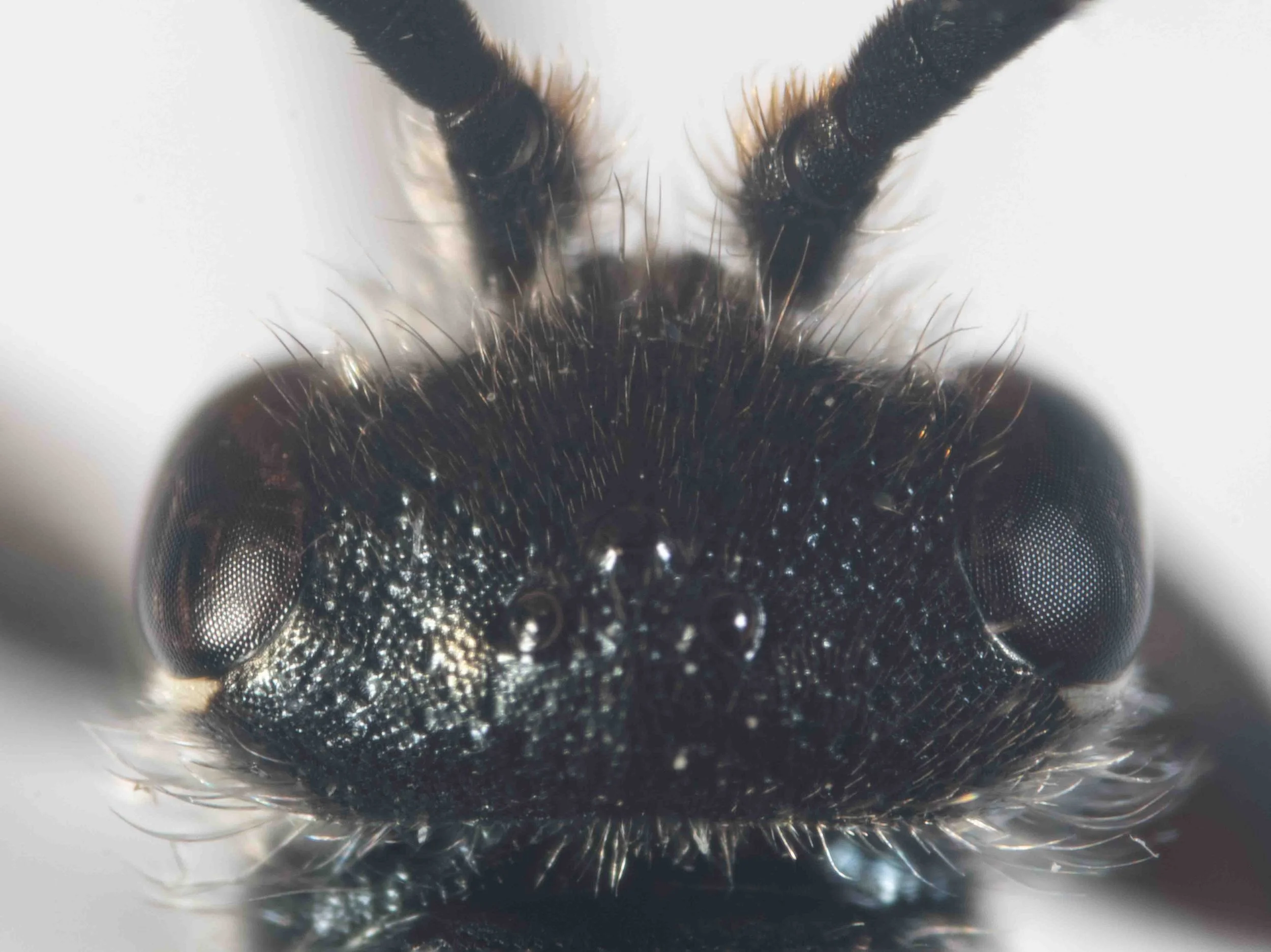
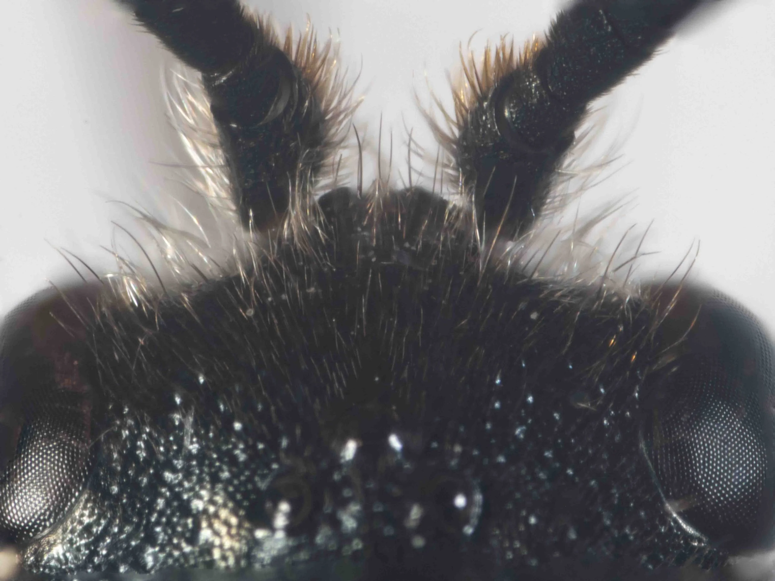
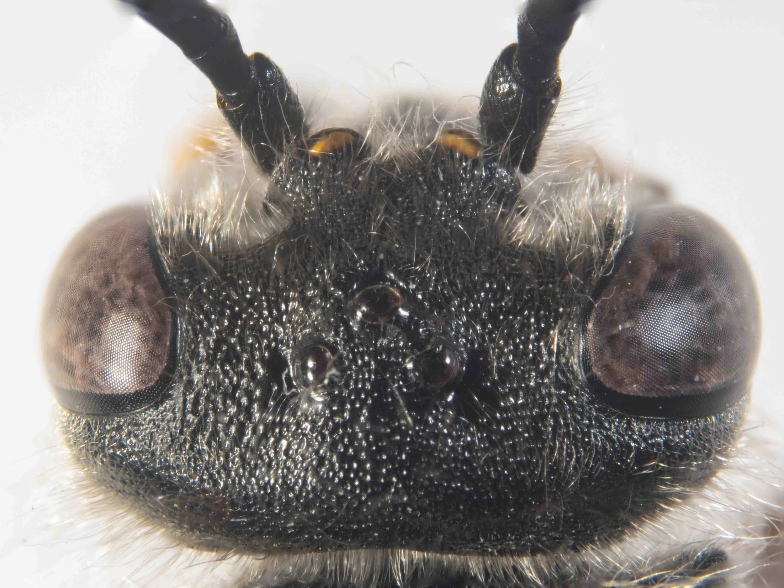
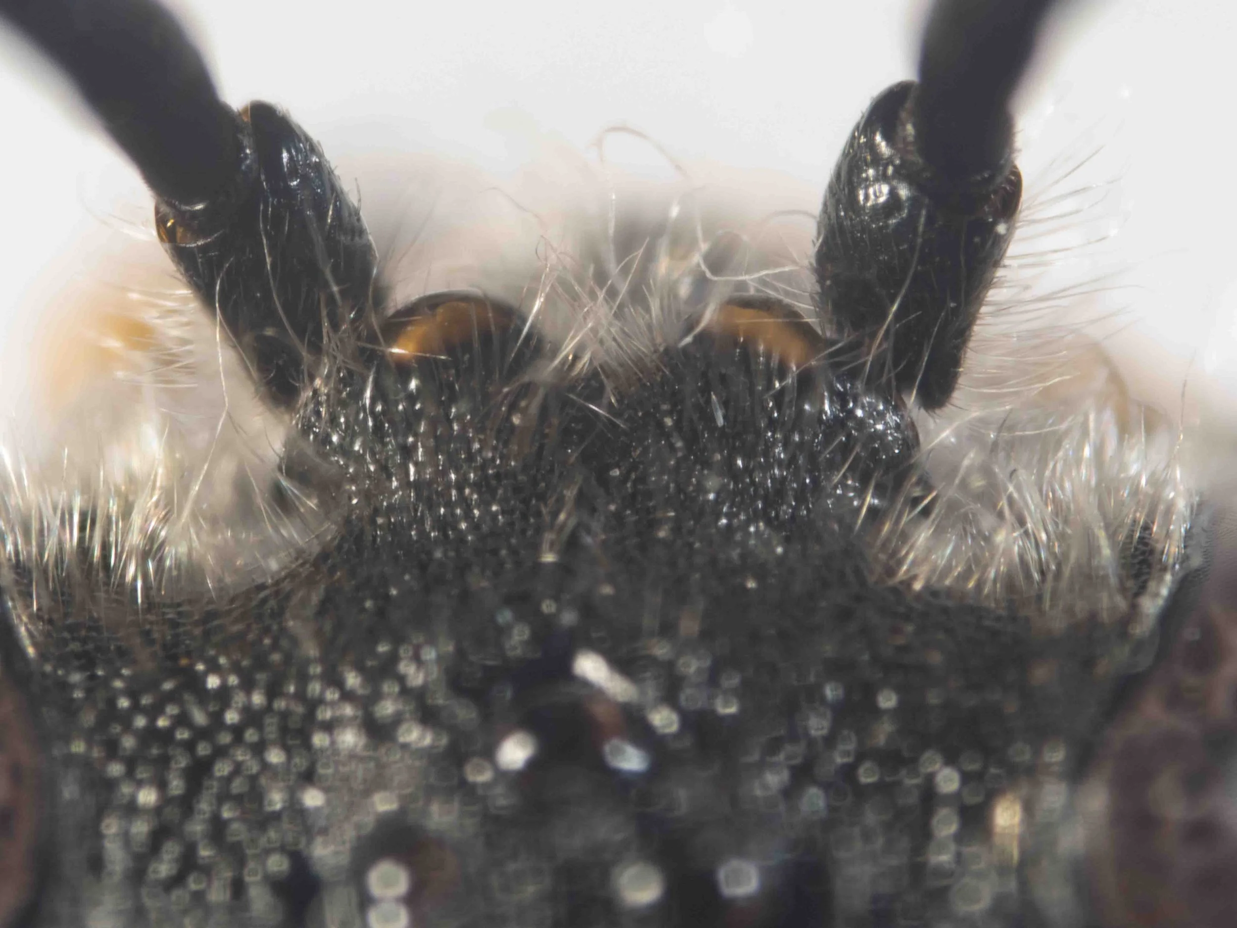
antennal prominence
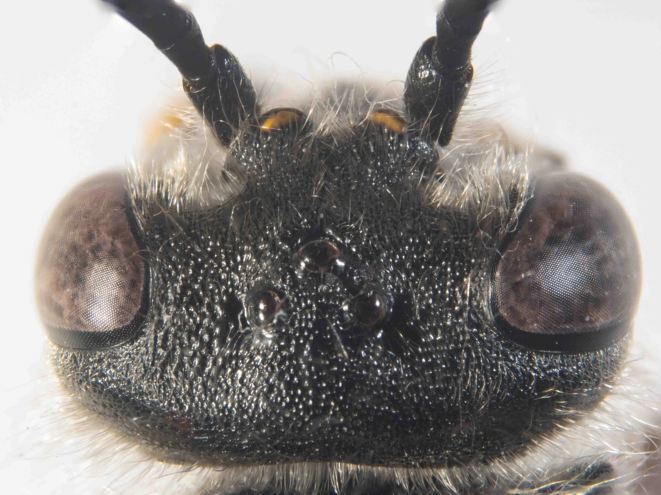
In many Thynnidae, the frons is produced above and between the antennal sockets. Viewed dorsally, this ventromedial region of the frons is therefore raised relative to the lateral regions.
The shape of then antennal prominence varies considerably between genera.
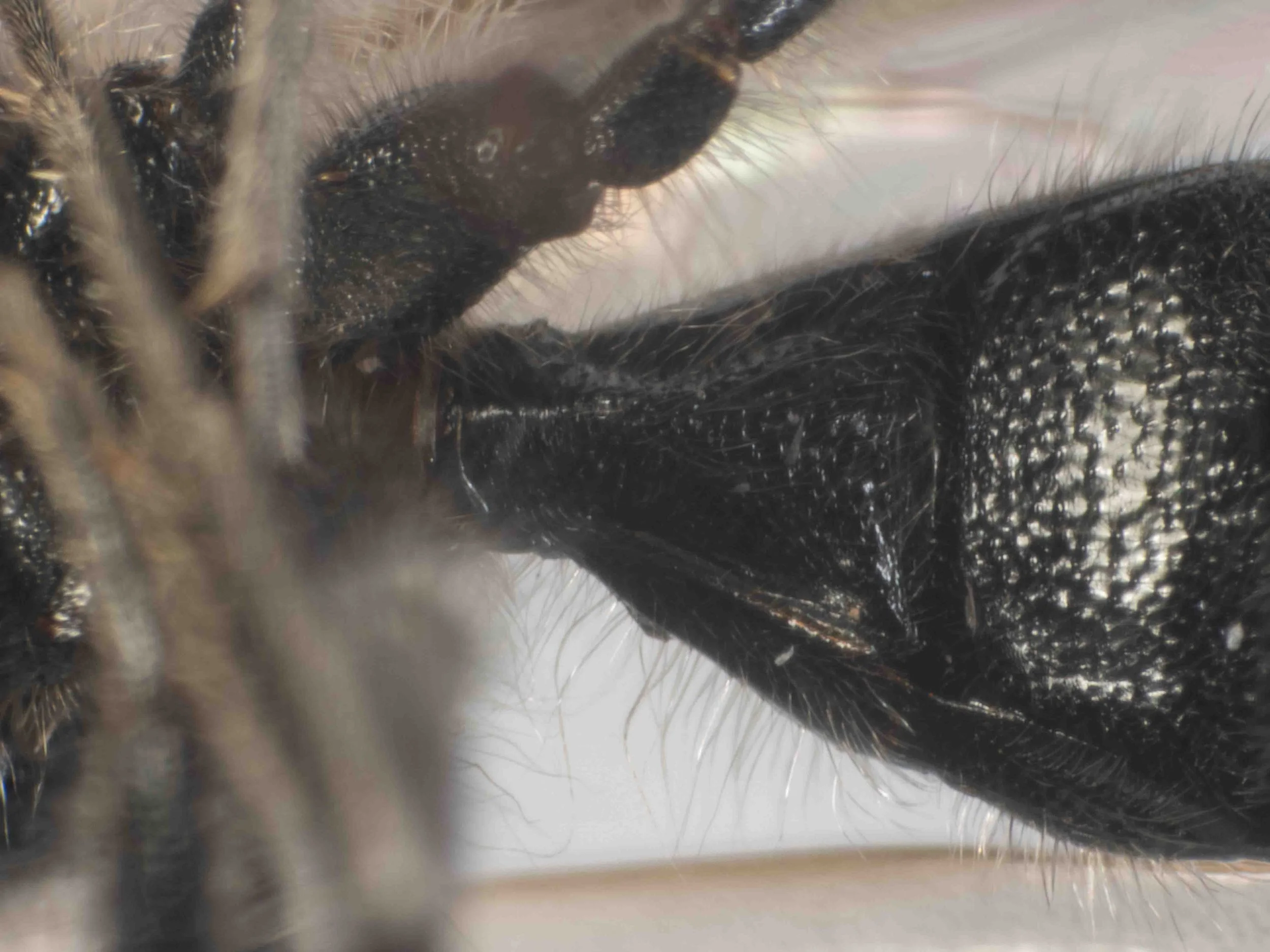
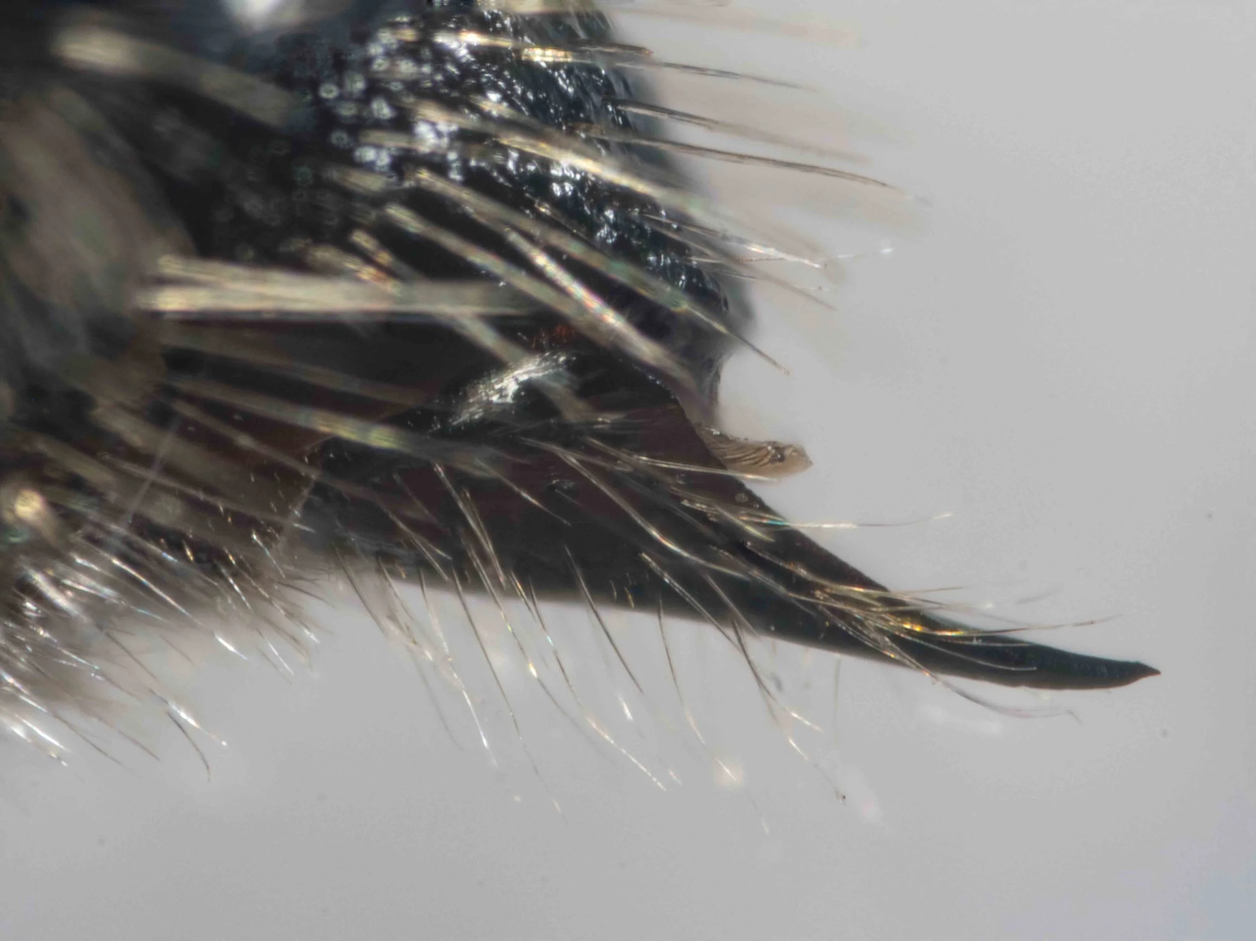
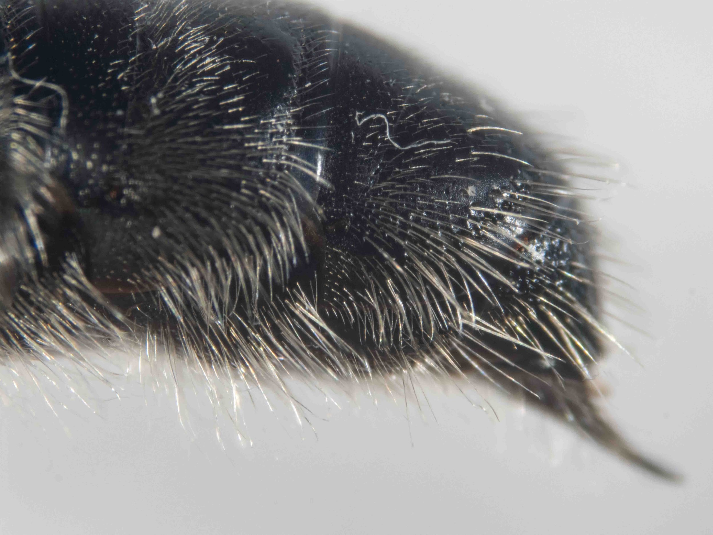
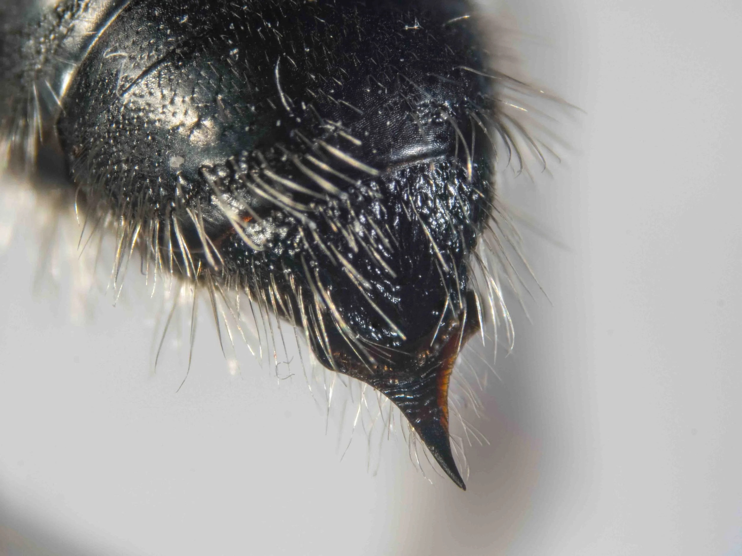
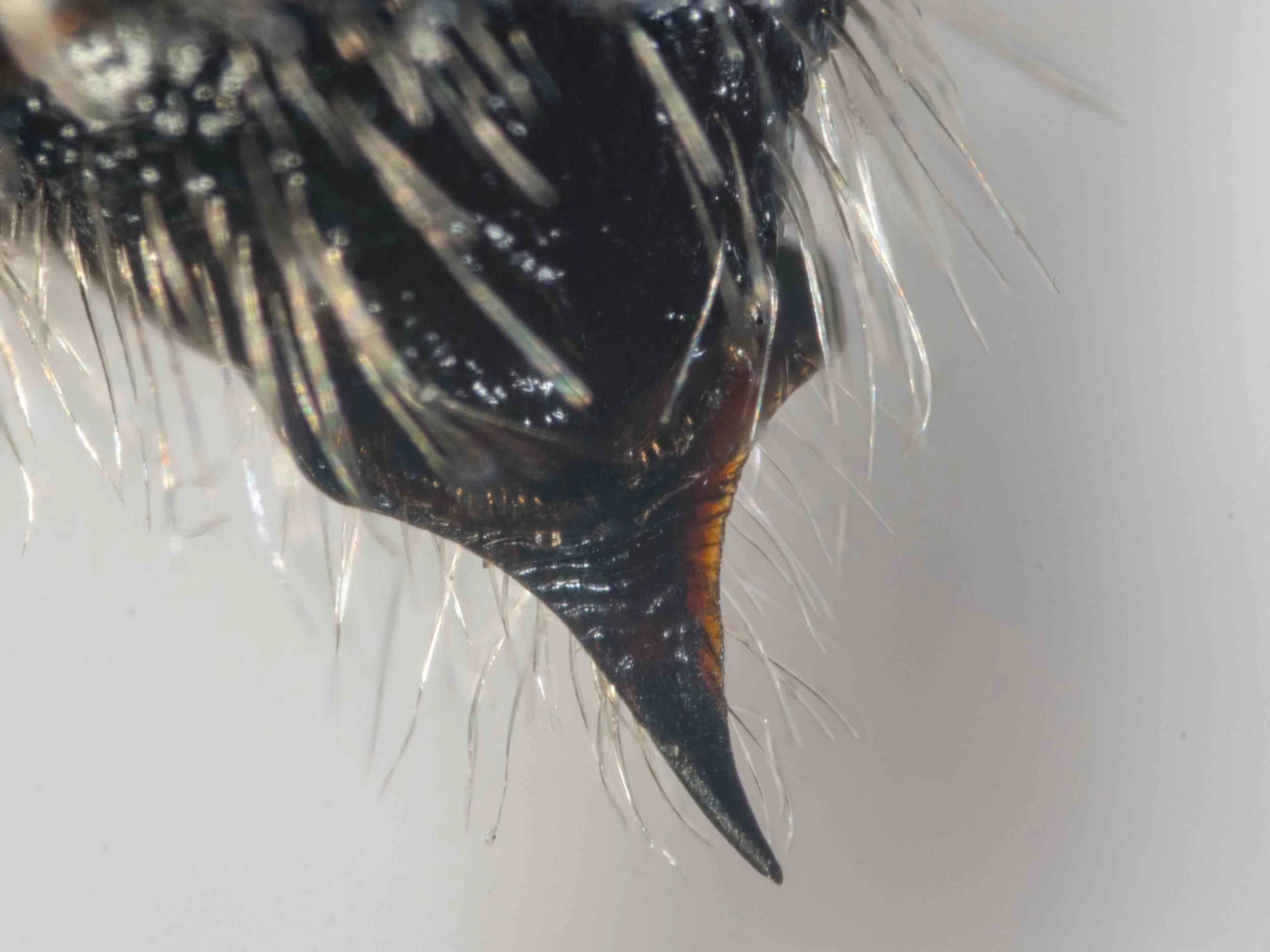
terminalia
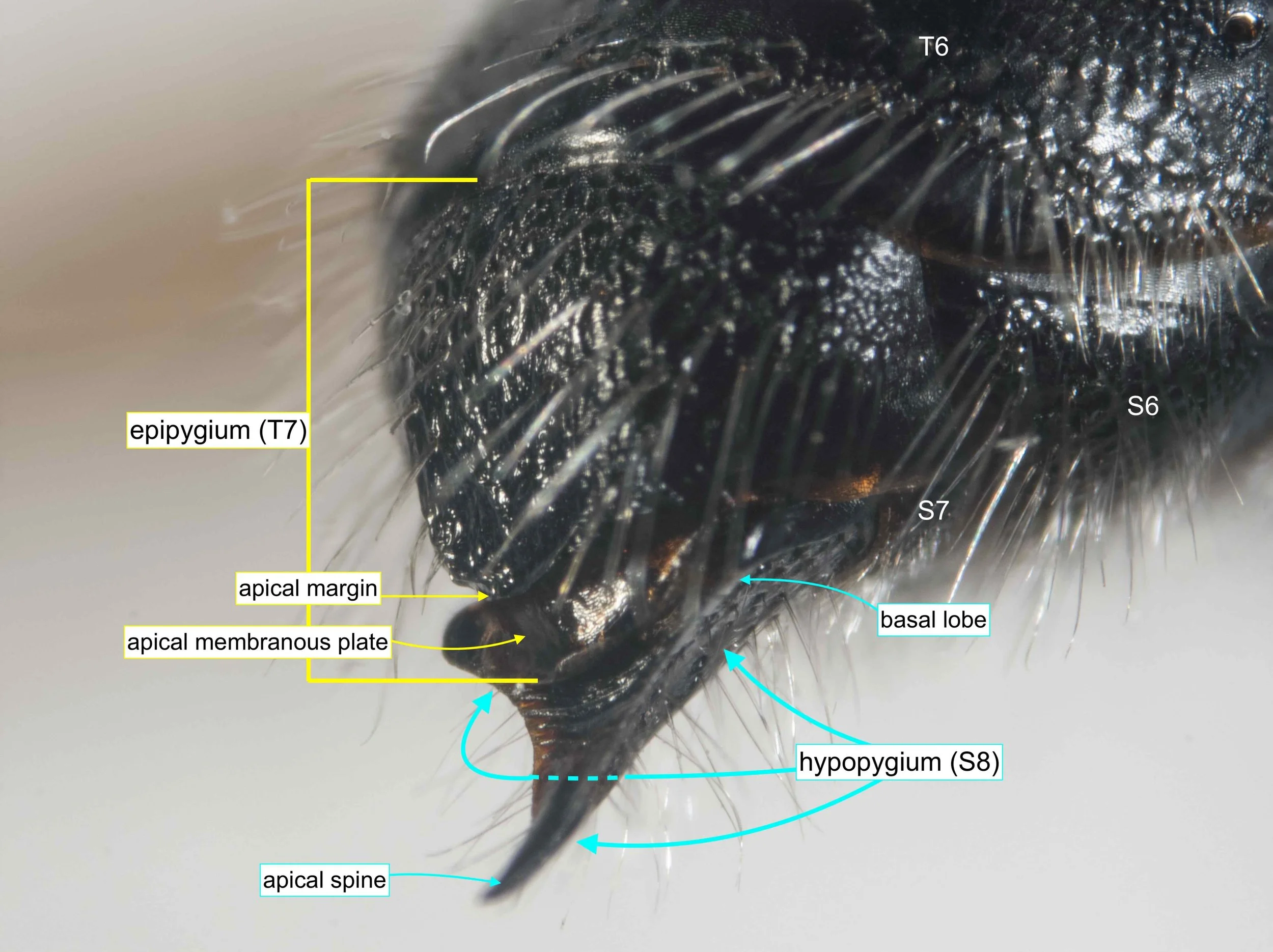
Both the epipygium (T7) and the hypopygium (S8) provide important clues to identification.
The epipygium is typically convex, as here, but in some genera it is produced apically into a plate before and above the apical margin. Note that such ‘subapical plates’ are quite distinct from the apical membranous plate, a widespread feature that may vary in width but is otherwise not particularly diagnostic.
The hypopygium is highly variable, particularly among the Thynninae.
forewing venation
2m-cu meets SCIII (so not Diamminae)
3rd interval of M: subequal to 2nd interval (3:3.5); more than half length 2r-m (3:3.8) (so neither Iswaroidini nor Agriomyini)
1r vein distinct (so neither Myzininae nor Iswaroidini)
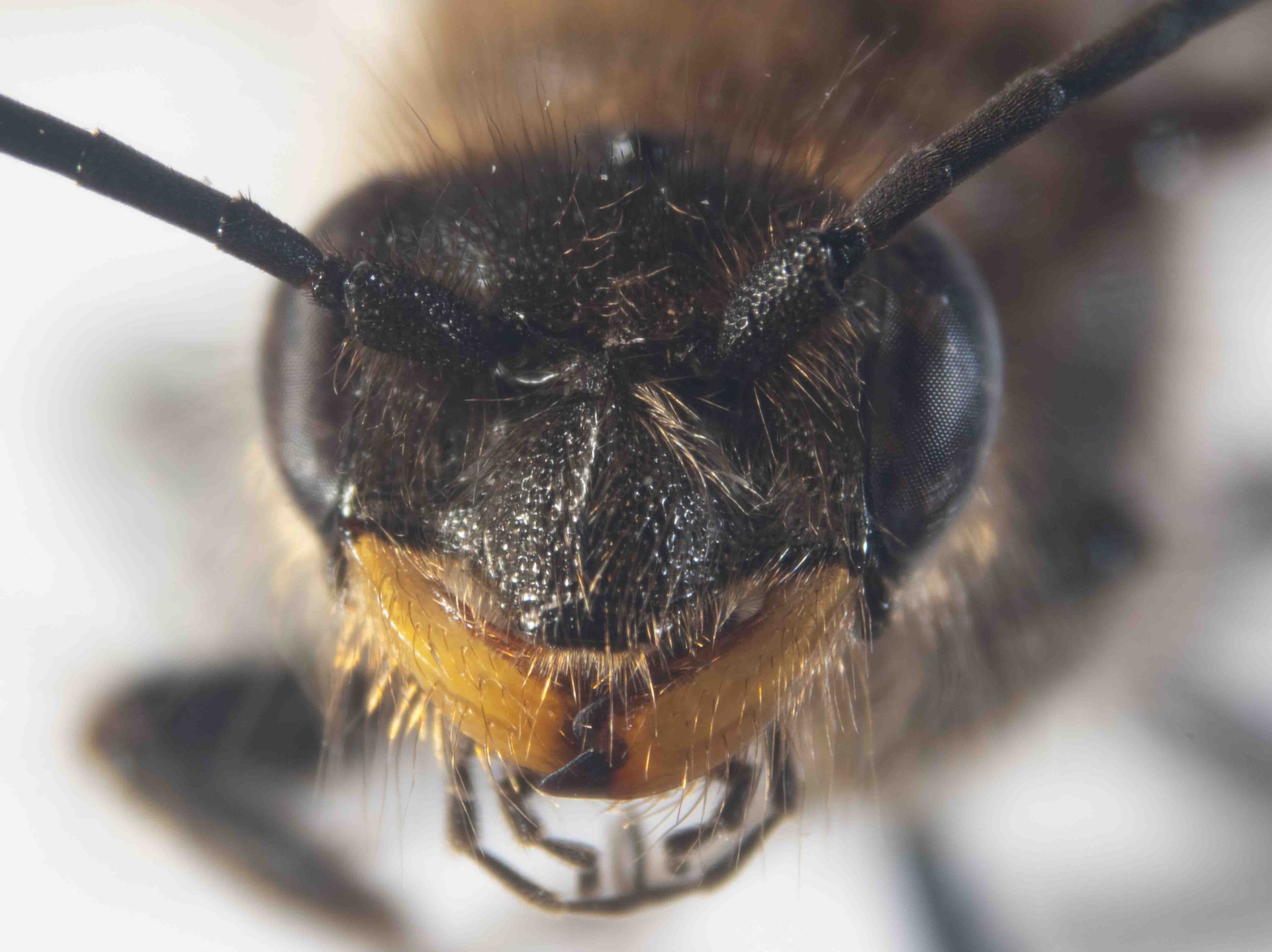
frons & clypeus
frons produced above antennal sockets (ie antennal prominence present) (so not Anthoboscinae)
clyeus with sagittal (ie median) carina, not obviously branched
clypeus (& mandibles)
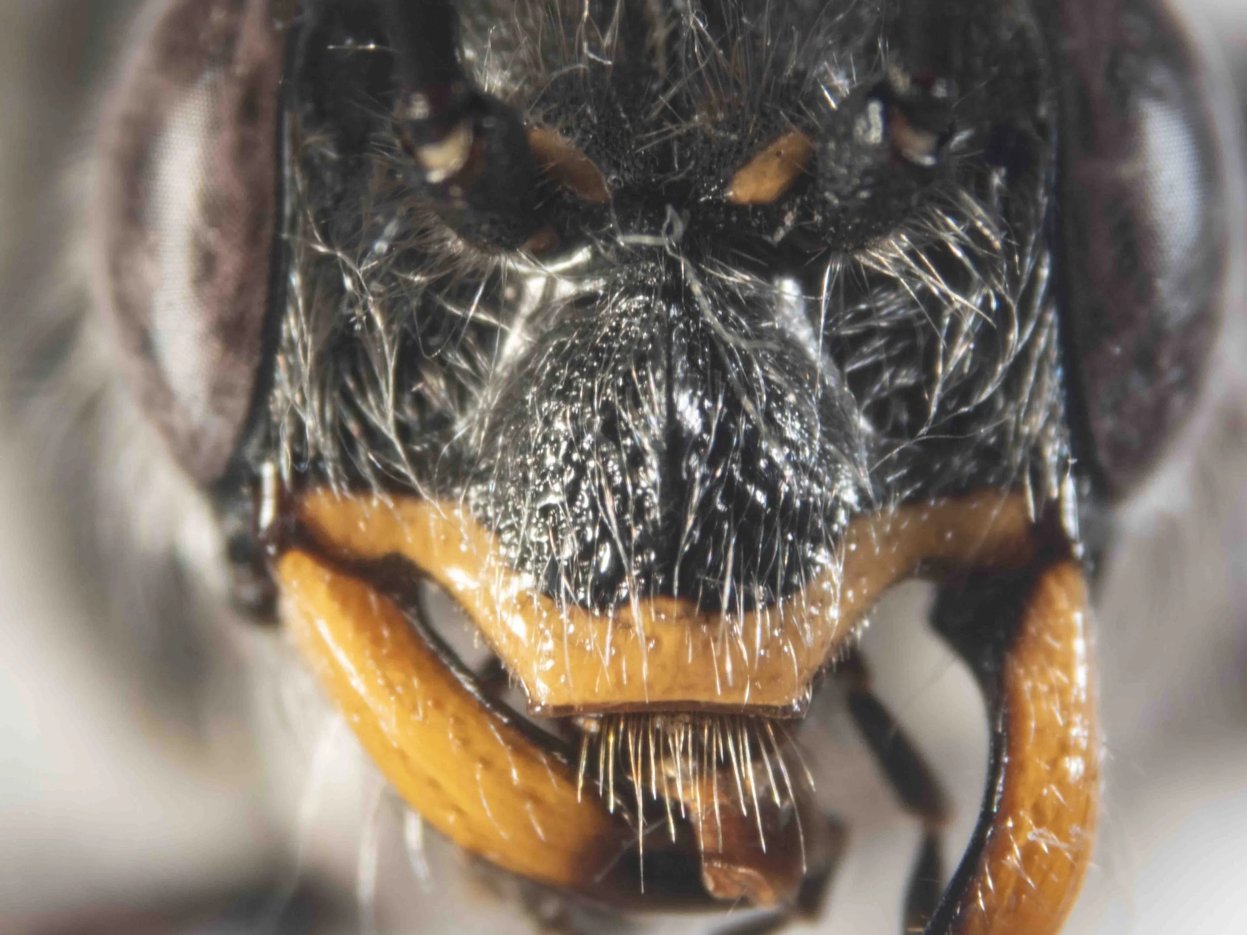
clypeus truncate apically
note broad mandibles!
antennal prominence
antennal prominence slightly raised above each antennal socket
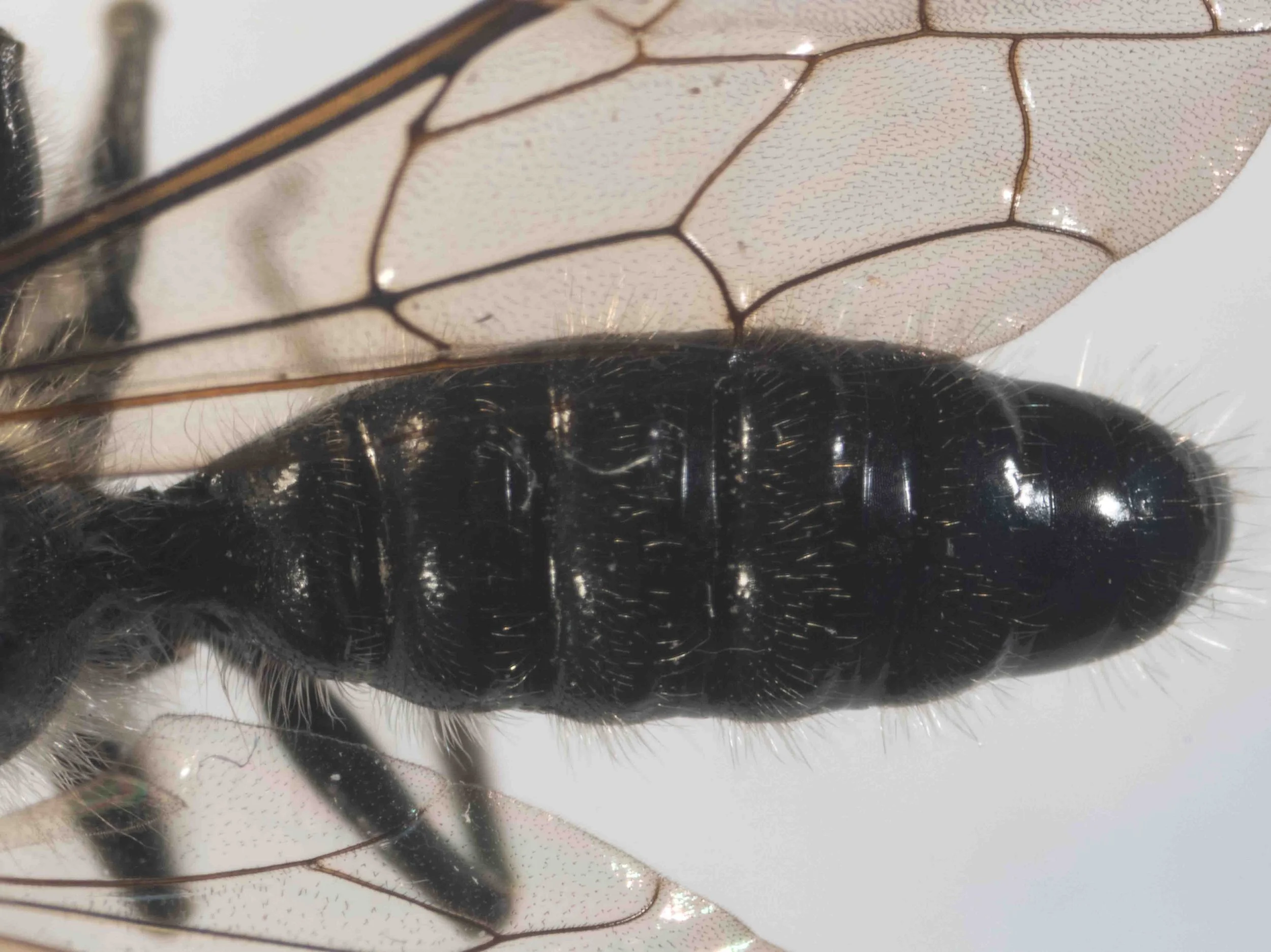
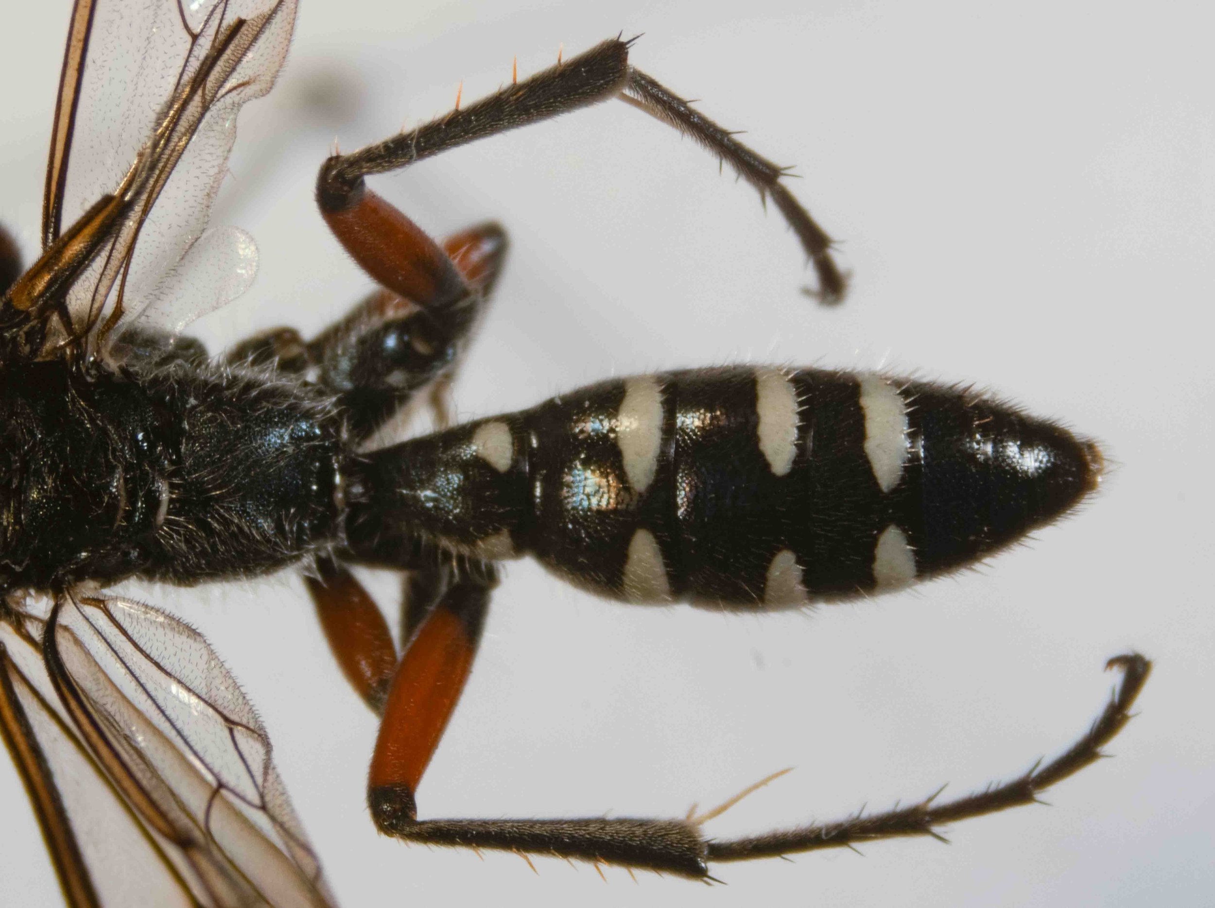
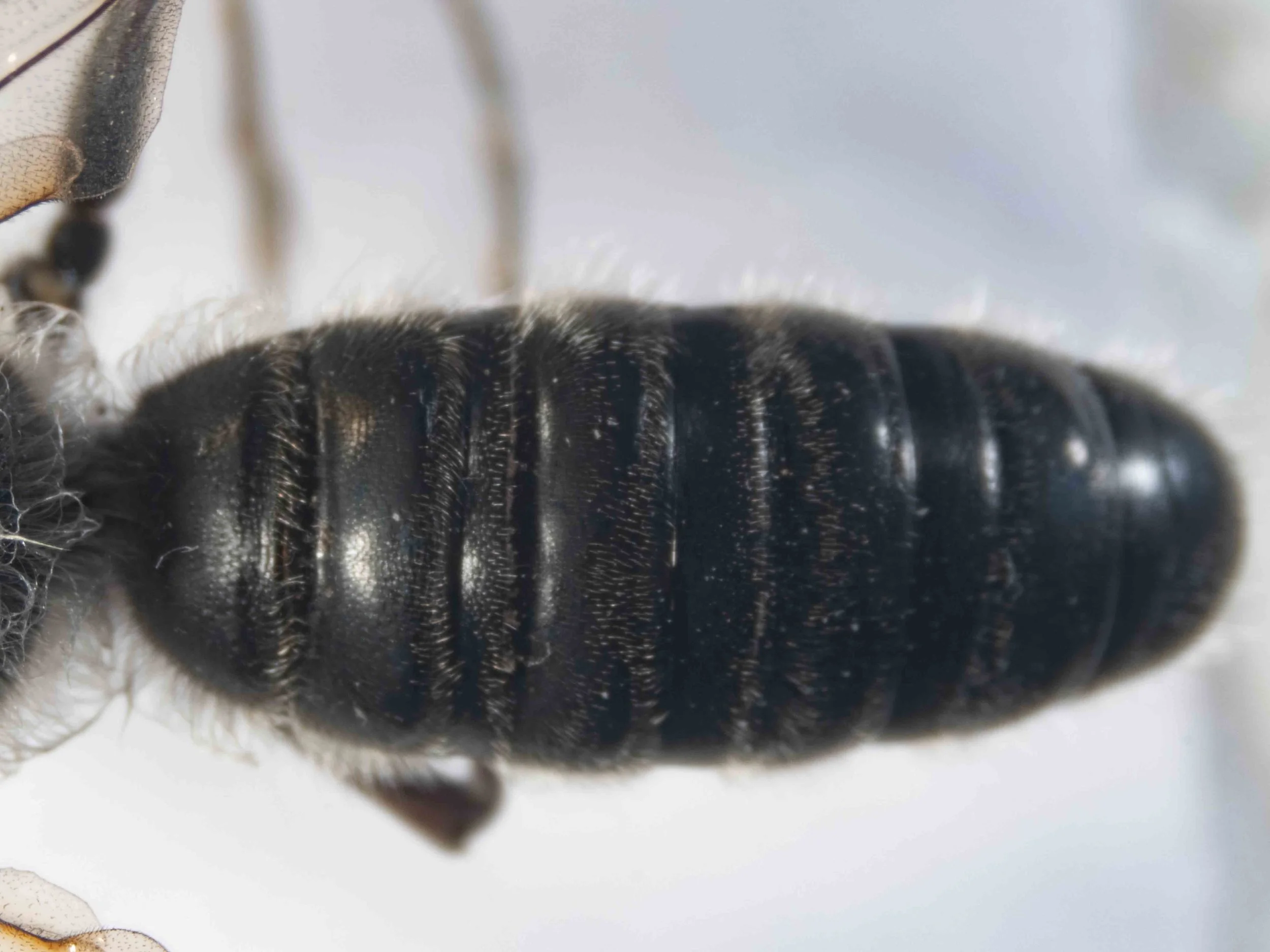
metasoma (dorsal view)
metasoma fusiform, without strong constrictions
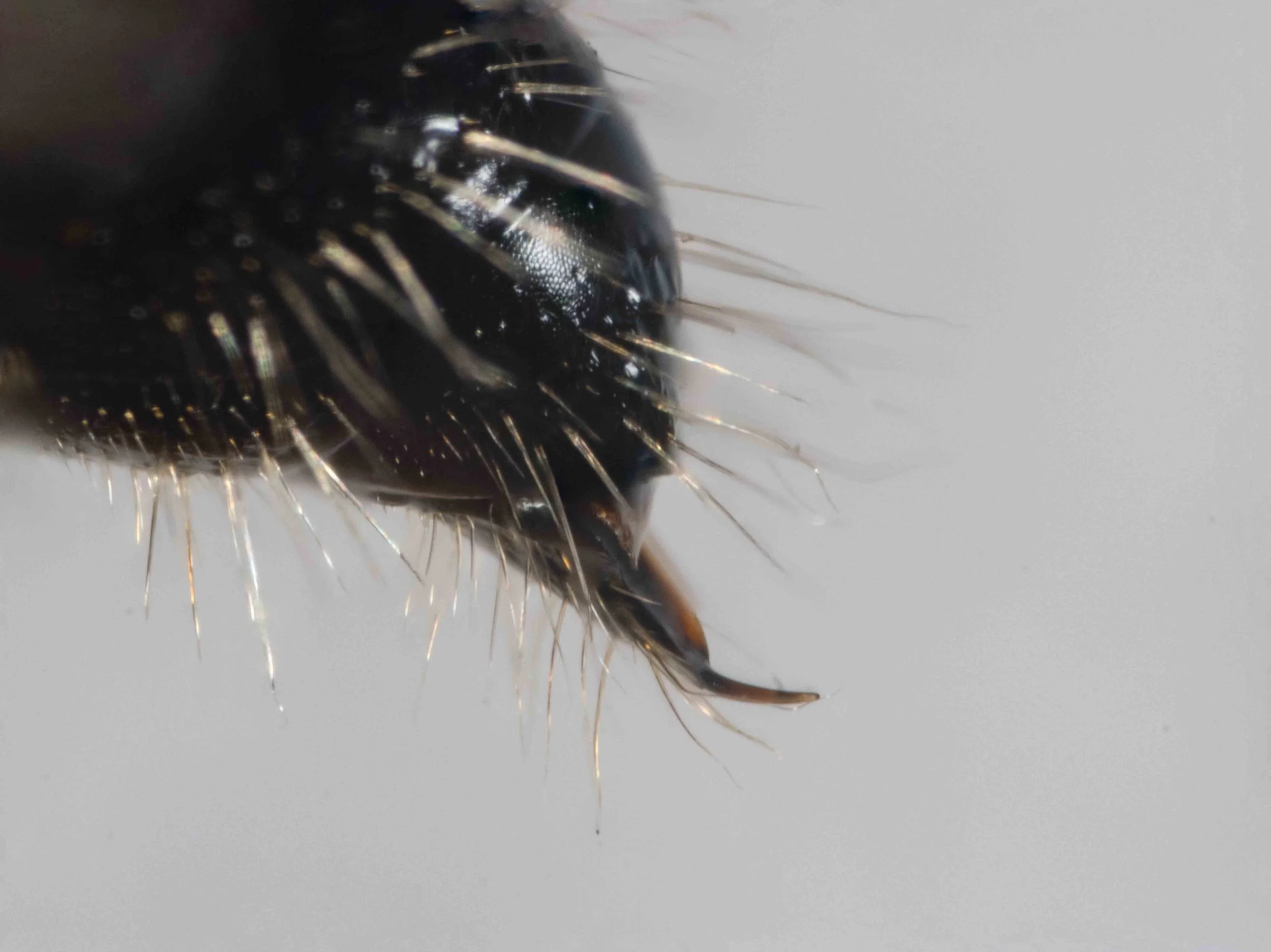
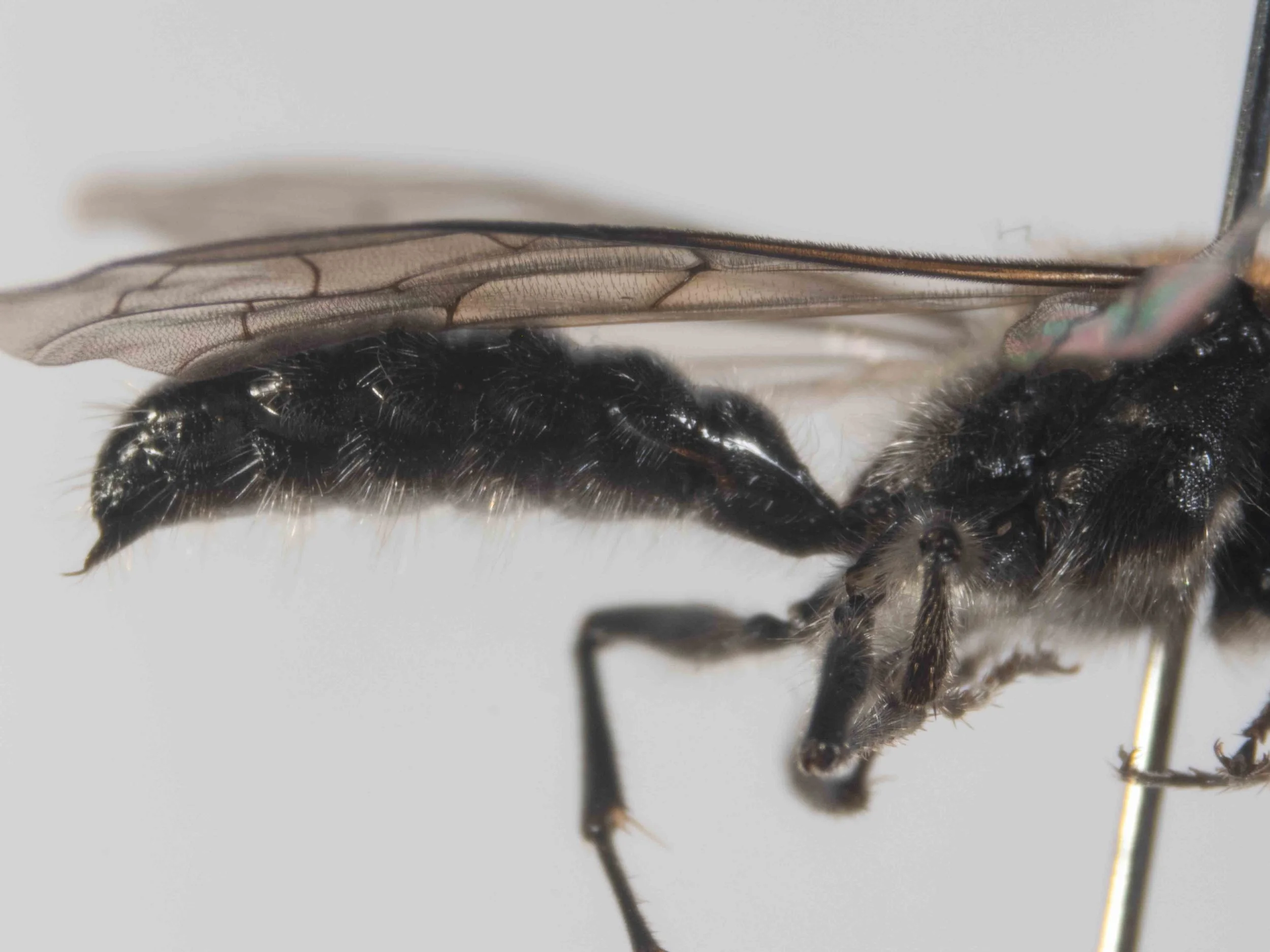
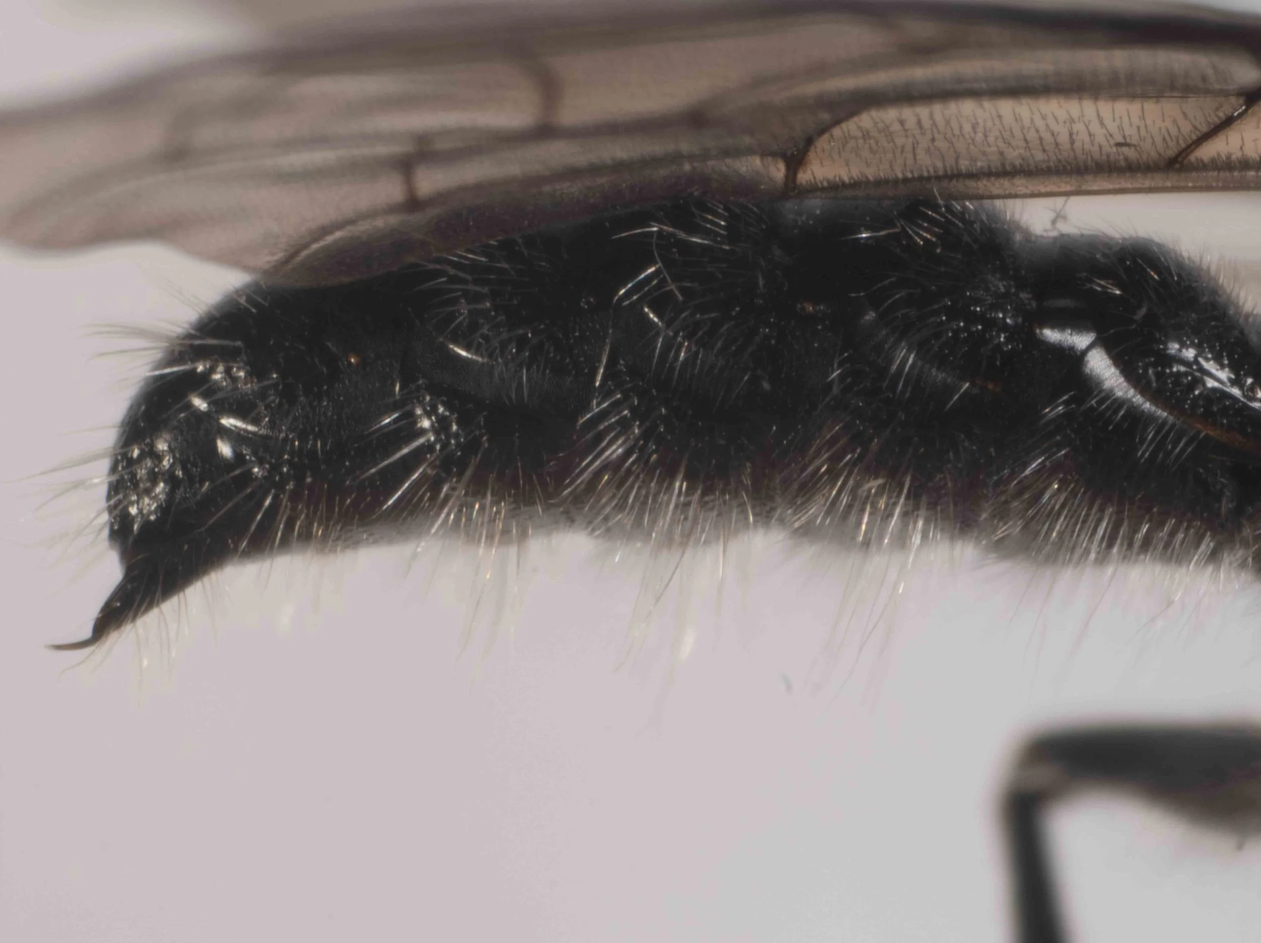
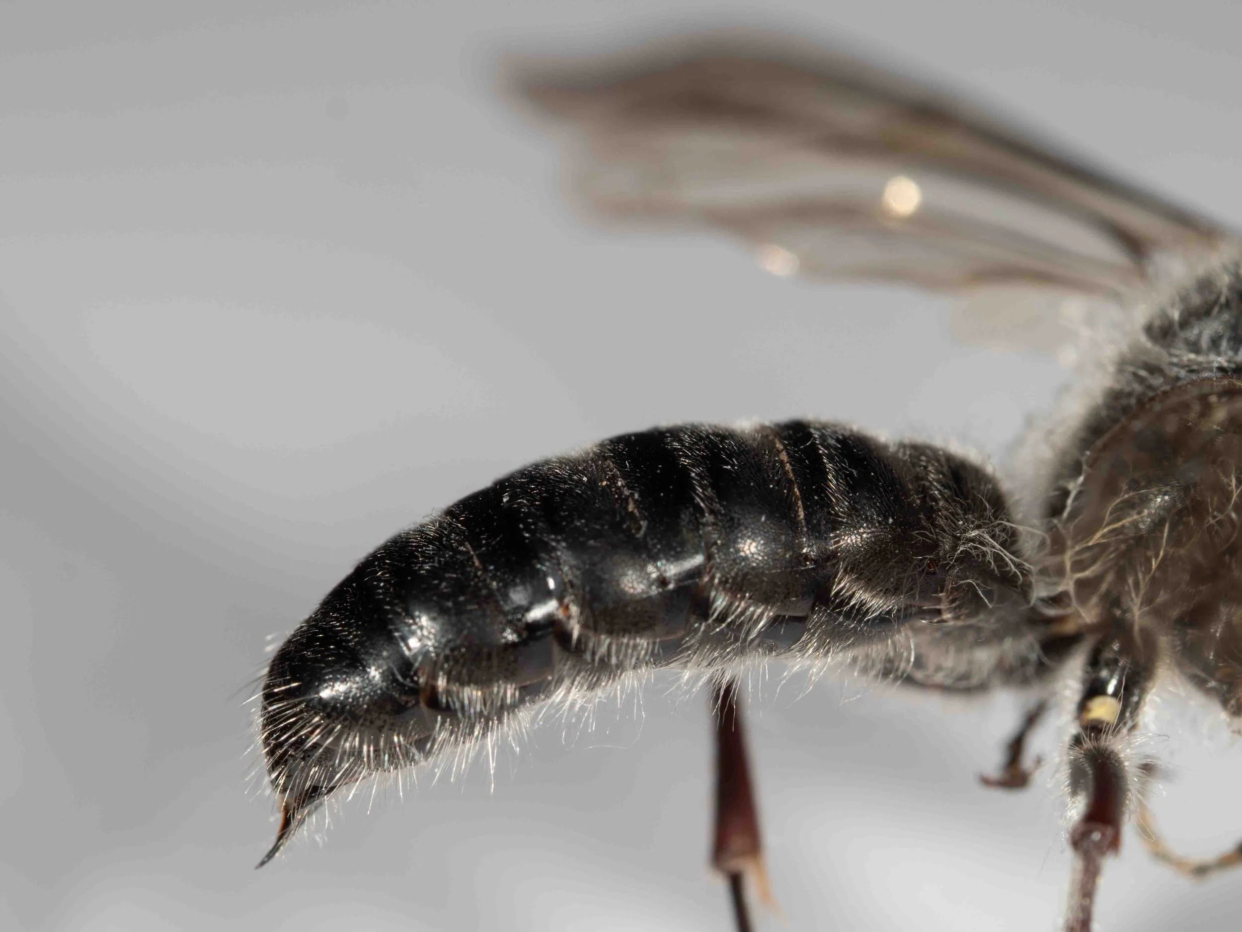
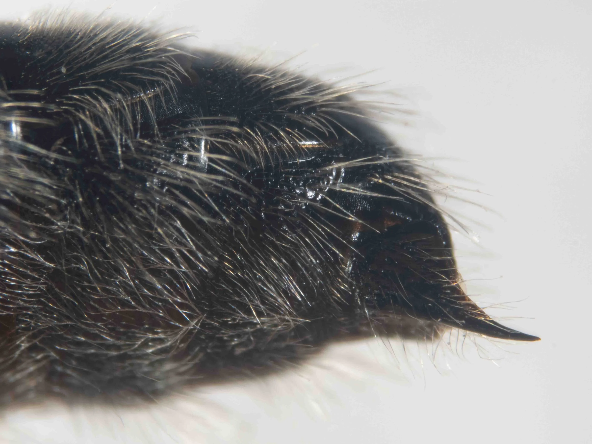
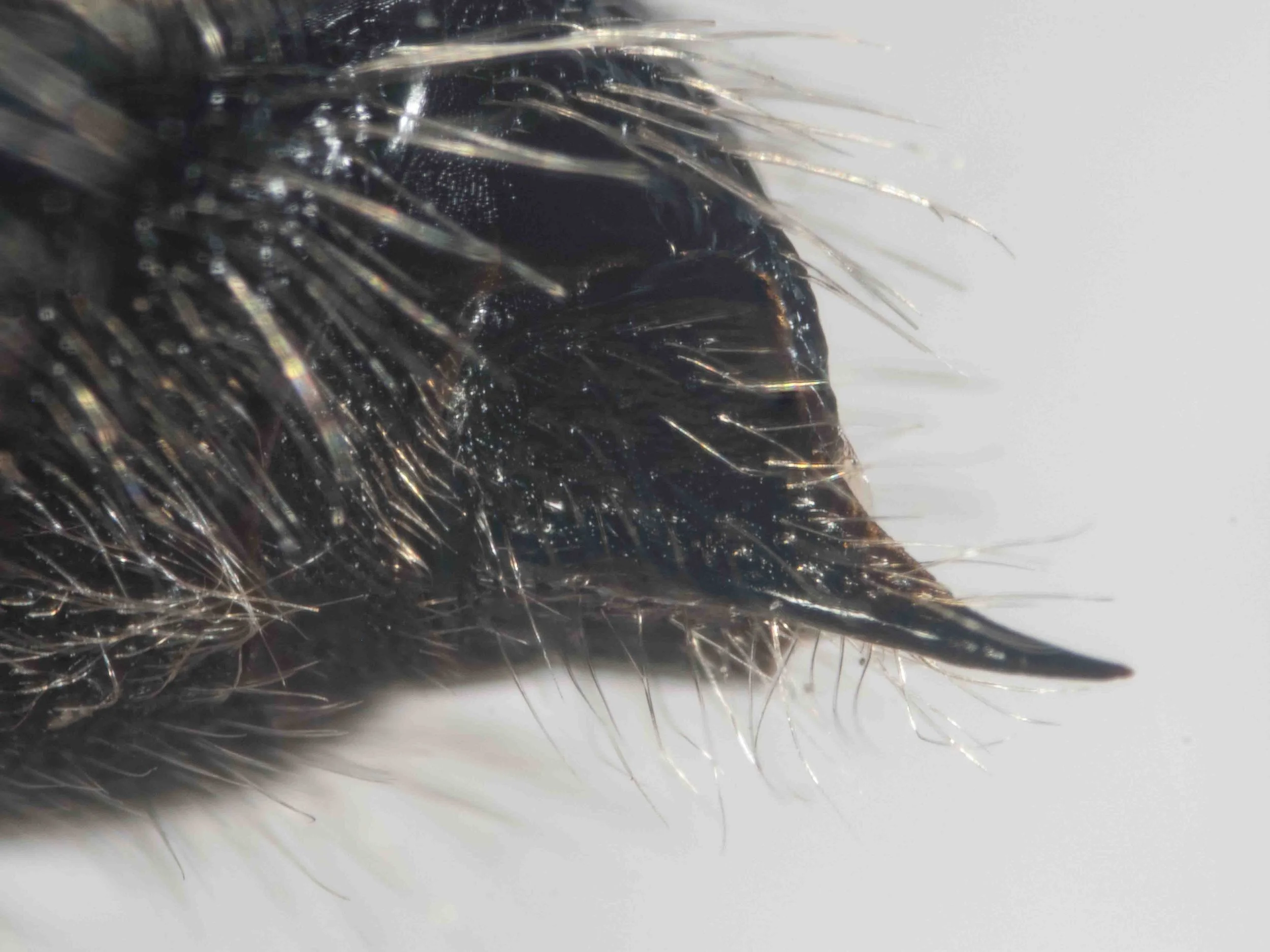
terminal segments (lateral view)
hypopygium with short, slightly upturned apical spine
epipygium (T7): strongly convex, not flat (unlike most Tachynomyini); without subapical plate
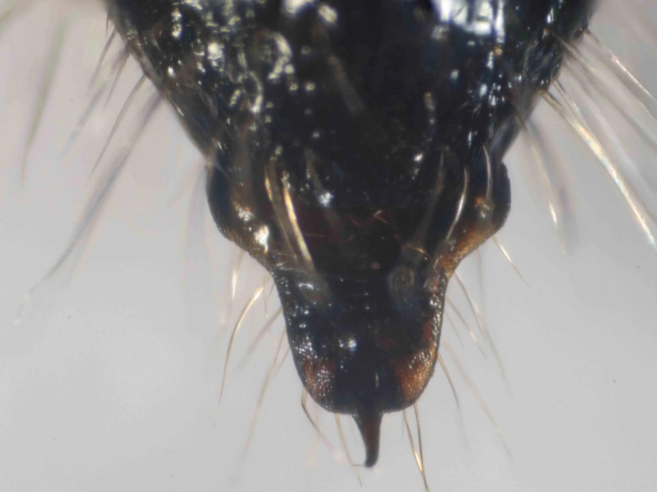
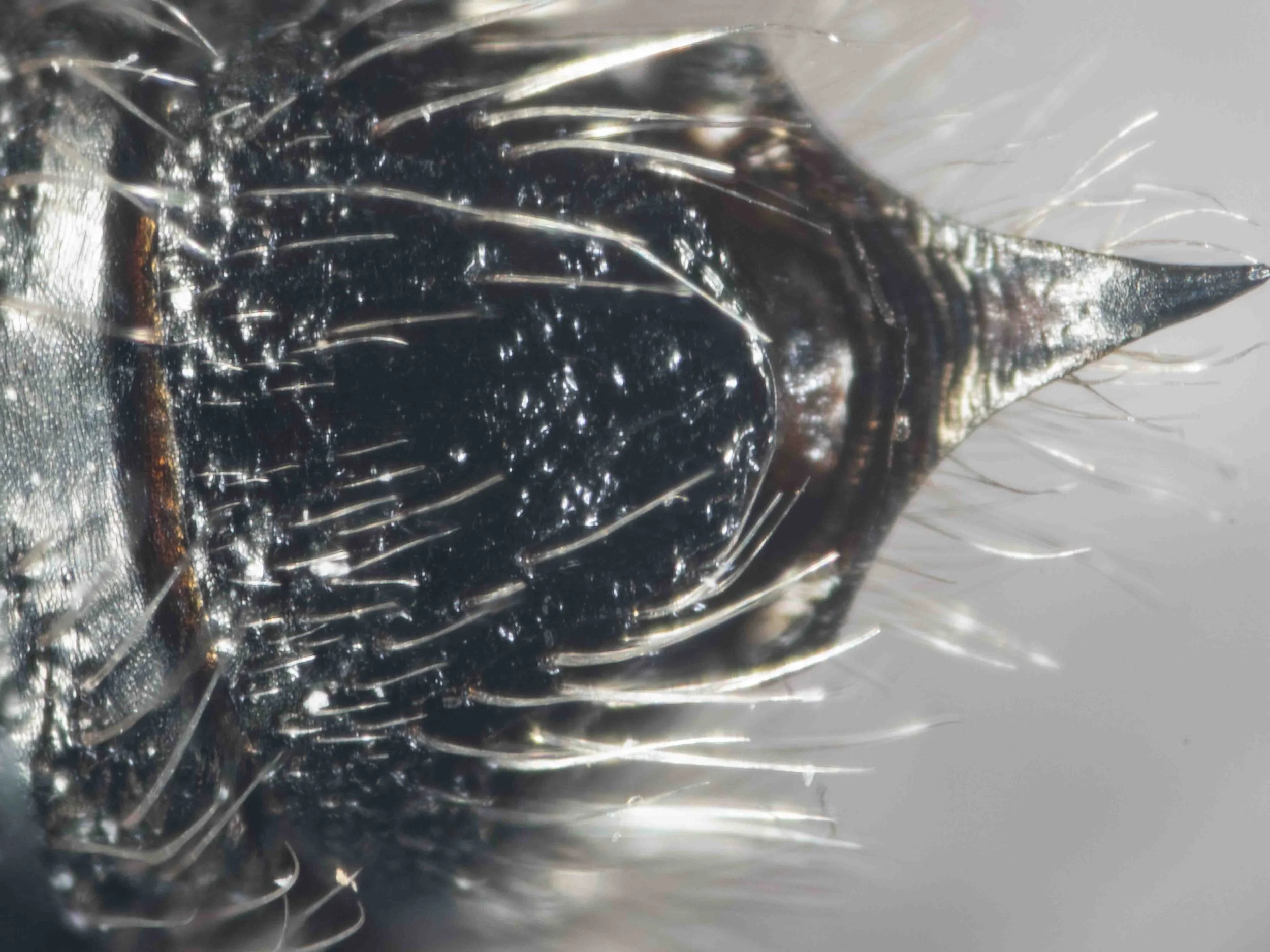
terminal segments (dorsal view)
hypopygium with: long, exposed dorsal surface that is multicarinate; rounded lateral lobes (‘basal angles’); short apical spine; apical margin rounded
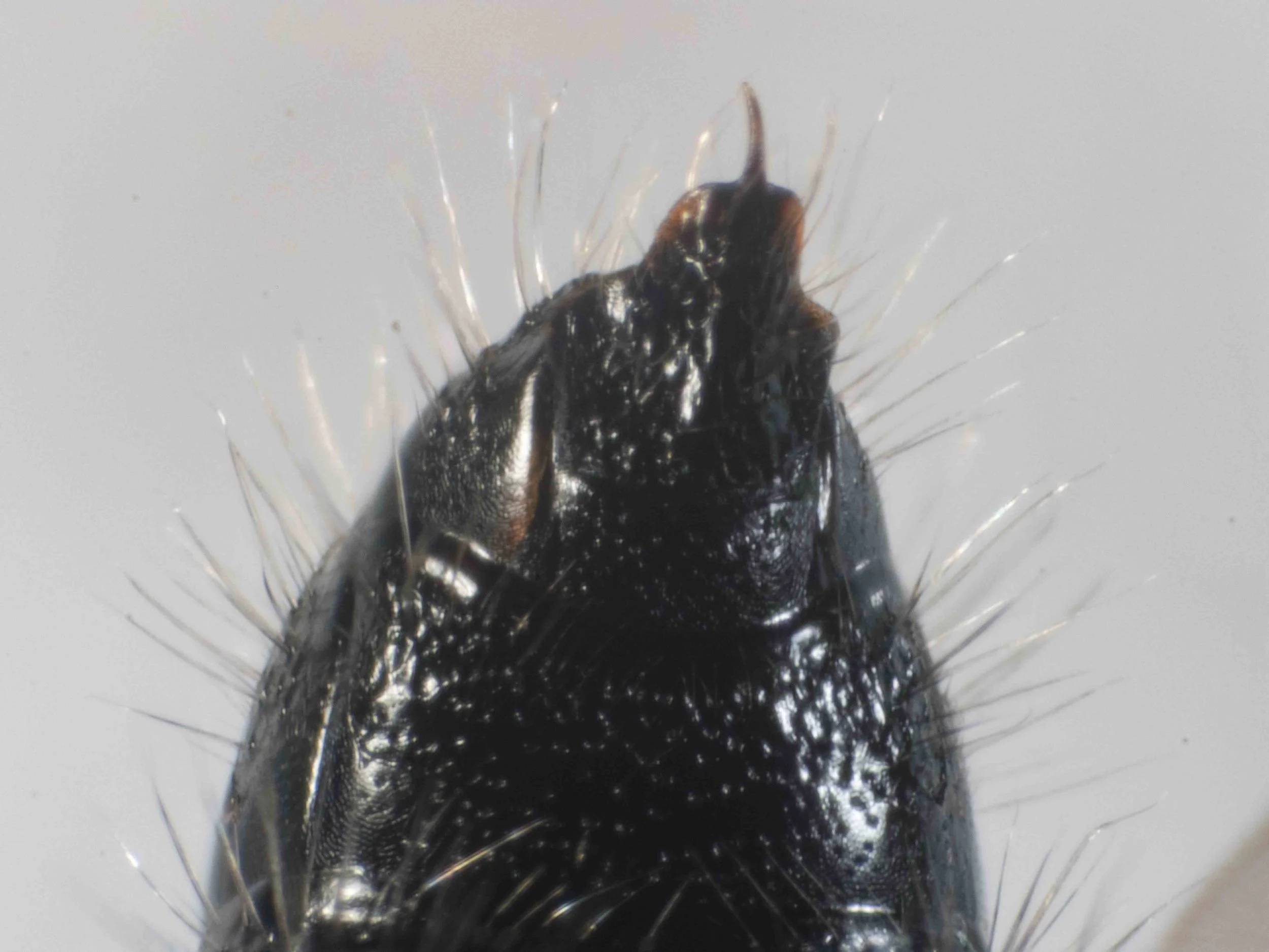
terminal segments (ventrolateral view)
metasoma (lateral view)
metasoma: moderately sclerotised; moderately constricted between segments
metasoma (lateral view)
S2-6 without posterolateral spines
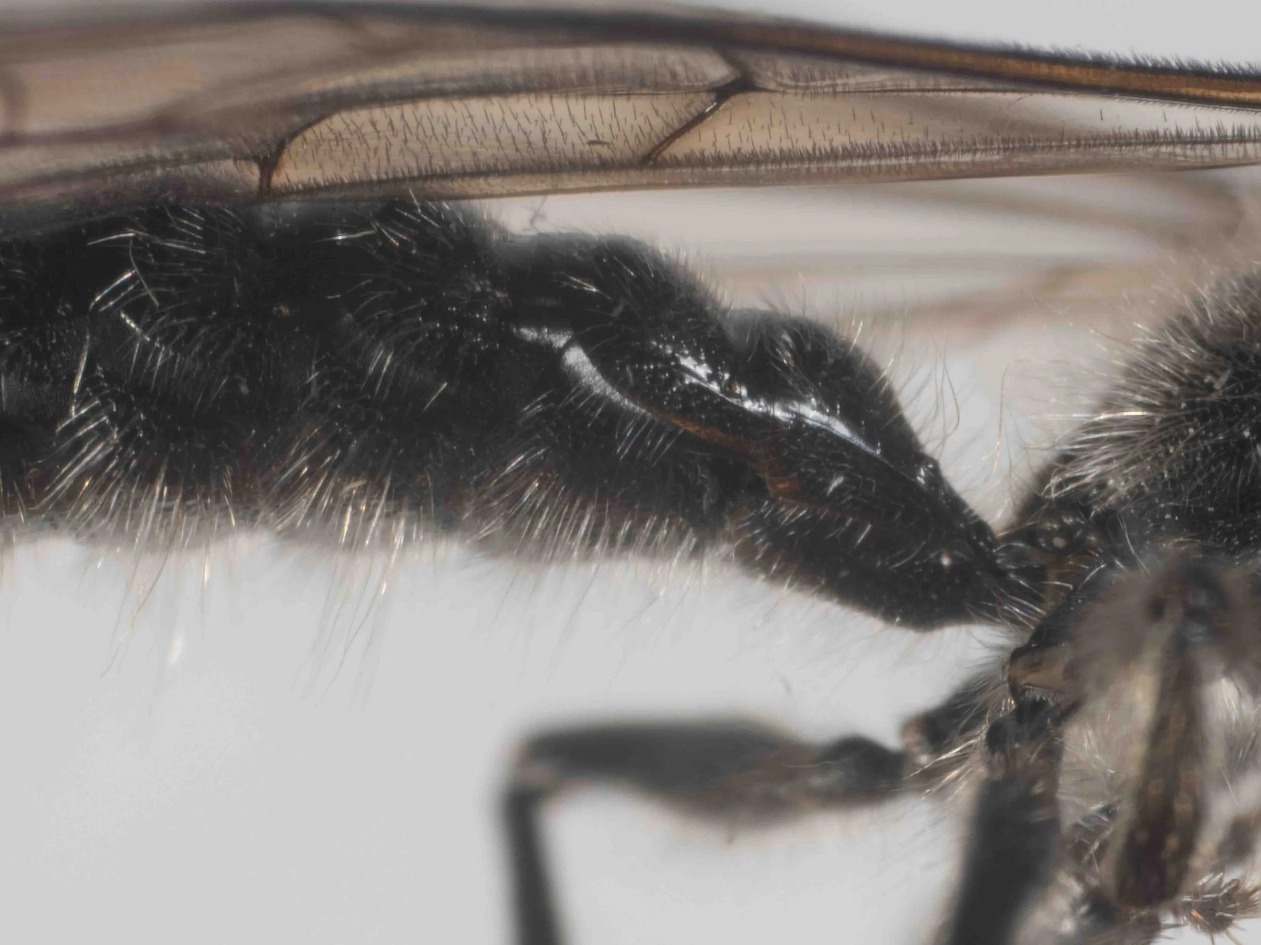
metasoma (lateral view)
S1 slightly angulate in profile
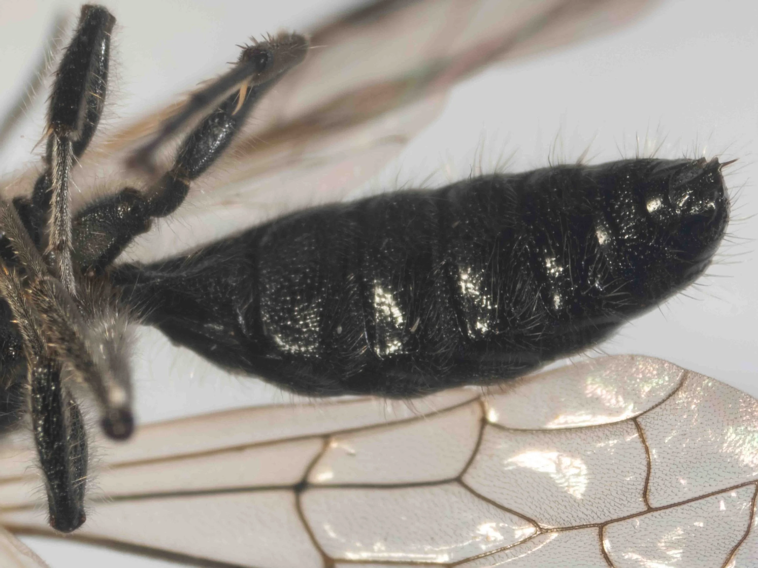
metasoma (ventrolateral view)
note that constrictions between segments are much less pronounced in this view (cf lateral view)
base of metasoma (ventrolateral view)
S1 weakly raised, without sagittal carina
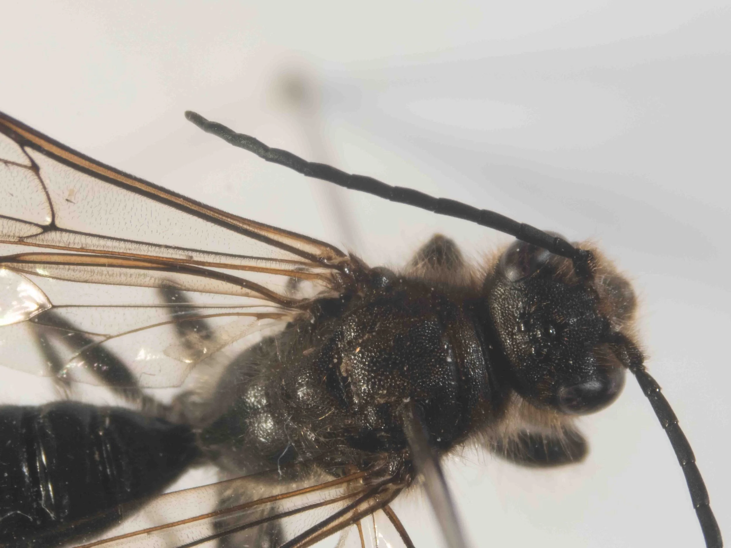
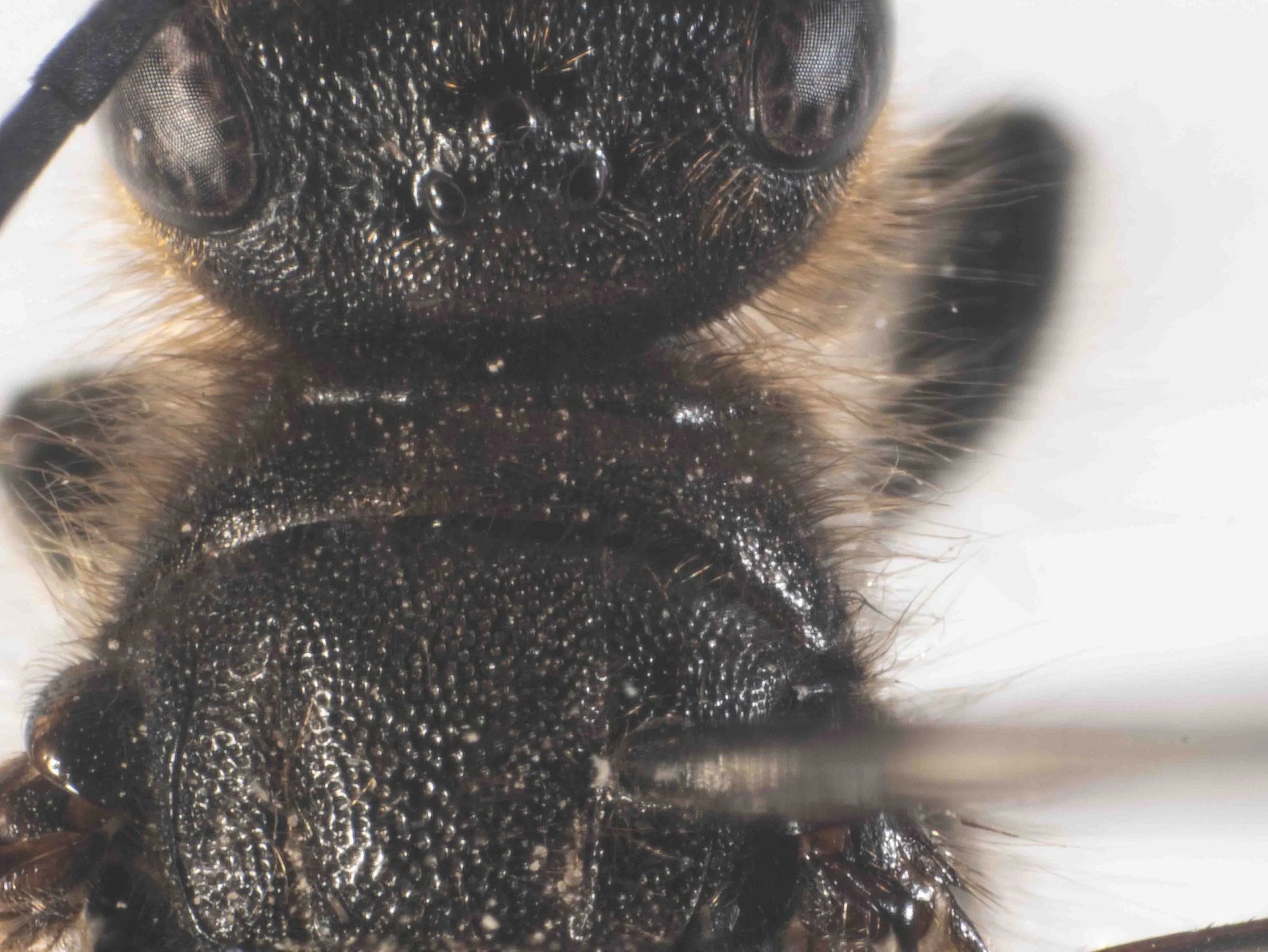
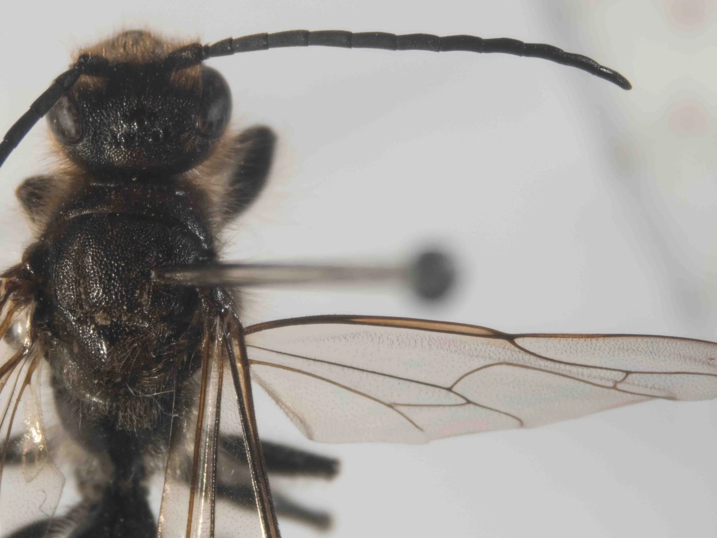
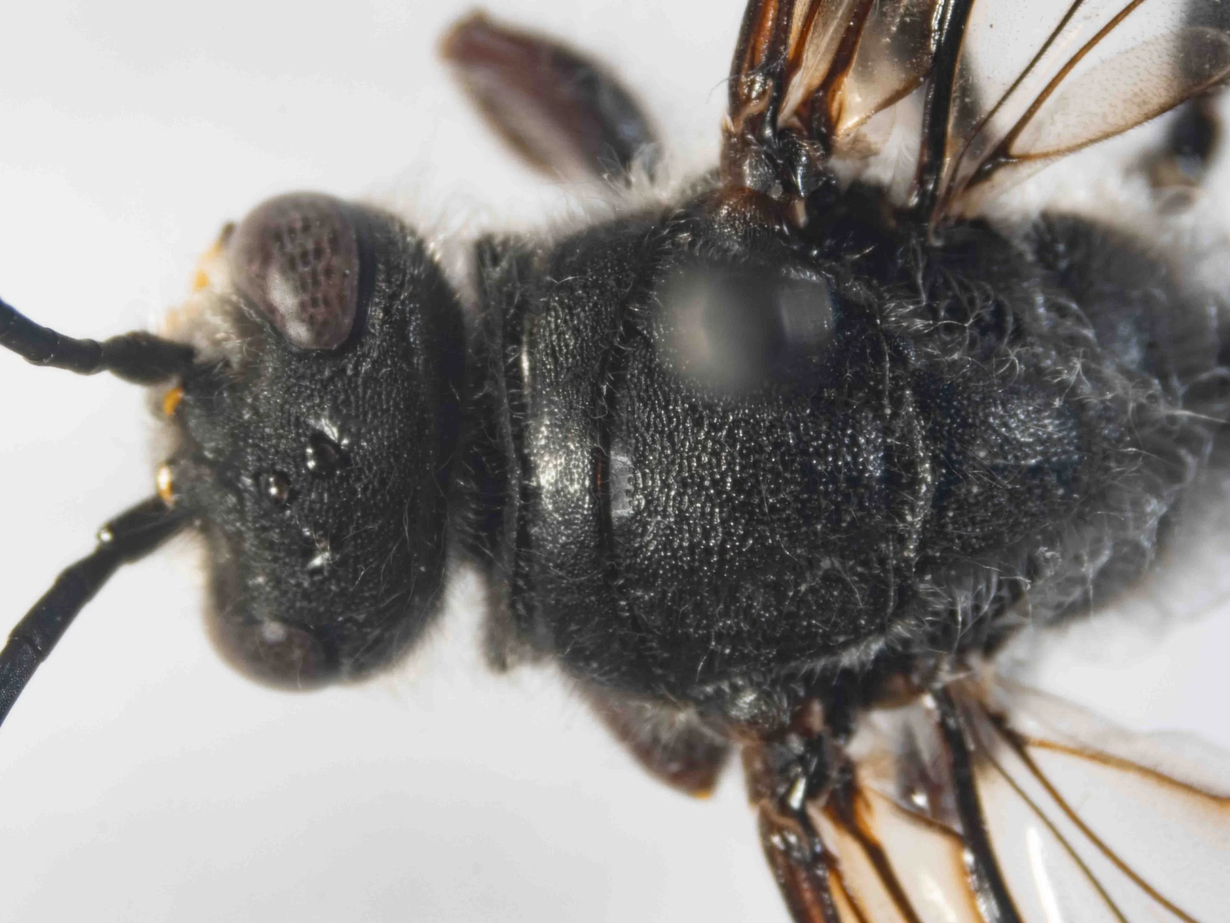
head & mesosoma (dorsal view)
pronotum: long dorsally; carinate anteriorly
propodeum rounded
vertex & pronotum (dorsal view)
head rounded posteriorly, not strongly concave (unlike most Tachynomyini)
pronotum: long (?) dorsally; carinate anteriorly
head & mesosoma (dorsal view)
pronotum: long dorsally; carinate anteriorly
antennae long
propodeum (dorsal view)
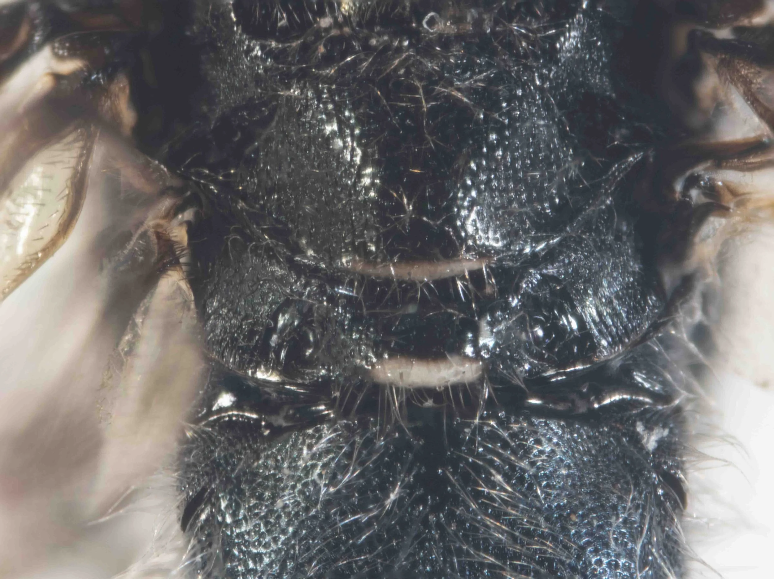
propodeum: evenly rounded, without distinct dorsal and vertical faces; without carina
head & pronotum (ventrolateral view)
hypostomal carina with fringe of setae of varying lengths
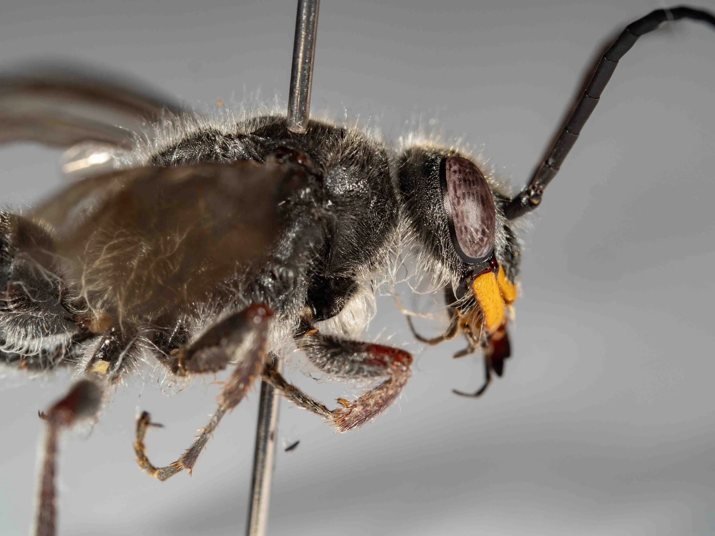
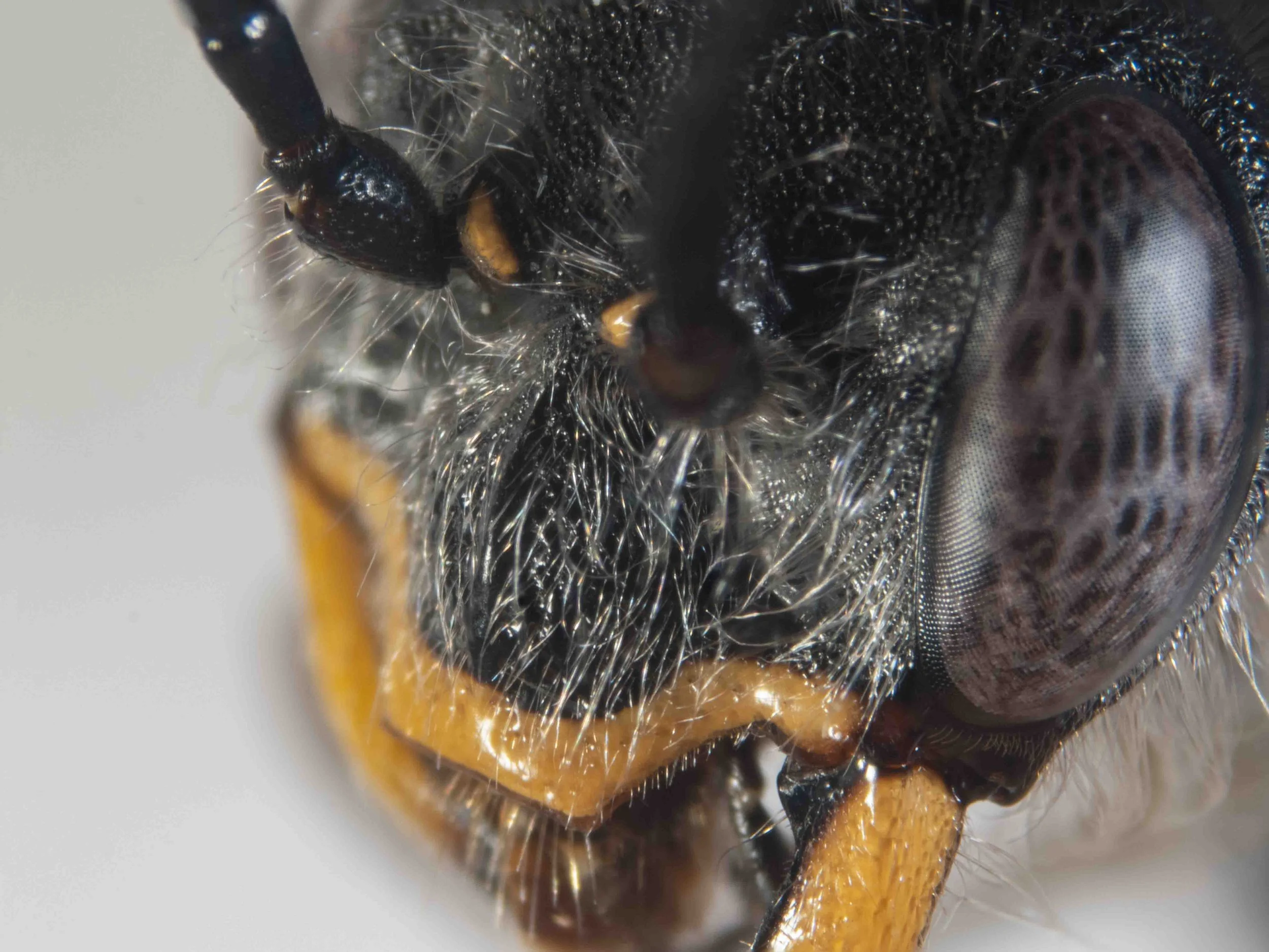
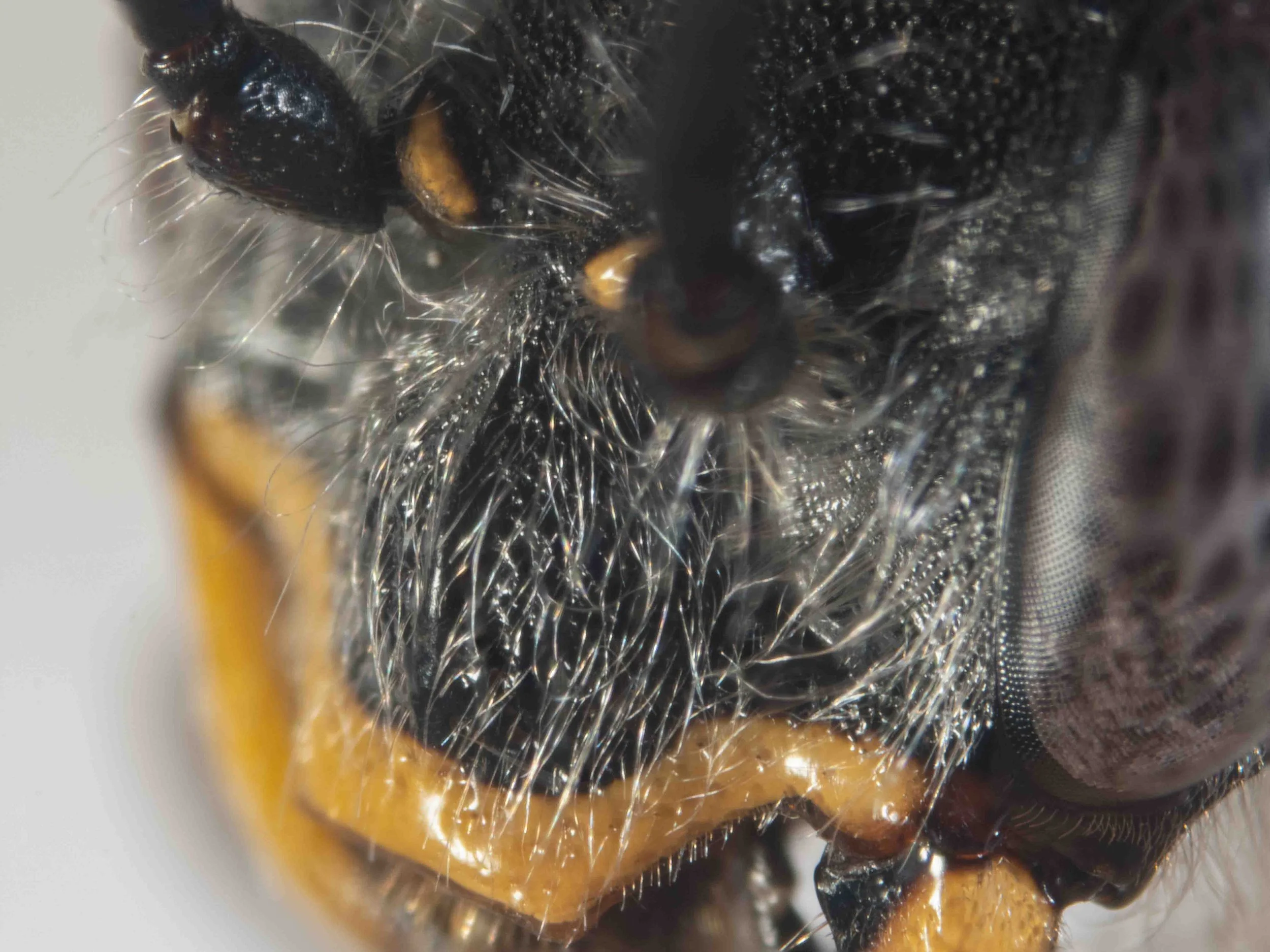
head (lateral view)
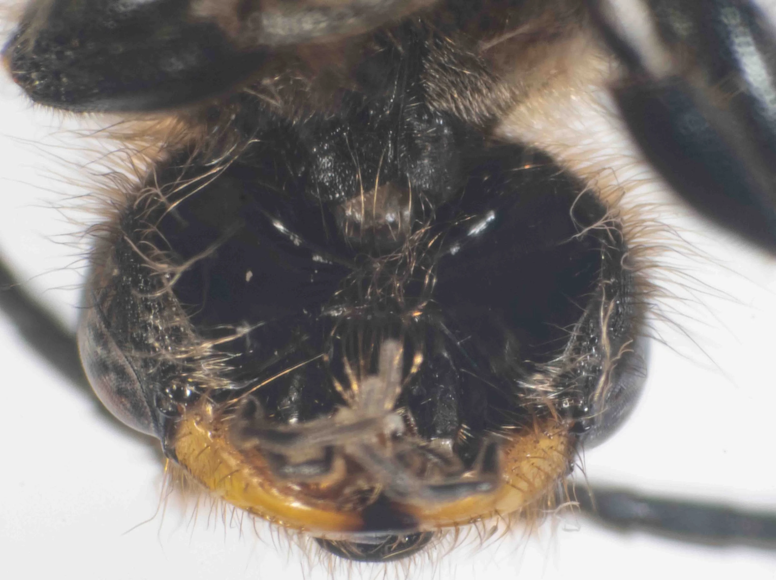
head (ventral view)
hypostoma broad, concave, polished
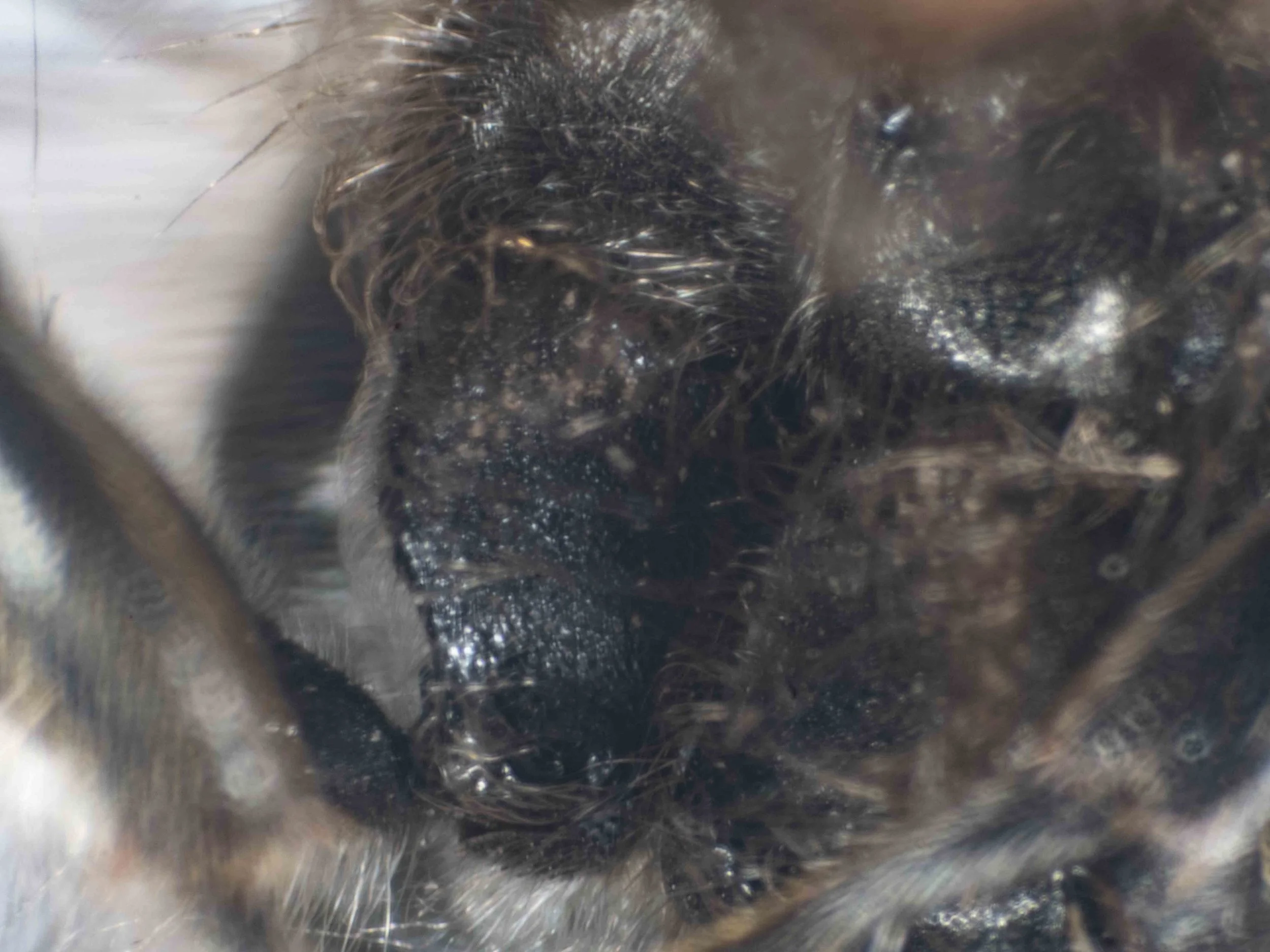
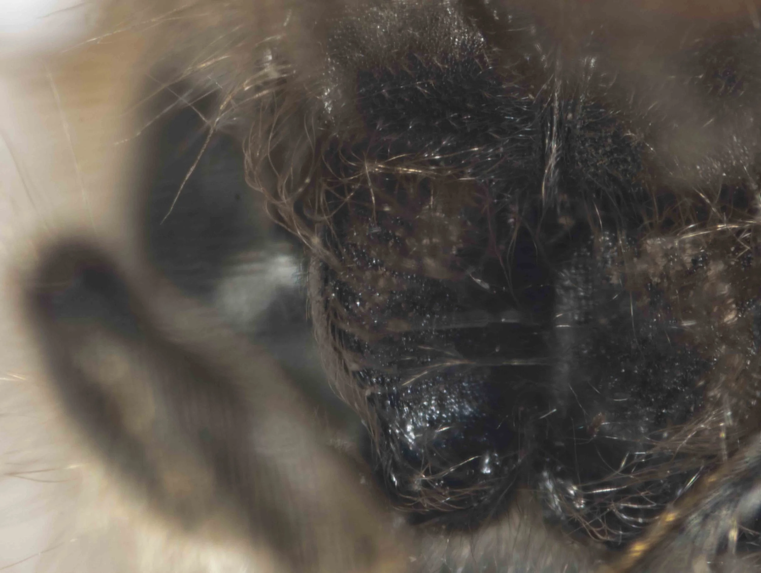
front coxa (ventral view)
front coxae are large and concave ventrally, with a sharp ridge between dorsal and ventral faces
front coxae (ventral view)
front coxae are large and concave ventrally
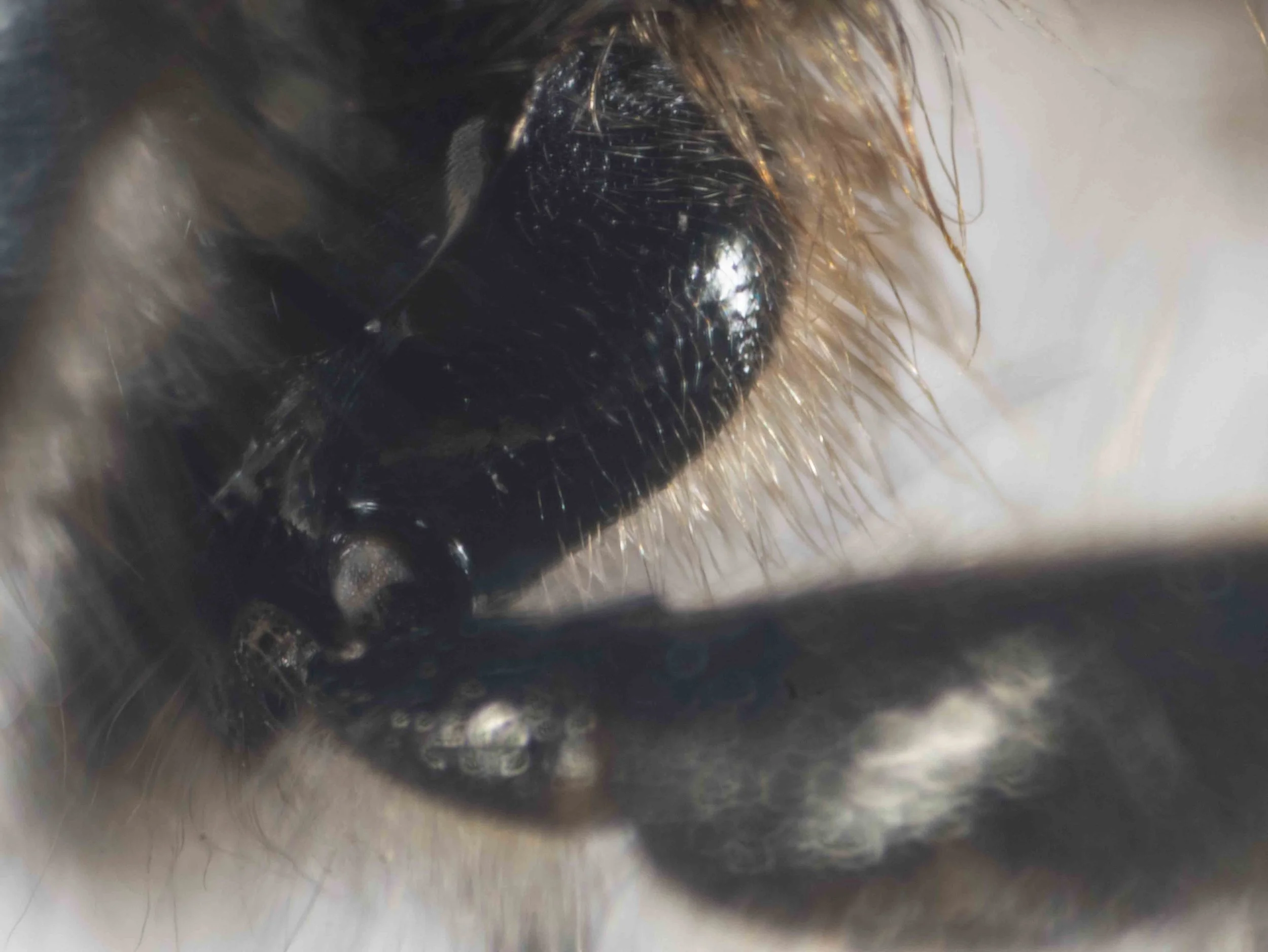
front coxa (dorsal view)
front coxa wide, with rounded (convex) dorsal surface
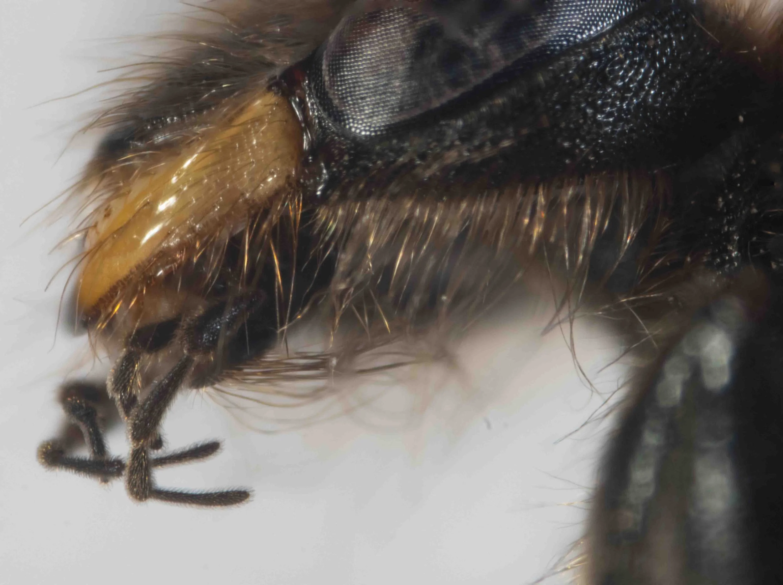
mouthparts (lateroventral view)
prementum with tuft of long, posteriorly directed setae
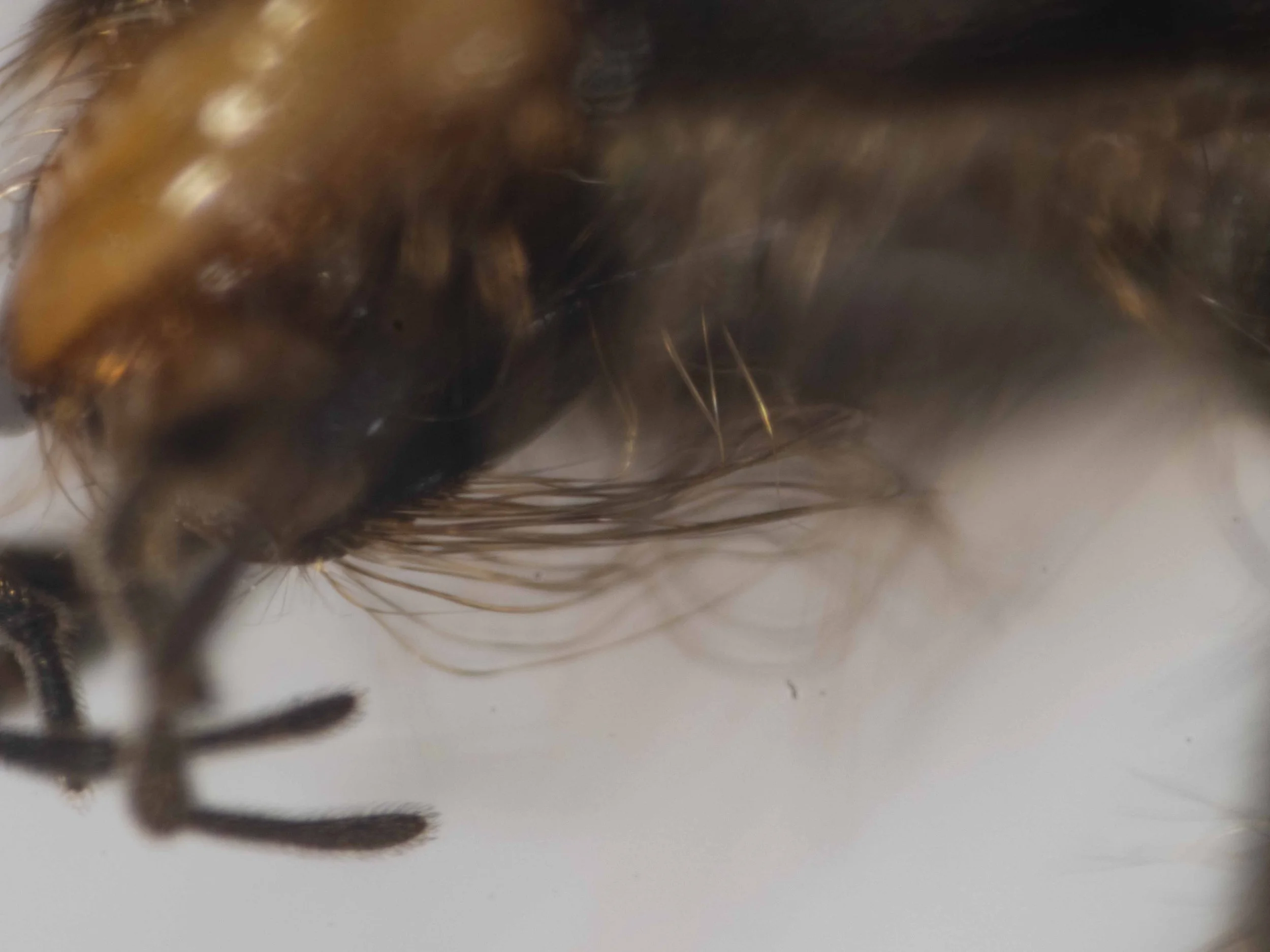
prementum (lateral view)
prementum with tuft of long, posteriorly directed setae
forewing (viewed dorsally)
both 1m-cu & 2m-cu join the M vein proximal to 2r-m (ie both recurrent veins meet SCII)
forewing (dorsal view)
without even a trace of 1r vein
forewing (dorsal view)
without even a trace of 1r vein
head (dorsal view)
frons strongly produced above antennal sockets (ie WITH antennal prominence)
metasoma (dorsal view)
metasoma strongly fusiform in dorsal view, glossy
without constrictions between segments (except between segments 1 & 2)
T1 longer than wide, considerably narrower than T2
antennal prominence (dorsal view)
antennal prominence (anterodorsally)
antennal prominence with narrow carina, V-shaped & contiguous with sagittal carina to clypeus
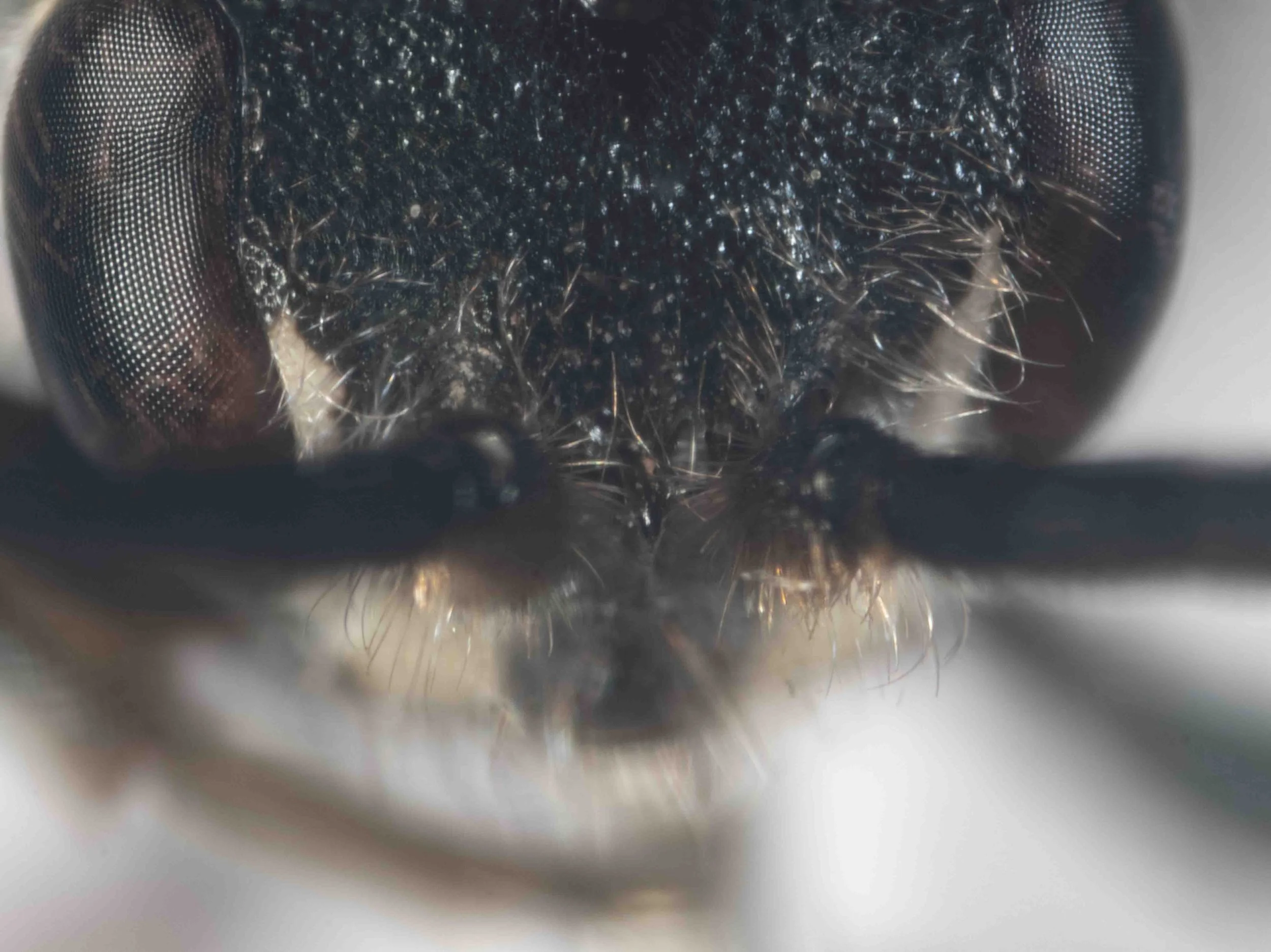
clypeus, antennal sockets, mandible
antennal sockets directed ventrolaterally
clypeus with sagittal carina, contiguous with V-shaped carina of antennal prominence
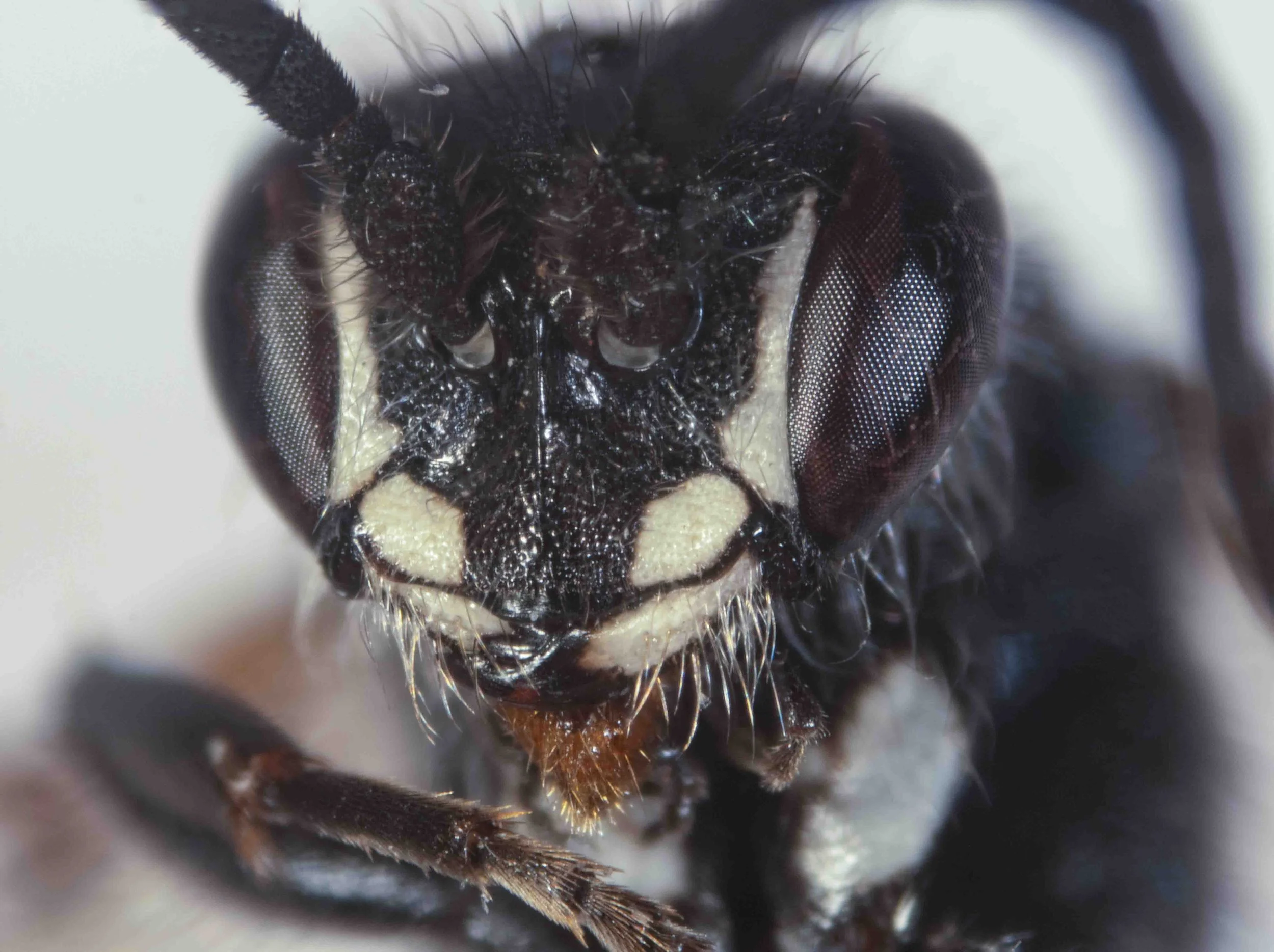
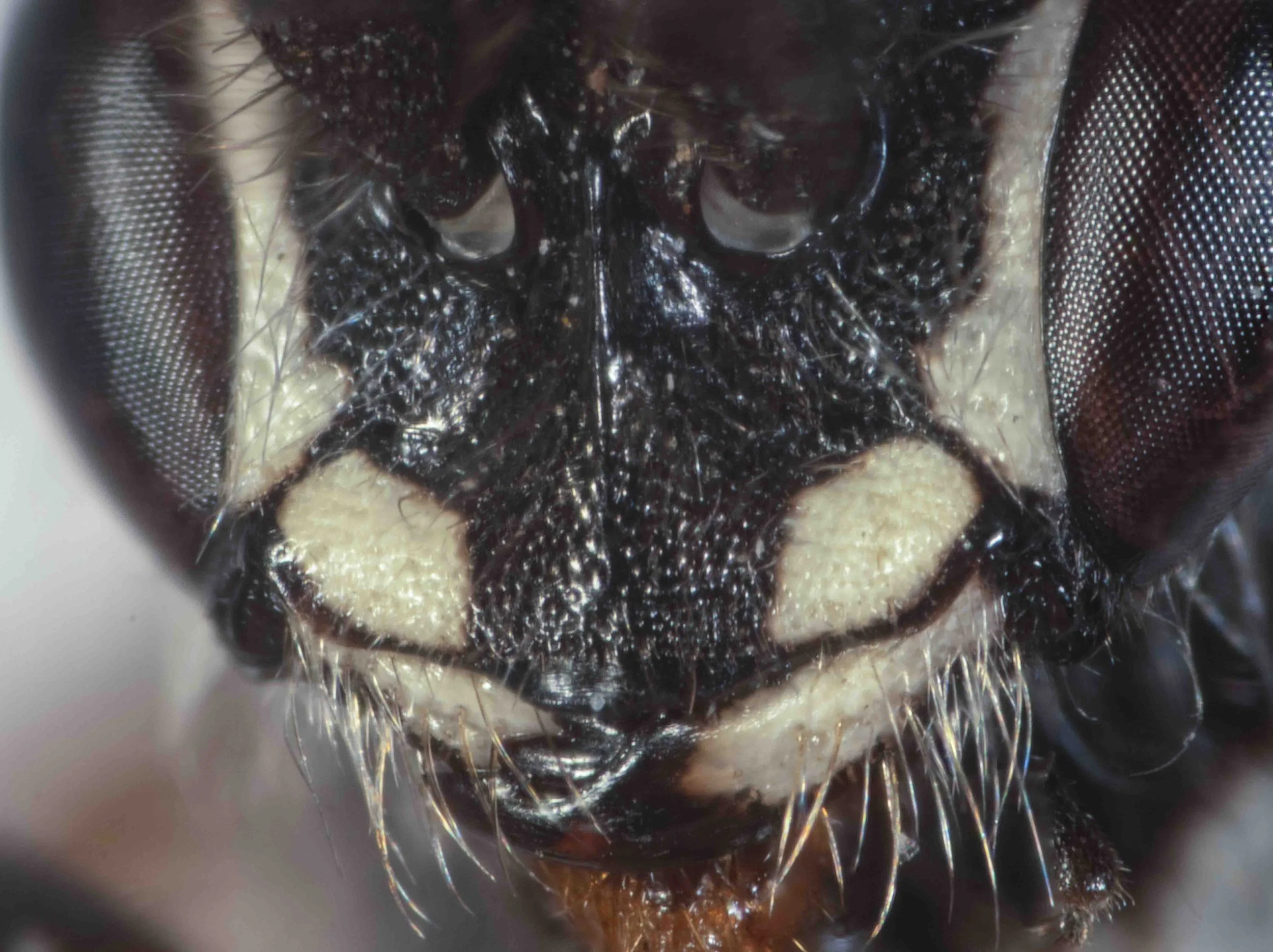
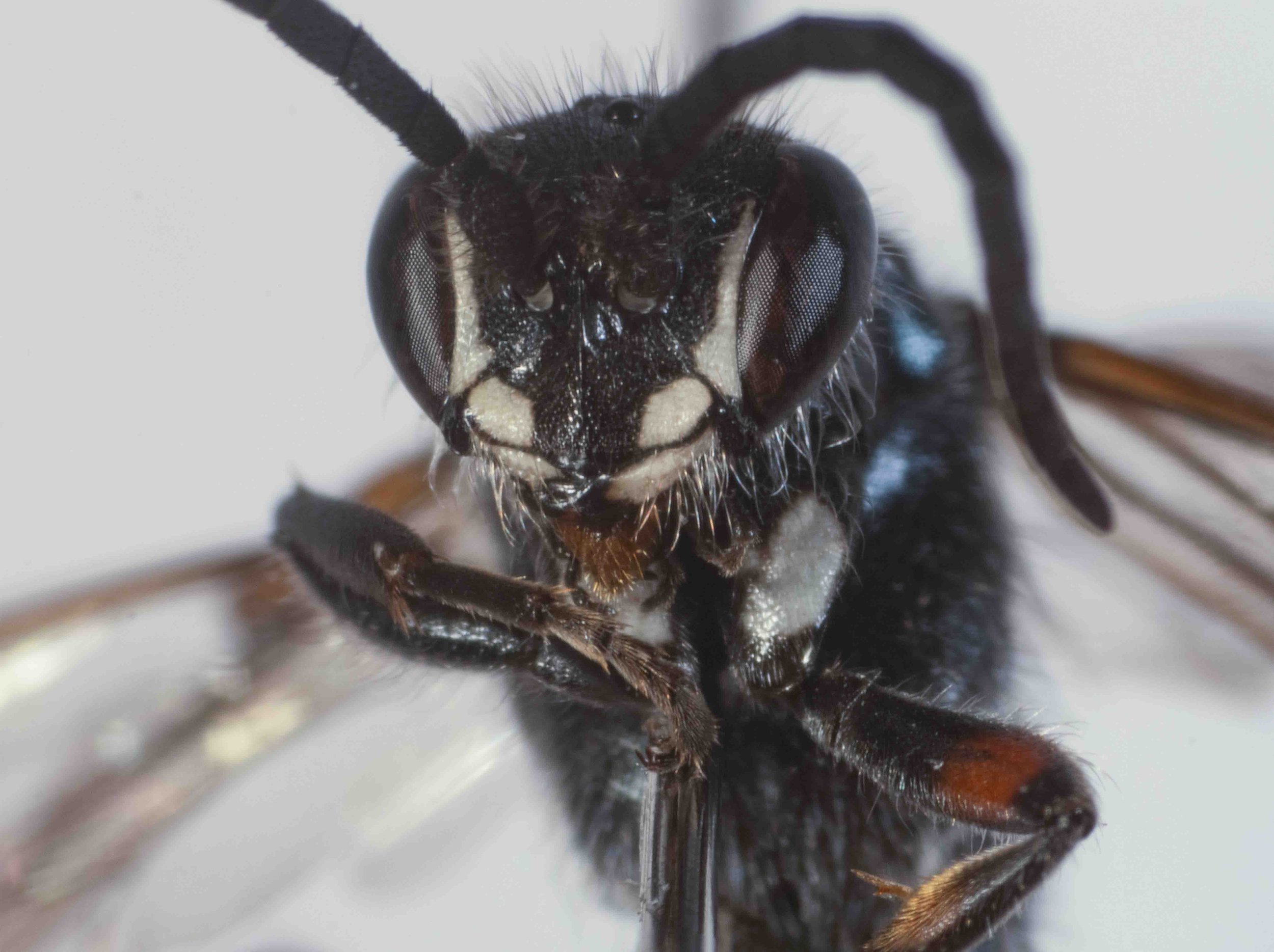
clypeus (frontal view)
lateral regions of clypeus long, rounded
apex of clypeus narrow, weakly trilobed, not strongly produced beyond lateral regions
pale colour: lateral lobes of clypeus; inner eye margins, mandibles for basal 2/3
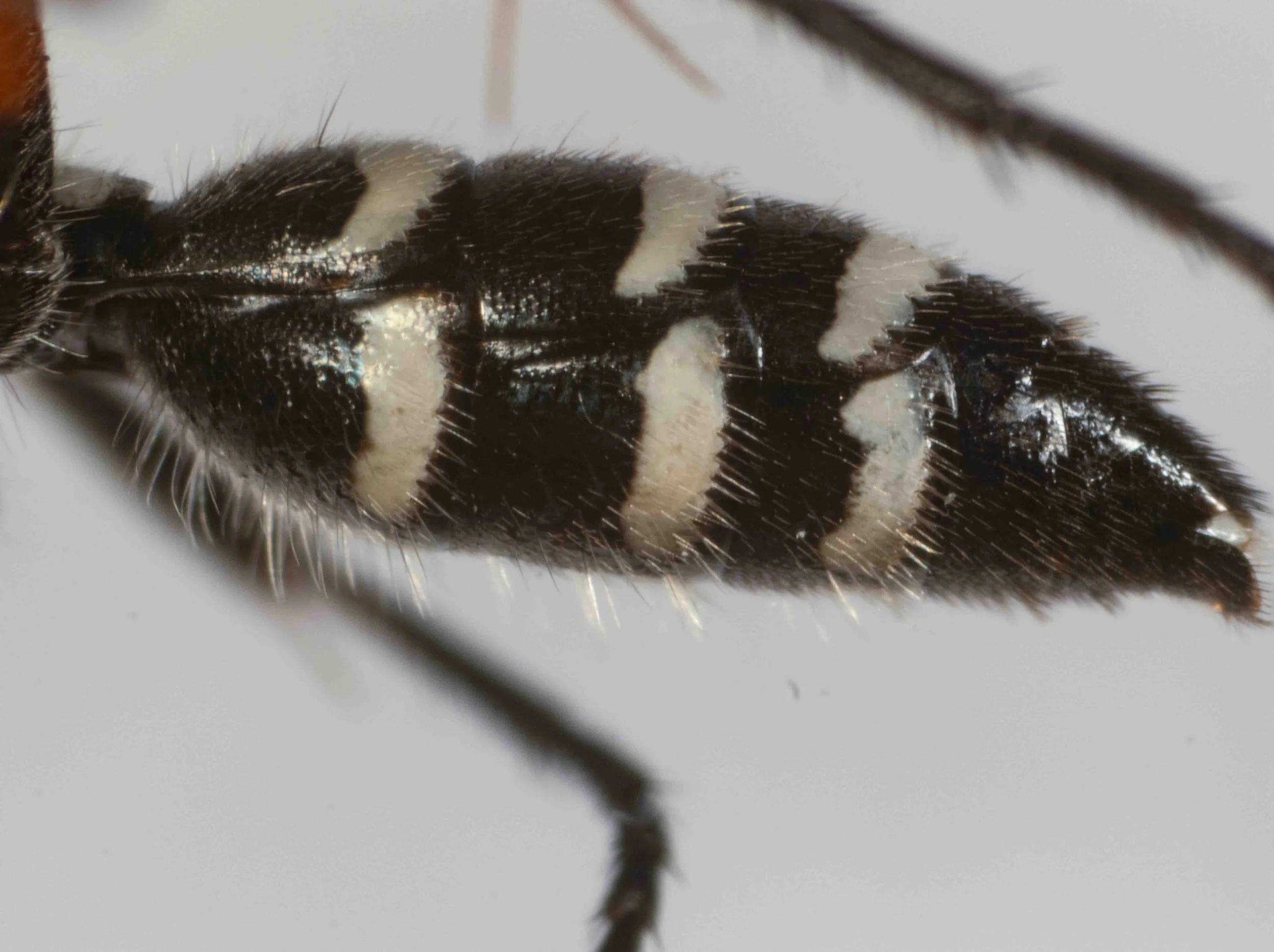
metasoma (lateral segments 2 to 7)
metasoma fusiform in lateral view
pale, lateral spots T1-4 & S2-4
propodeum & T1 (dorsal view)
propodeum rounded (without delineated dorsal and lateral surface), with shallow sagittal depression
T1 near triangular, with pair of white marks posterolaterally
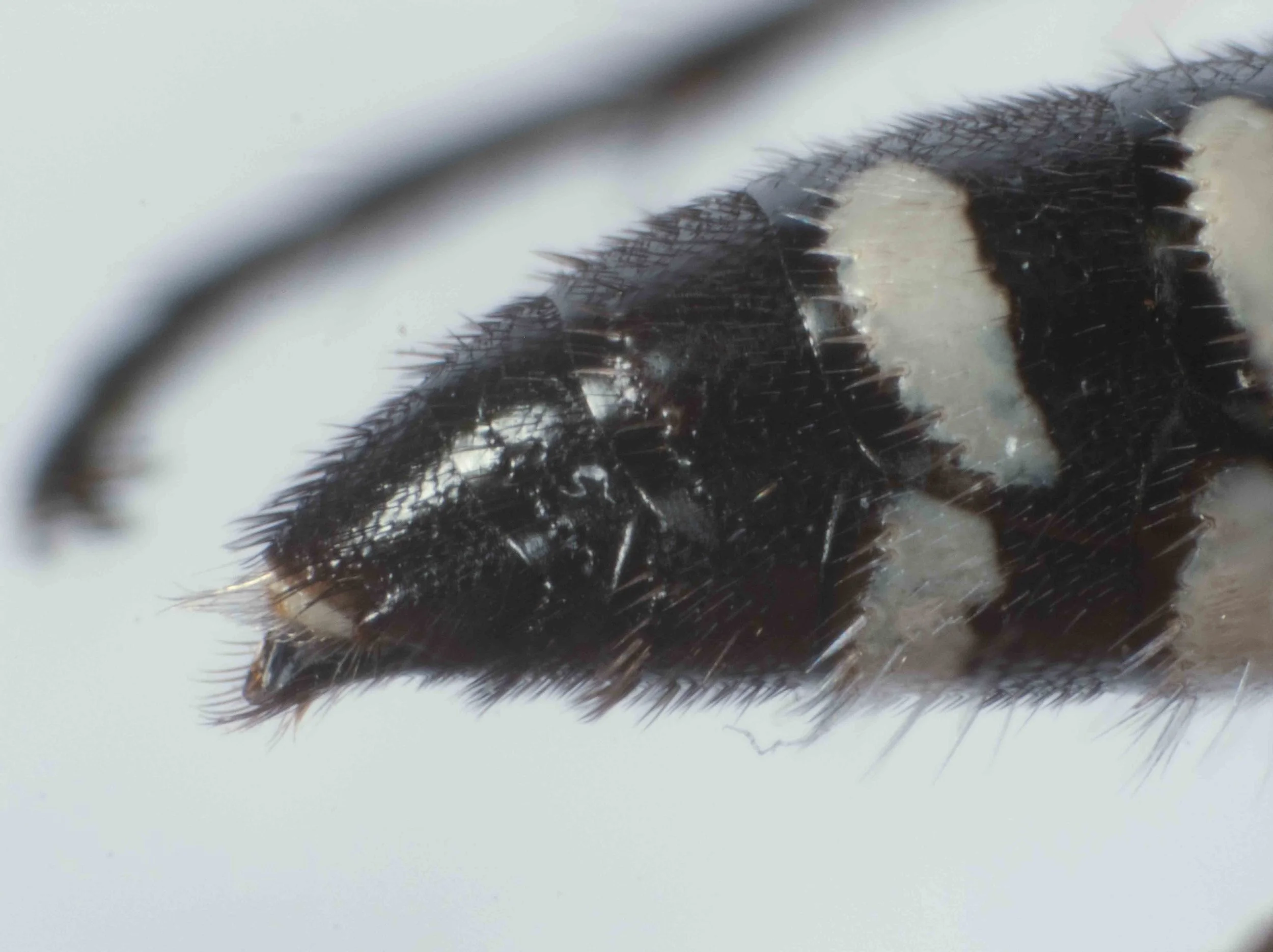
metasoma apex (lateral view)
terminalia (viewed dorsally)
epipygium convex, without subapical plate
apices of parameres (pale) exposed
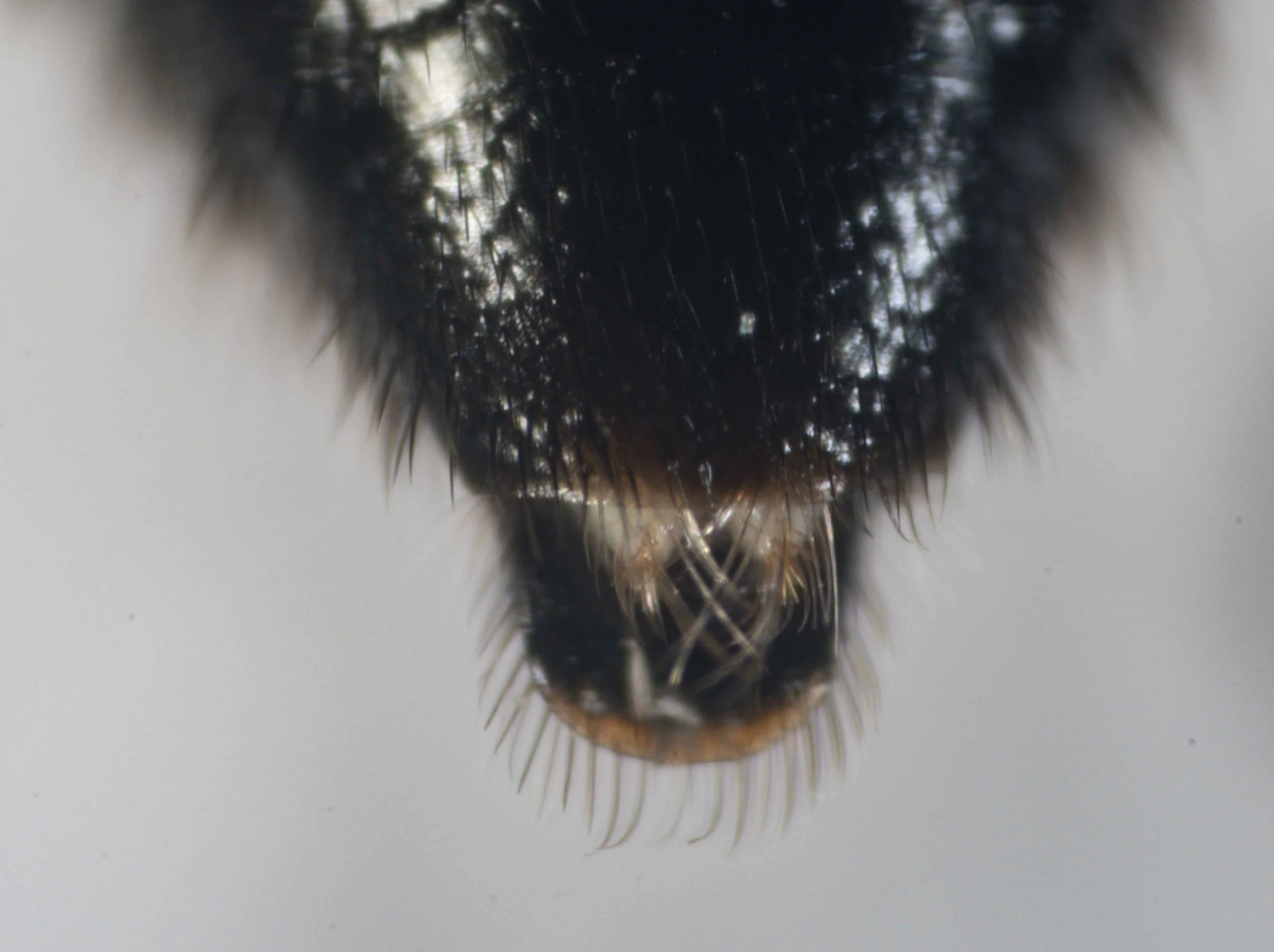
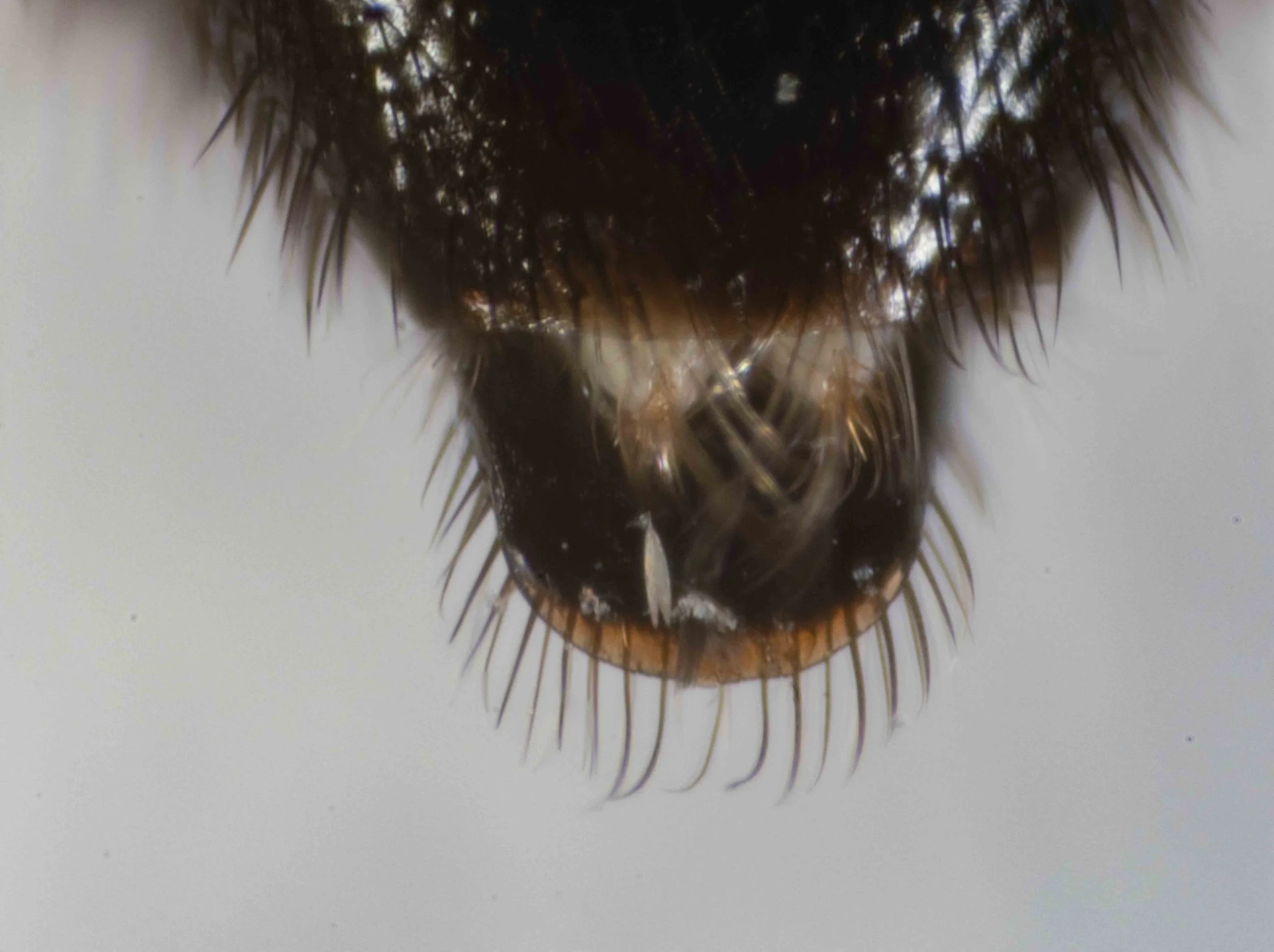
terminalia (viewed dorsally)
hypopygium rounded, without spines or teeth, WITH fringe of stout setae arising subapically
apices of parameres (pale) exposed
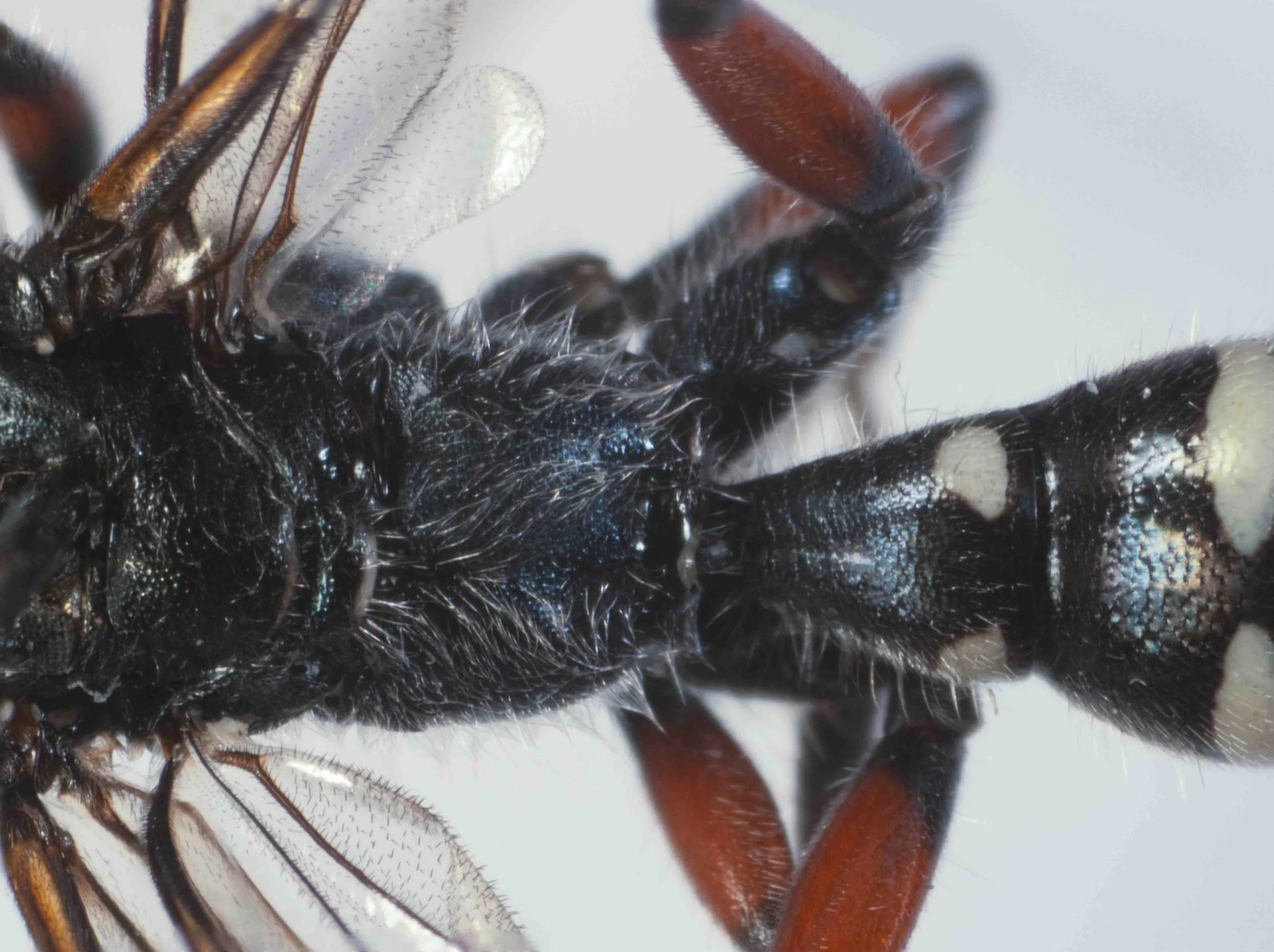
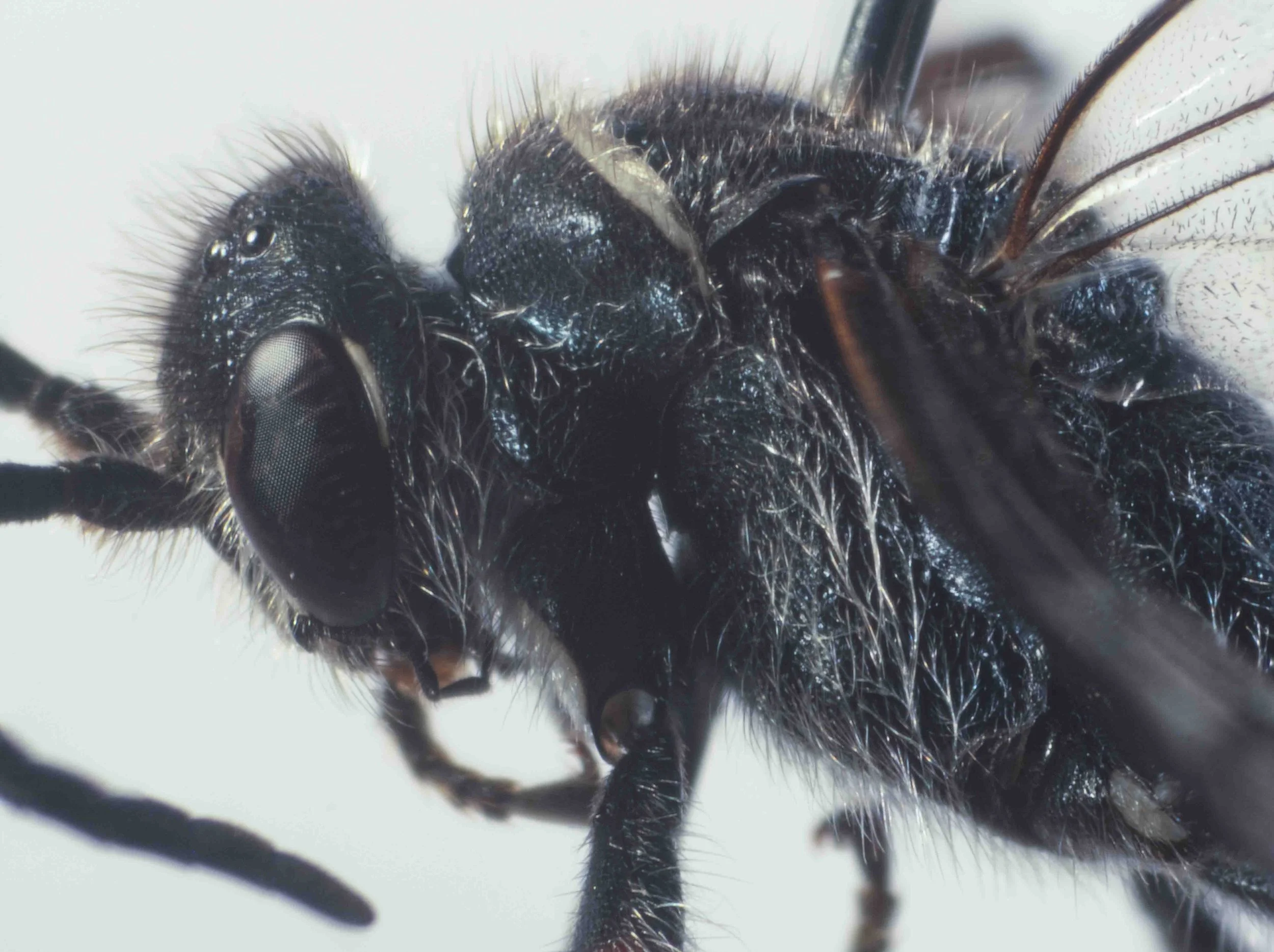
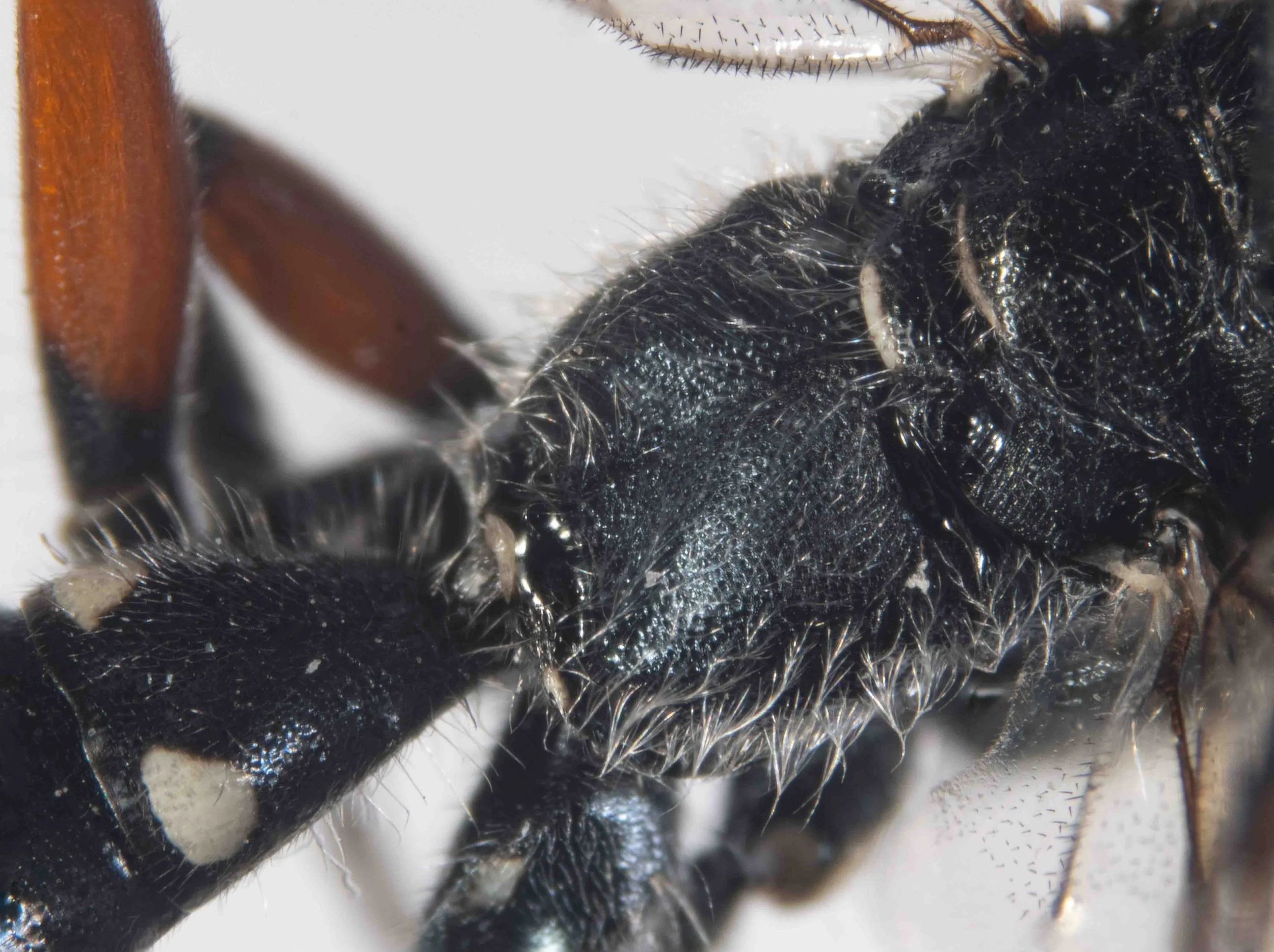
pronotum (viewed laterally)
pronotal disc rounded anteriorly, without carina
pronotum (viewed dorsally)
pronotum widest posteriorly, lateral edges rounded, converging anteriorly, without transverse carinae or groove
leg colour
front coxae with large, pale spots anteriorly
femora partly red/orange
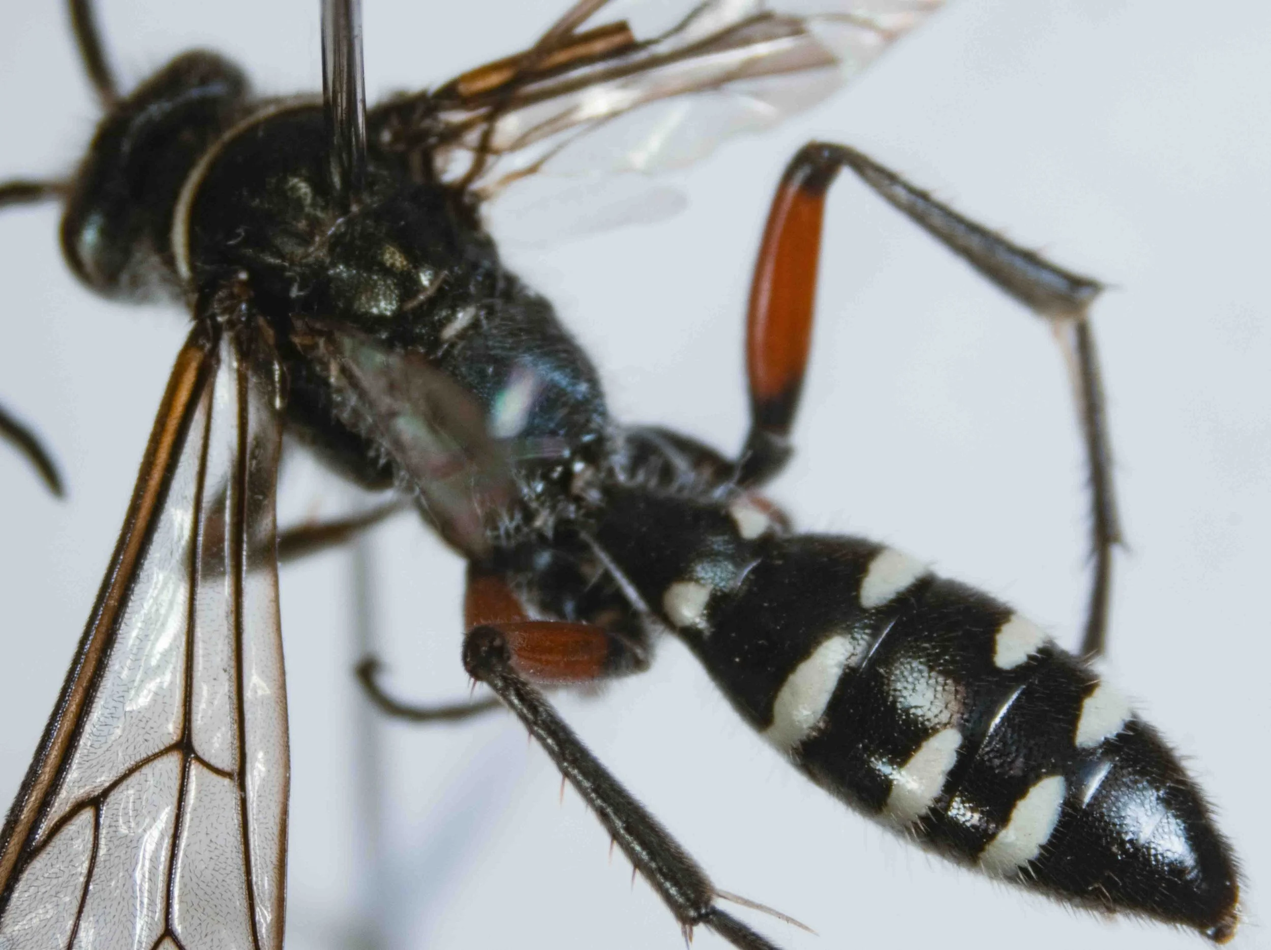
leg & body colour
pale, lateral spots T1-4
hind femora red (except black basally & apically)
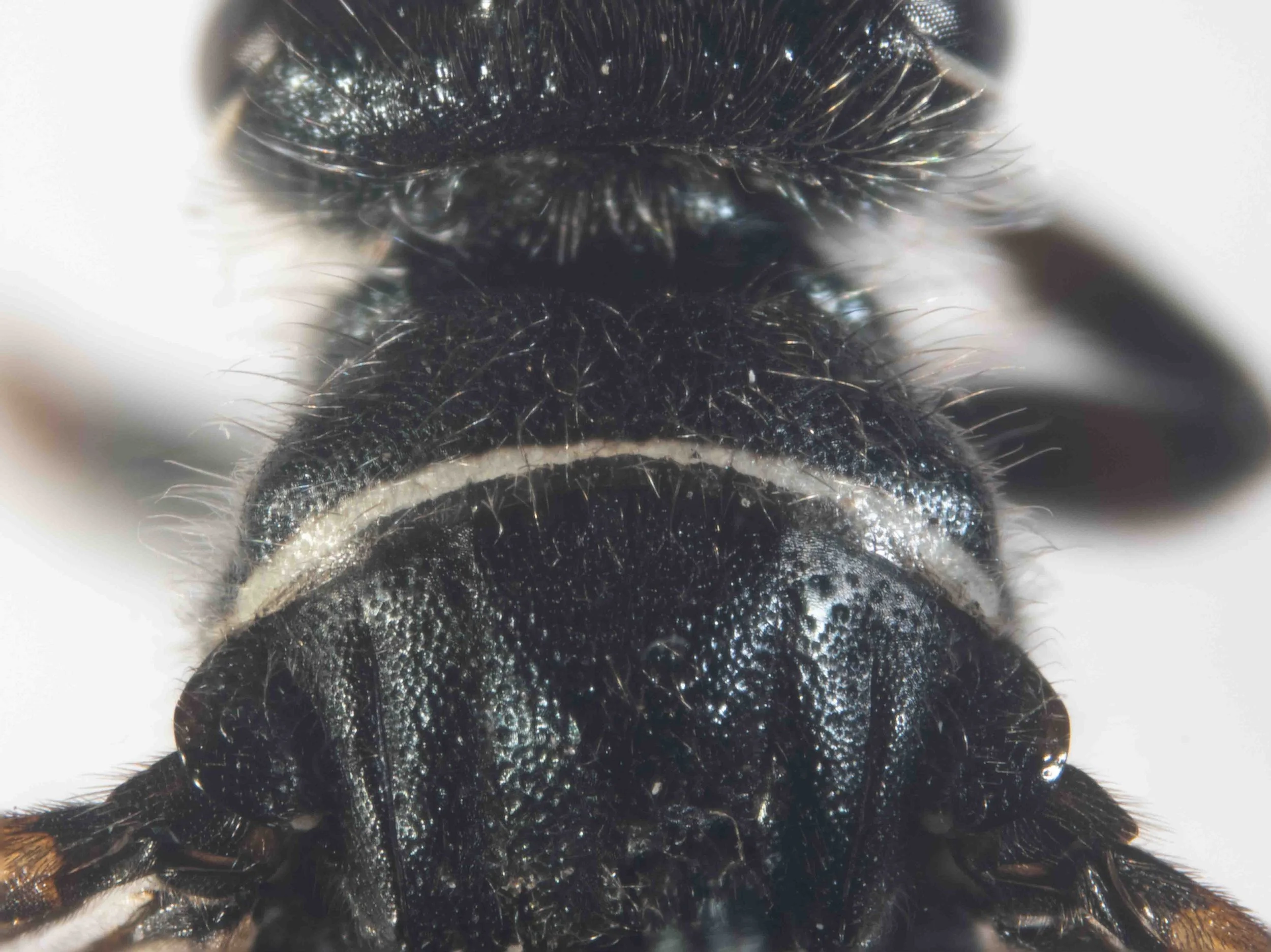
scutellum & metanotum (viewed dorsally)
scutellum & metanotum with narrow, transverse pale bands mediapically
propodeum (viewed dorsolaterally)
medial shallow depression, broadened posteriorly
forewing venation (viewed dorsally)
2m-cu meets SCIII, 1r distinct
forewing, showing SCI (viewed dorsally)
1r distinct
frons shape (viewed dorsally)
frons strongly produced above antennal sockets
metasoma & hypopygium
metasoma fusiform, not strongly constricted
hypopygium with apical spine, not strongly upturned
metasoma (viewed dorsally)
metasoma fusiform
clypeus (viewed anteriorly)
clypeus long medially, broadly truncate, with sagittal carina
S1 (viewed laterally)
S1; roundly raised medially, but without sharp carina; highest posteriorly, then with short oblique posterior surface; without tubercle
terminal sternites (viewed ventrolaterally)
S5 & S6 without posterolateral spines
terminalia (viewed ventrolaterally)
hypopygium with broadly rounded basal angles, raised above level of spine
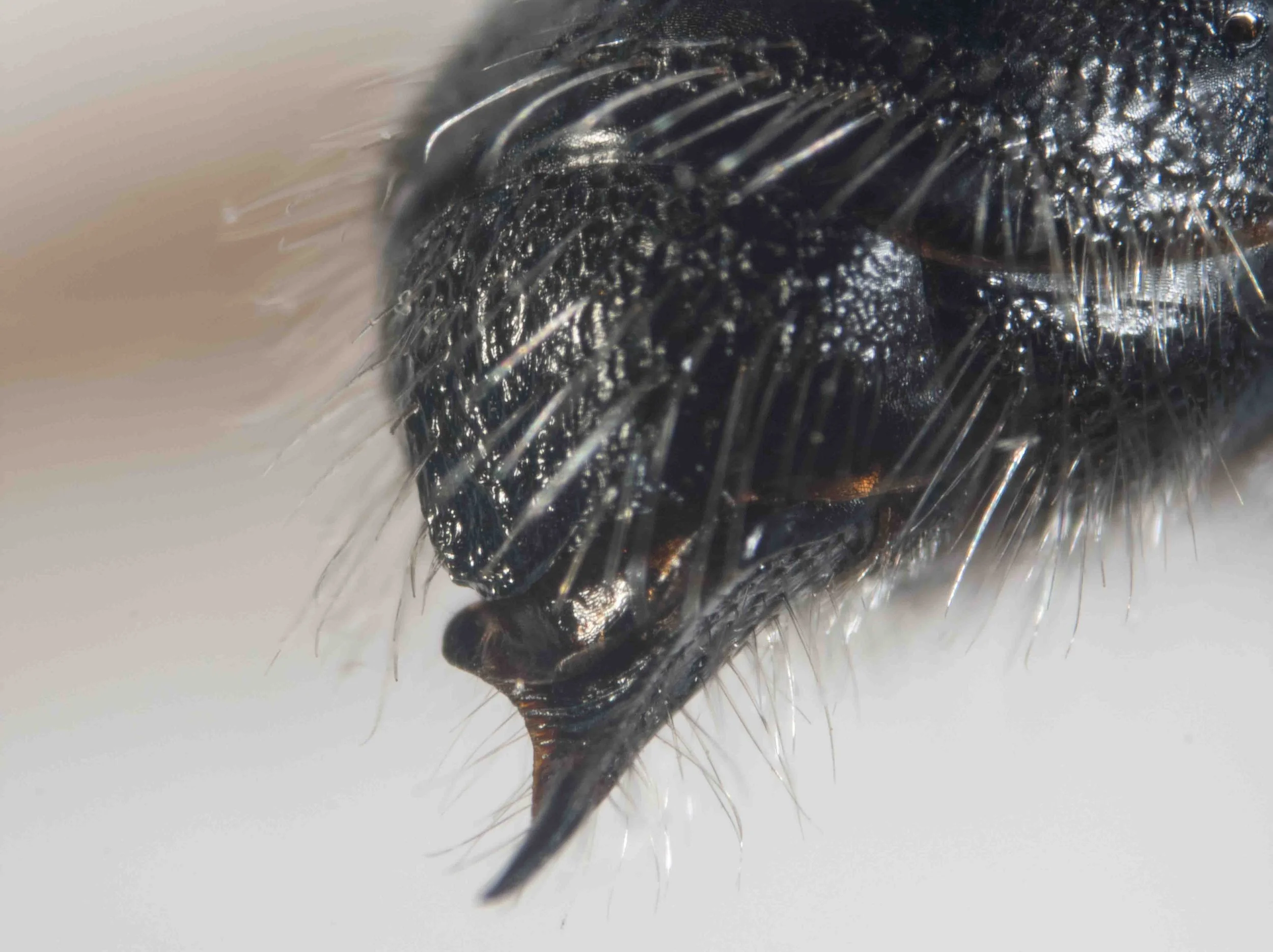
terminalia (viewed posterolaterally)
hypopygium with broadly rounded basal angles, raised above level of spine
epipygium without subapical plate