female as found dead on ground, antennae intact
body length = 13mm, Benson (1938) reports female body length 11-14mm

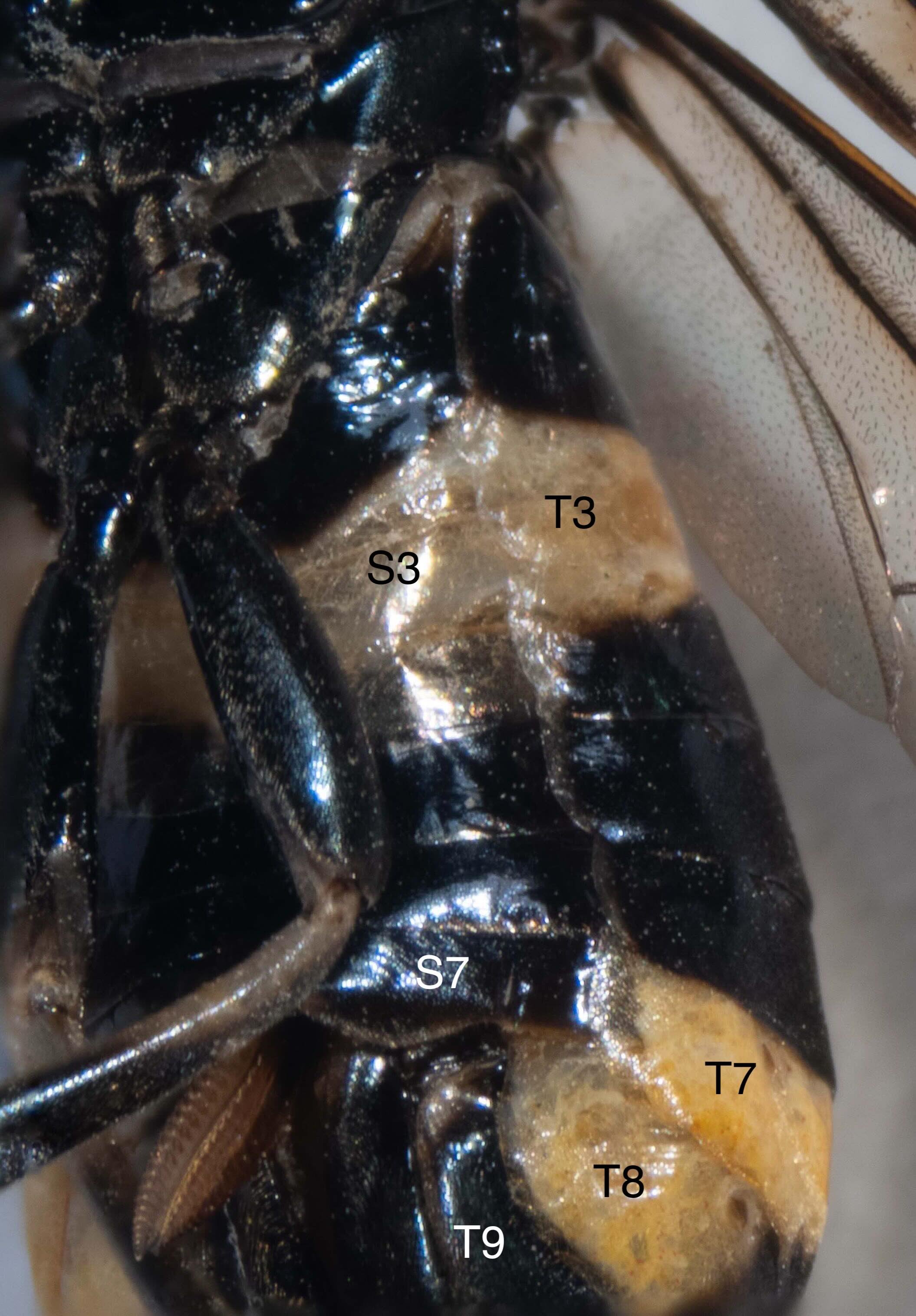
Identity of terga
shows bulge at anterior border of T7 - Benson (1938) lists this as a diagnostic character separating Pterygophorus from Lophyrotoma

Abdomen with two yellow bands
anterior yellow band confined to tergum 3 dorsally
posterior yellow band confined dorsally to a narrow bulge at the anterior border of tergum 7

Abdomen with two yellow bands
anterior yellow band extends laterally on tergum 3, crossing into anterior half of tergum 4 in its lateral region
posterior yellow band extends ventrally, expanding to posterior edge of tergum 7 and crossing into tergum 8

Abdomen with two yellow bands
anterior yellow band expands into sternum 3, making a complete band around segment 3
posterior yellow band expands to ventral border of tergum 7 and 8
tergum 10, pygidium is also yellow

Abdomen with two yellow bands
anterior yellow band expands into sternum 3, making a complete band around segment 3
posterior yellow band expands to ventral border of tergum 7 and 8

Abdomen with two yellow bands
anterior yellow band expands onto sternum 3, making a complete band around segment 3
posterior yellow band expands to ventral border of tergum 7 and 8 - left side

Abdomen with two yellow bands
posterior yellow band expands to ventral border of tergum 7 and 8 - left side

Abdomen with two yellow bands
posterior yellow band expands to ventral border of tergum 7 and 8 - right side
terminal segment, tergum 10 (pygidium) is yellow
extended saw visible

Abdomen morphology and colour
view of tergum 10, pygidium

Abdomen morphology
shows saw extended from sheath

distal end of saw
arrow shows tooth #10

photomicrograph of tooth #10
43 denticulations present on this tooth - Benson (1938) reports 28-40 denticulations present in P. cinctus

drawing of 10th saw tooth of P. cinctus
from Benson (1938) fig. 6

Antennal morphology
scape and pedicel black/dark brown
8 basal antennomeres yellow
remaining 13 apical antennomeres black
most apical antennomere is clubbed
total 23 antennomeres. antenna length = 0.4 length of forewing

Antennal morphology
scape black, pedicel dark yellowish-brown

Antennal morphology
8 basal flagellomeres yellow. pecten directed antero-ventrally on each flagellomere, becoming longer in apical flagellomeres.

Head morphology

P. cinctus head frontal view
shows eyes converging in front so that length of eye is about equal to distance between eyes in front - Benson (1938) fig. 1

Head morphology
apex of clypeus emarginate (vs. truncate, separating P. cinctus from P. distinctus according to Morice, 1919)
diagnostic characters of Pterygophorus vs. Lophyrotoma (according to Benson, 1938)
distance between eyes in front subequal to (1.1x) length of eye (vs. at most 2/3)
breadth of clypeus 2/3 length of eye (vs. about equal)
breadth of clypeus at 2/3 length of eye (vs. as long as)

Head morphology
black; short, fine pale pubescence; shining, impunctate

Head morphology

Head morphology
ligula same length as 3 basal segments of labial palp
labium 1.1x greatest breadth of clypeus

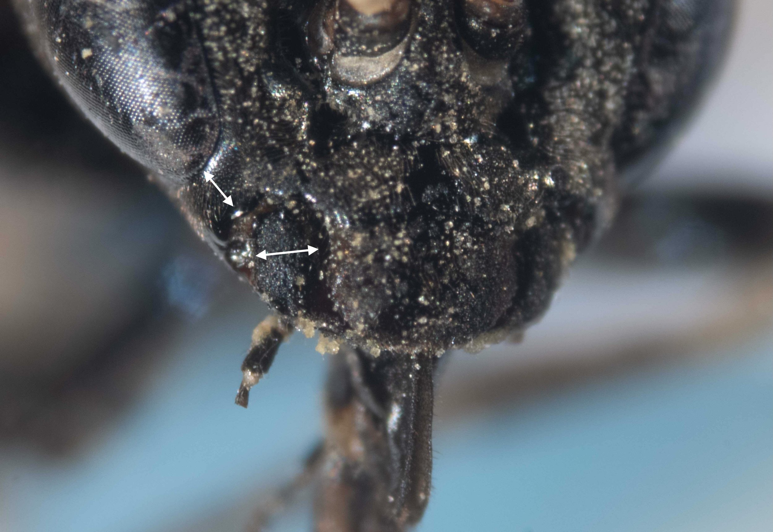
Head morphology
malar space (arrowed) = 0.42 breadth of mandible at base (arrowed) (vs. more than 0.5 in P. insignis and P. facielonga according to Benson, 1938)

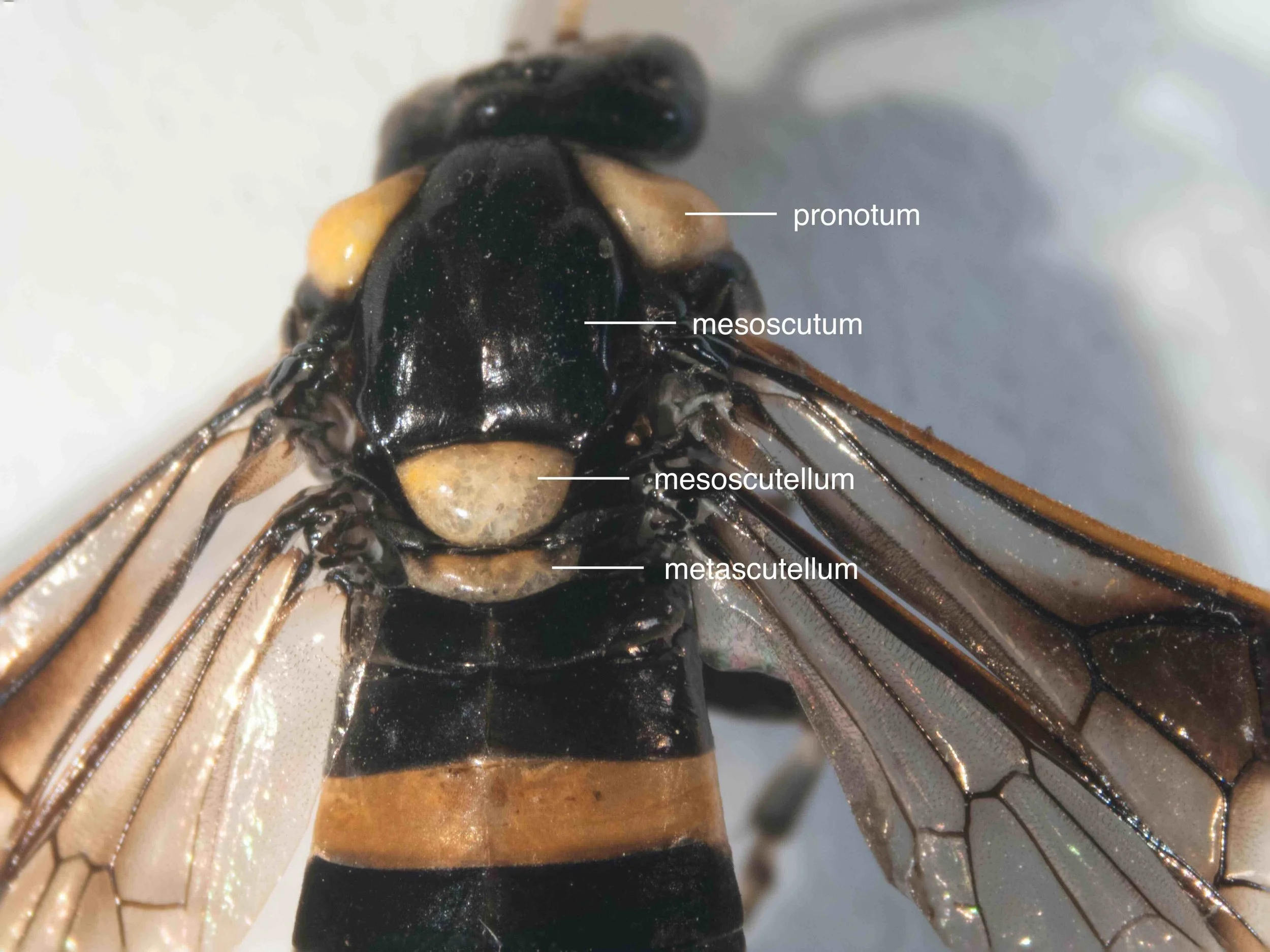
Thorax morphology and colour
pronotum, mesoscutellum and metascutellum pale yellow and shiny between sparse punctures; mesoscutum black

Thorax morphology and colour
thorax covered by short, fine, pale pubescence

Thorax morphology and colour
short, fine, pale pubescence over thorax
accessory furrow on pronotum (arrowed) subparallel with side-margin of pronotum - diagnostic feature separating Pterygophorus from Lophyrotoma spp. except for L. zonalis and L. cygna
pronotum colour yellow with black anterior border and black behind accessory furrow
mesepisternum black with large central yellow area

Thorax morphology
side portion of pronotum cut off by accessory furrow broader than length of malar space (vs. about half length of malar space) - separates P. cinctus, P. distinctus, P. insignis and P. facielonga from P. turneri

P. cinctus pronotum lateral view
shows accessory furrow on pronotum - Benson (1938) fig. 5

Thorax morphology
ventral view of mesepisternum showing large yellow patch and short, fine, pale pubescence

Forewing morphology and colour - ventral view
costa and stigma yellowish-brown
hyaline with dark brown band extending along anterior border to wing apex, covering 1st discoidal, 1st cubital and radial cells completely and half of 2nd and 3rd cubital cells
venation black in basal and anterior region, brown in apical region

Leg colour
coxa, trochanter, femur - all legs black
tibia - all legs black except for whitish-yellow on ventral side of basal 1/3rd
tarsi - all legs black except for whitish-yellow on dorsal side of basal 1/3rd of all tarsomeres

Forewing dorsal view
costa and stigma yellowish-brown
hyaline with dark brown band extending along anterior border to wing apex, covering 1st discoidal, 1st cubital and radial cells completely and half of 2nd and 3rd cubital cells
venation black in basal and anterior region, brown in apical region

Leg colour
hind leg removed showing black colour in all parts of all segments except for whitish-yellow on ventral side of basal half of tibia and on dorsal side of basal 1/3rd of all tarsomeres

Leg morphology
hind basitarsus no longer than following tarsal segment


