Hylis australis (Eucnemidae: Melasinae: Epiphanini)
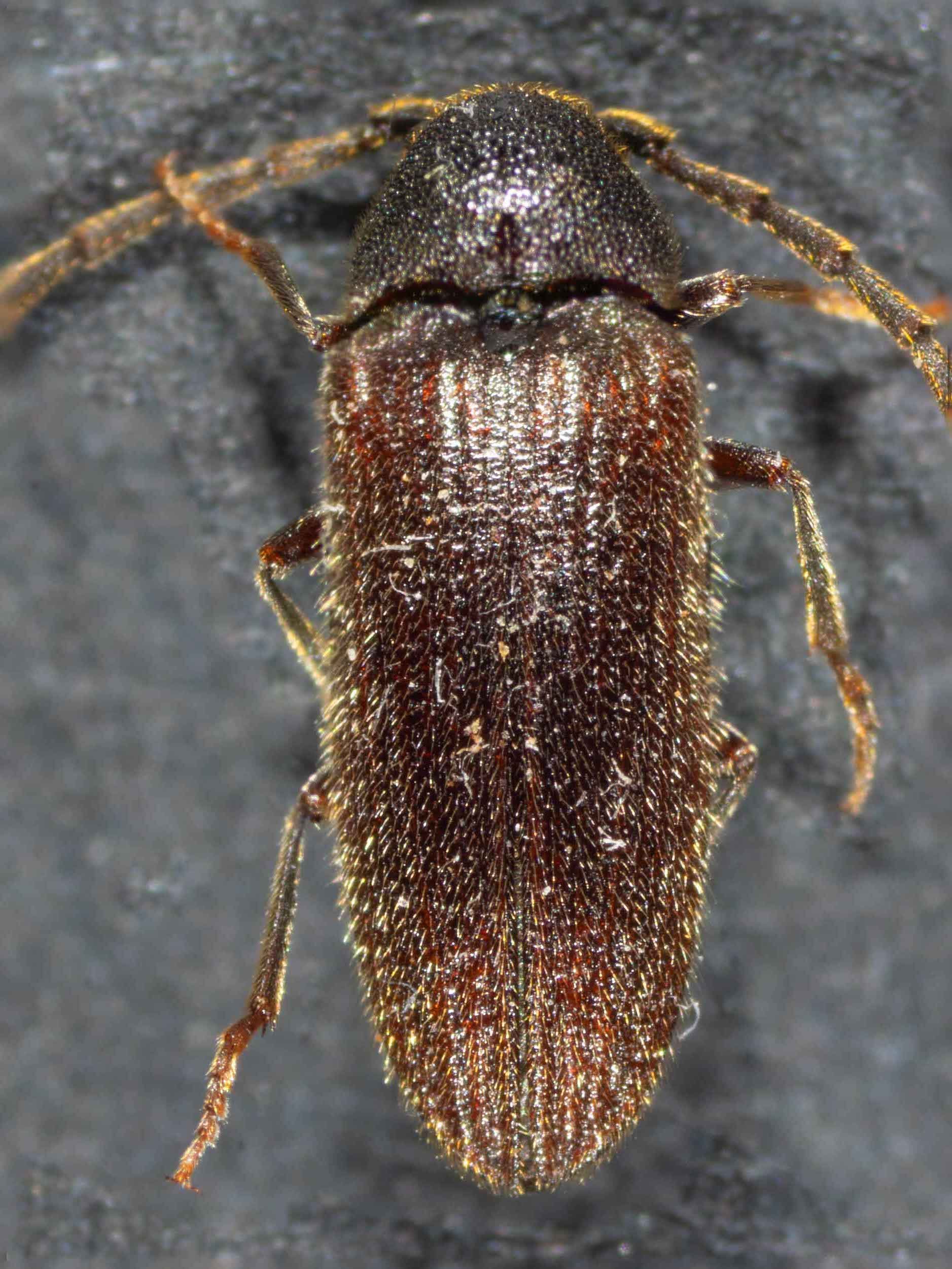
Workbook
Identification of click beetle found on vegetation on/around 21st October, 2023.
Family
I initially identified it as a Eucnemidae sp. - iNaturalist observation here.
Features that separate Eucmenidae from Elateridae include:
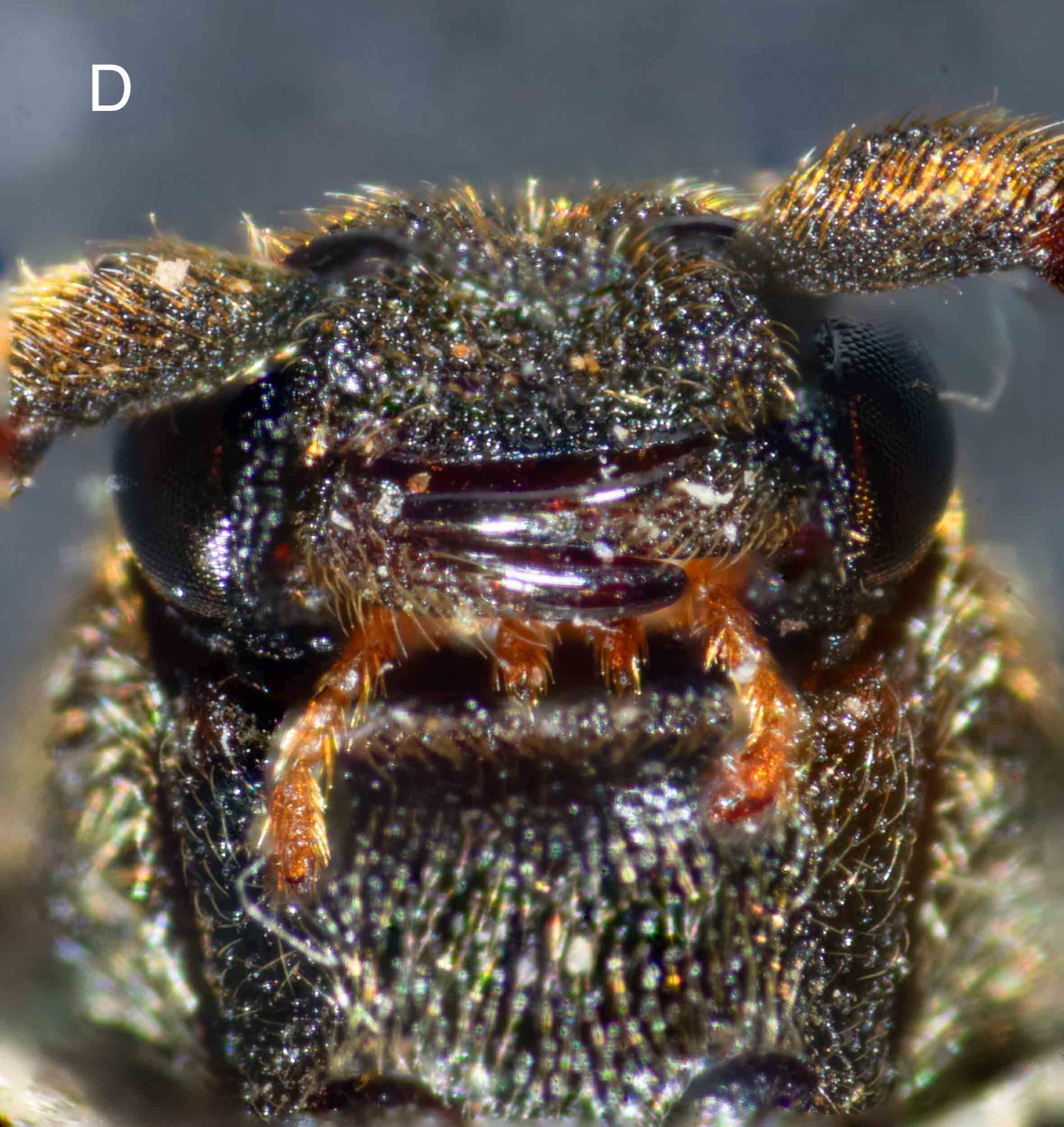
antennae not close to eyes (fig. B, C, D)
pronotum trapezoidal (quadrilateral with no or two sides parallel) (fig. A, B)
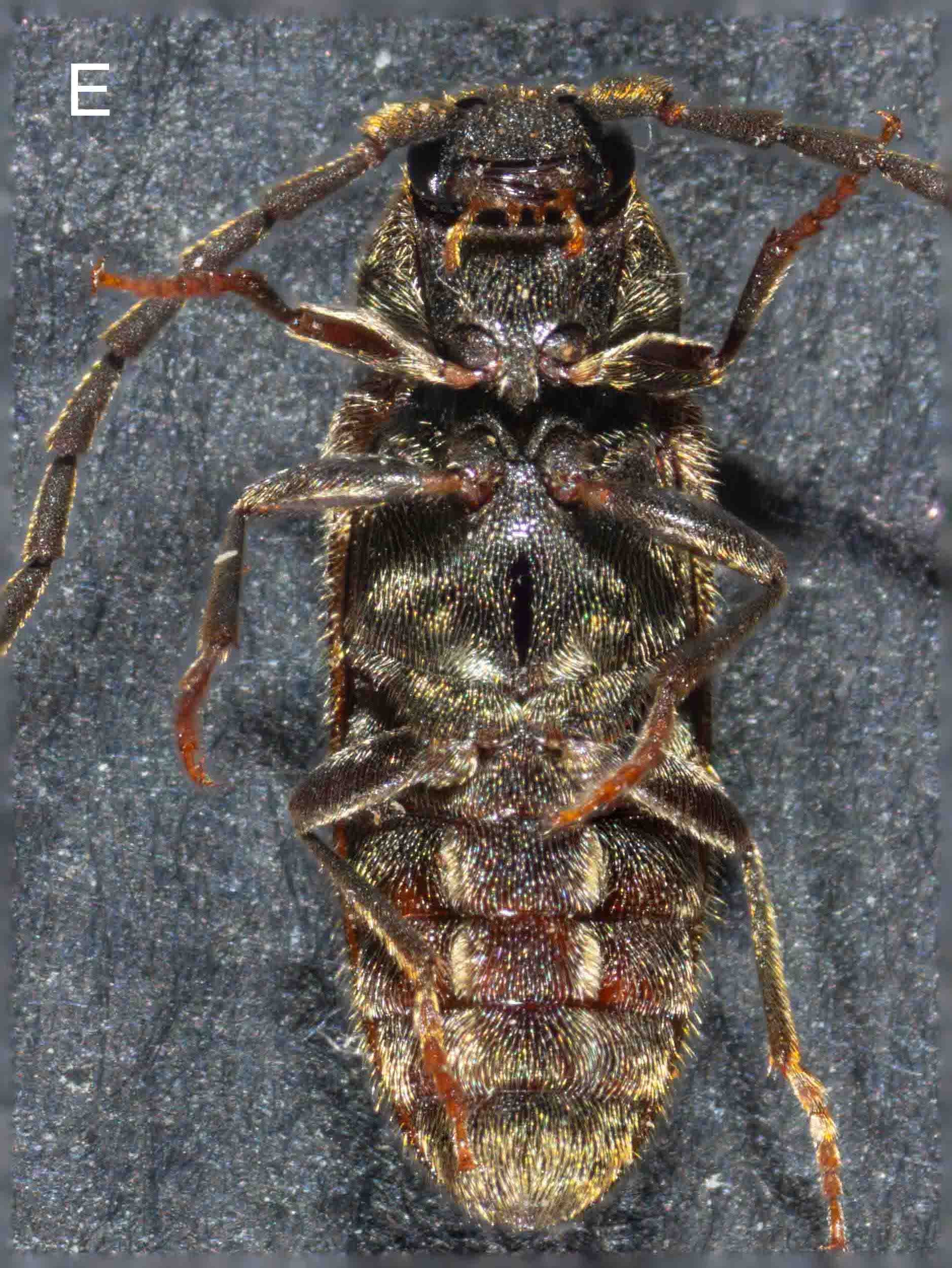
front end of prosternum is straight, not lobed (fig. C, E)
labrum concealed beneath clypeus (fig. D)
all five ventrites are connate (fig. C, E)
tarsi usually simple (fig. E, F)
Subfamily and Tribe
AFD lists 5 subfamilies of Eucnemidae - Anischiinae, Eucneminae, Macraulacinae, Melasinae and Pseudomeninae.
For identification to subfamily and tribe, I used the key in Muona, J. (1993) “Review of the phylogeny, classification and biology of the family Eucnemidae (Coleoptera)”. Entomologica Scandinavia Suppl. 44 pp. 55-57. (Available on ResearchGate).
step 1. Protibia with 1 or 2 well developed apical spurs? - see fig. G
tibia of right proleg - only one apical spur
–> step 8. Antennomeres 9-11 enlarged, hypomera unmodified? or
Antennomeres 9-11 rarely enlarged, and then hypomera with lateral antennal grooves?
Antennomeres 9, 10 are not enlarged compared to more proximal ones. Only antennomere 11 is longer - figs, H, I.
Hypomeron is unmodified (simple) - fig. J, medial edges of hypomera indicated with white arrows.
–> step 13 Hypomera simple or with notosternal or basally open lateral antennal grooves, metatibiae with rounded angle between lateral and caudal surfaces, median lobe usually fused to lateral lobes? or
Hypomera with basally closed lateral antennal grooves, metatibiae with sharp angle between lateral and caudal surfaces, median lobe always free?
Hypomera simple - fig. J
Metatibiae with rounded angle between lateral and caudal surfaces - see figs. K, L, M.
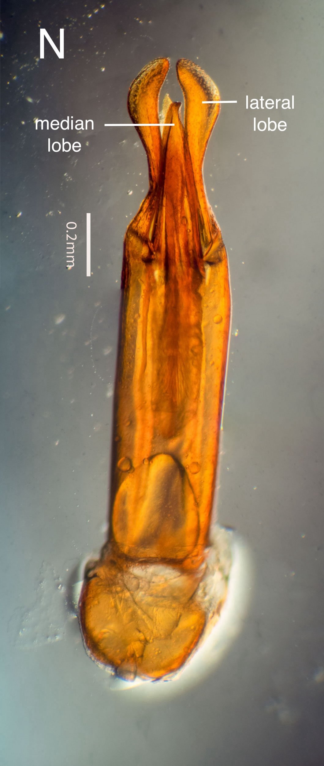
Median lobe of aedeagus fused to lateral lobes - see fig. N
–> step 14 meso- and metatibiae without spines or spine combs on lateral surfaces, hypomera simple? or
meso-and metatibiae with spines or spine combs on lateral surfaces, hypomera with notosternal antennal grooves?
Meso- and metatibiae without spines or spine combs on lateral surfaces - see figs. A, C, E, K, L, M.
Hypomera simple - see fig. J.
–> step 15 Hypomera parallel sided (–> Melasinae: Melasini)? or
Hypomera more or less triangular?
Hypomera more or less triangular - see fig. J
–> step 16 Lateral pronotal ridge minutely serrate, hypomera often with notosternal antennal grooves (–> Melansinae: Dirhagini)? or
Lateral pronotal ridge smooth, hypomera simple?
Lateral pronotonal ridge smooth - see figs. A, B
Hypomera simple - see figs. C, E, J
–> step 17 Metacoxal plates parallel-sided or widening laterad? or
Metacoxal plates widening mediad?
Metacoxal plates widening mediad - see fig. J (metacoxal plates indicated with red arrows)
–> step 20 Frons usually produced, head frequently with medial or transverse keels, dorsal surface usually densely punctate, male protarsomere 1 simple, median lobe deeply or widely bifurcate apically? or
Frons never produced, head evenly convex without keels, dorsal surface with moderate punctation, shiny, antennomeres simple, male protarsomere 1 with apical sex comb, medial lobe entire apically?
Frons produced - fig. D
Head not with medial or transverse keels - fig. B, D
Dorsal surface densely punctate - fig. B, D
male protarsomere 1 simple - fig. F
median lobe of aedeagus deeply and widely bifurcate apically - fig. N
–> Melasinae: Epiphanini
Muona lists following characters of tribe Epiphanini. Most are evident in this specimen:
form narrowing craniad and caudad (fig. A)
labrum attached underneath frontoclypeal region (fig. D)
mandibles short (fig. D), with secondary ventral tooth (fig. D’-dissected from female in this iNat observation)
sensory pegs concentrated on apex and sides of single antennomeres? (need SEM to visualise)
protibia with one spur (fig. G)
hypomera narrowing craniad (fig. J)
mesepimera fused with mesepisterna
metacoxal plates narrowing laterad (fig. J)
ventrites connate (fig. C, E)
hindwing with reduced apical fields
frontoclypeal region usually expanded (fig. J)
head often with median keel (not in this case)
microsculpture usually strongly developed; punctation usually dense (figs. A, B, D)
male protarsomere 1 simple (fig. F)
hypomera without antennal grooves (fig. J)
aedeagus morphology - median lobe fused with lateral lobes, distinct, without dorsal basal struts; lateral lobes bilobed; median lobe deeply and widely bifurcate (fig. N)
AFD lists one genus in this tribe - Hylis, which is monotypic - Hylis australis (Fleutiaux, 1896)
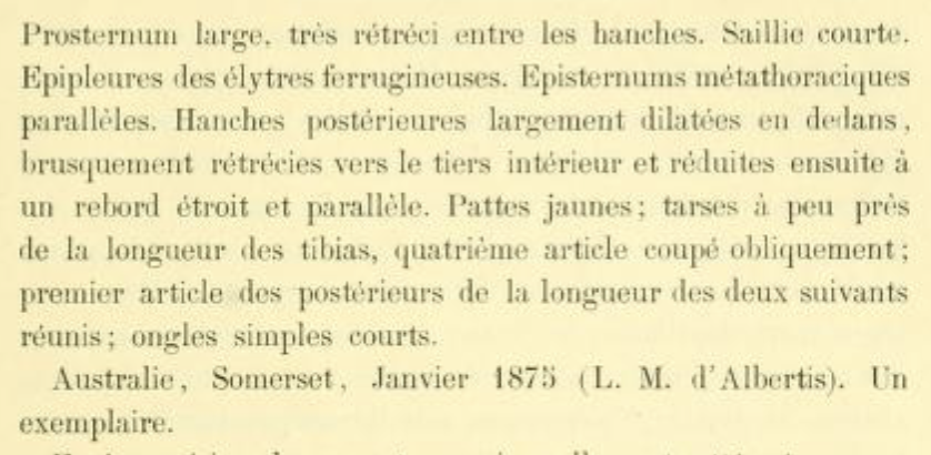
Does this specimen match the original description of Hylis australis?
Hypocoelus australis, n. sp. 4 1/2 mill. Body oblong, a little convex; reddish-brown, ferruginous on the edge of the epistome, most of the pronotum, the base and suture of the elytra; yellowish pubescence. Head with a rather strong and little uneven punctation, no carina between the eyes. Epistome narrowed at its base, a little wider than the distance between it and the eyes. Antennae longer than the base of the prothorax, quite thick, with a brownish-reddish colour; first antennomere longer than the others, as long as the following two together; second very short; third a little toothed; the rest cylindrical, about the same length, connected by a peduncle; tenth less thick and shorter than the preceding; eleventh missing. Pronotum wider than long, trapezoidal; sides slightly rounded anteriorly; sinuous base; posterior angles a little extended; surface marked with a smooth line in the midline; strong punctation, becoming more closely spaced and rougher on the sides. Scutellum ferruginous, semicircular. Elytra subparallel, becoming narrower in their posterior third, weakly striated. Ventral side a clear reddish colour; strong punctation on the prosternum, tight and rough on the propleura, finer on the metasternum, indistinct on the anterior part of the abdomen, stronger more posteriorly. Hypomera longer than wide, triangular, becoming narrower anteriorly, widely but weakly furrowed along the lateral border. Prosternum wide, narrowed between the coxae. Epipleura of the elytra ferruginous. Metathoracic episterna parallel. Posterior femora widely dilated on the inside, suddenly narrowing towards the inner third and then reduced to a narrow and parallel edge. Yellow legs; tarsi about the length of the tibiae, fourth tarsomere cut obliquely; first hind tarsomere as long as the next two combined. Claws simple and short.
The current specimen appears to match this description quite well.
Eucnemidae have been sighted on the block on a couple of occasions previously. These look similar to this species.
One individual, probably a male based on the length of its antennae, in 2019.
A female with a male in copula together with a second male in 2020.
This is a workbook page … a part of our website where we record the observations and references used in making species identifications. The notes will not necessarily be complete. They are a record for our own use, but we are happy to share this information with others.
