
mystery mygalomorphs
Workbook
A. Timeline & observations
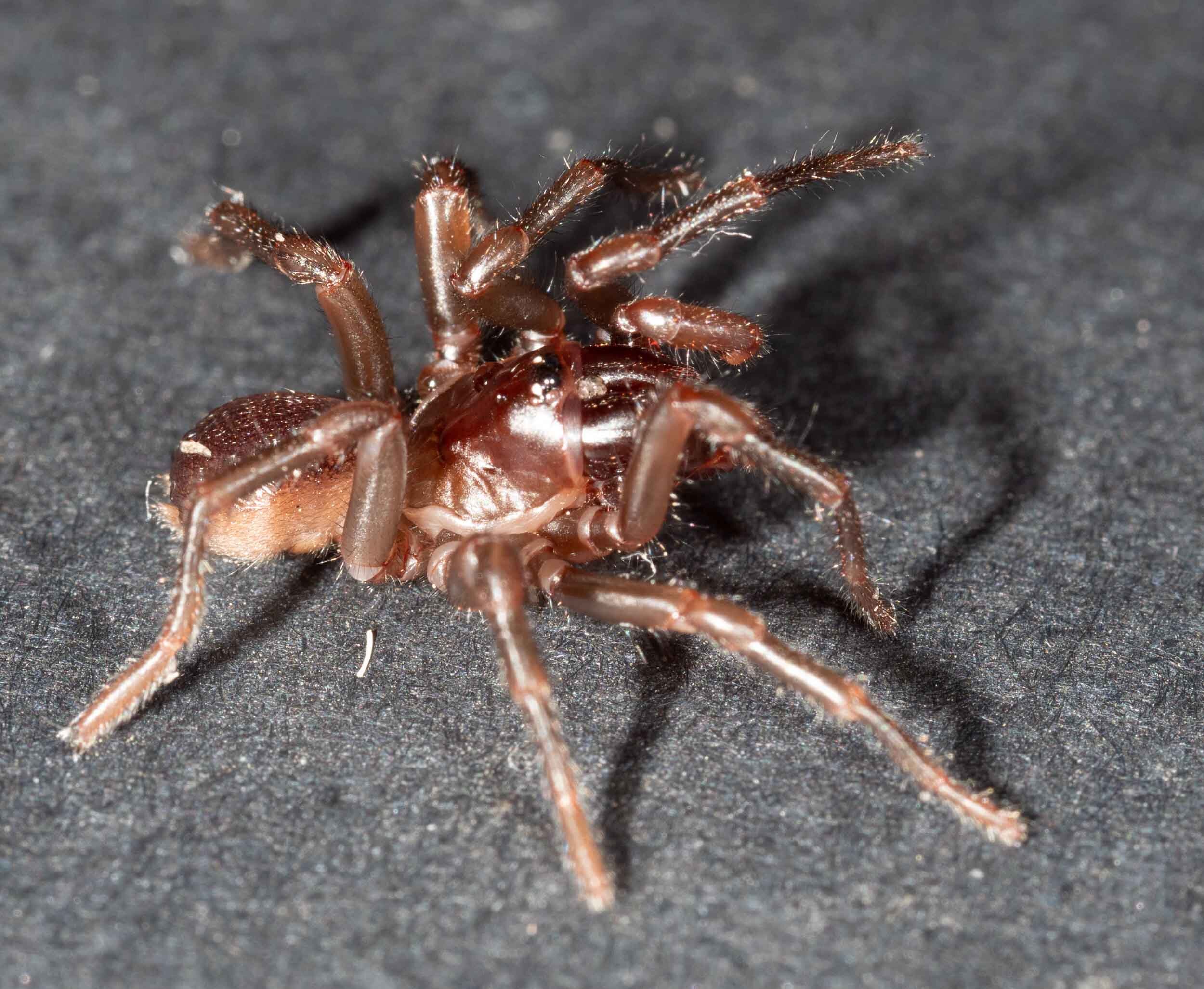
On 22nd May, 2020 we found 4 spiders wandering in the grass and climbing up the outside wall of the house. They were all within a few square metres of one another. It was 11:24pm, raining steadily, and the ambient temperature was 11ºC.


We collected all four. They appeared to be mygalomorphs, and so of particular interest to us – sightings are not common and we were keen to confirm the species identity for our records.
They are all mature males. Each pedipalp bears a well-developed embolus. It is our understanding that this structure only appears after the final moult to sexual maturity (Foelix, 2011) … although perhaps there are exceptions (?).
Initially they were pale coloured and passive. Immediately after collection we photographed the spiders. We noted that they were pale in colour, not the dark colour we associate with funnel webs. They were also quite passive, with 2 out of 4 rolling over in the ‘play-dead’ pose. This is a behaviour we normally associate with Idiopidae. Anthelidae, on the other hand, normally assume a defensive pose with fangs displayed. One spider did so, but only after considerable provocation.


We killed one spider for closer examination on Day 1. Soon after collection, we placed one spider (#3) in the freezer for 2 hours. He was then examined and photographed over the following 2 days. During this time he was kept at room temperature, in a petri dish. The other 3 spiders were kept alive in petri dishes supplied with wet paper towel for humidity.
By Day 3 (24/5) the living spiders had changed colour and behaviour. They became noticeably darker and apparently larger. Their spinnerets were also more obvious, visible from above. Most notably, they were all more reactive. None would play dead. At the least provocation they would rear back with front legs raised and fangs exposed. They did not jump, but would hold the pose for extended periods.
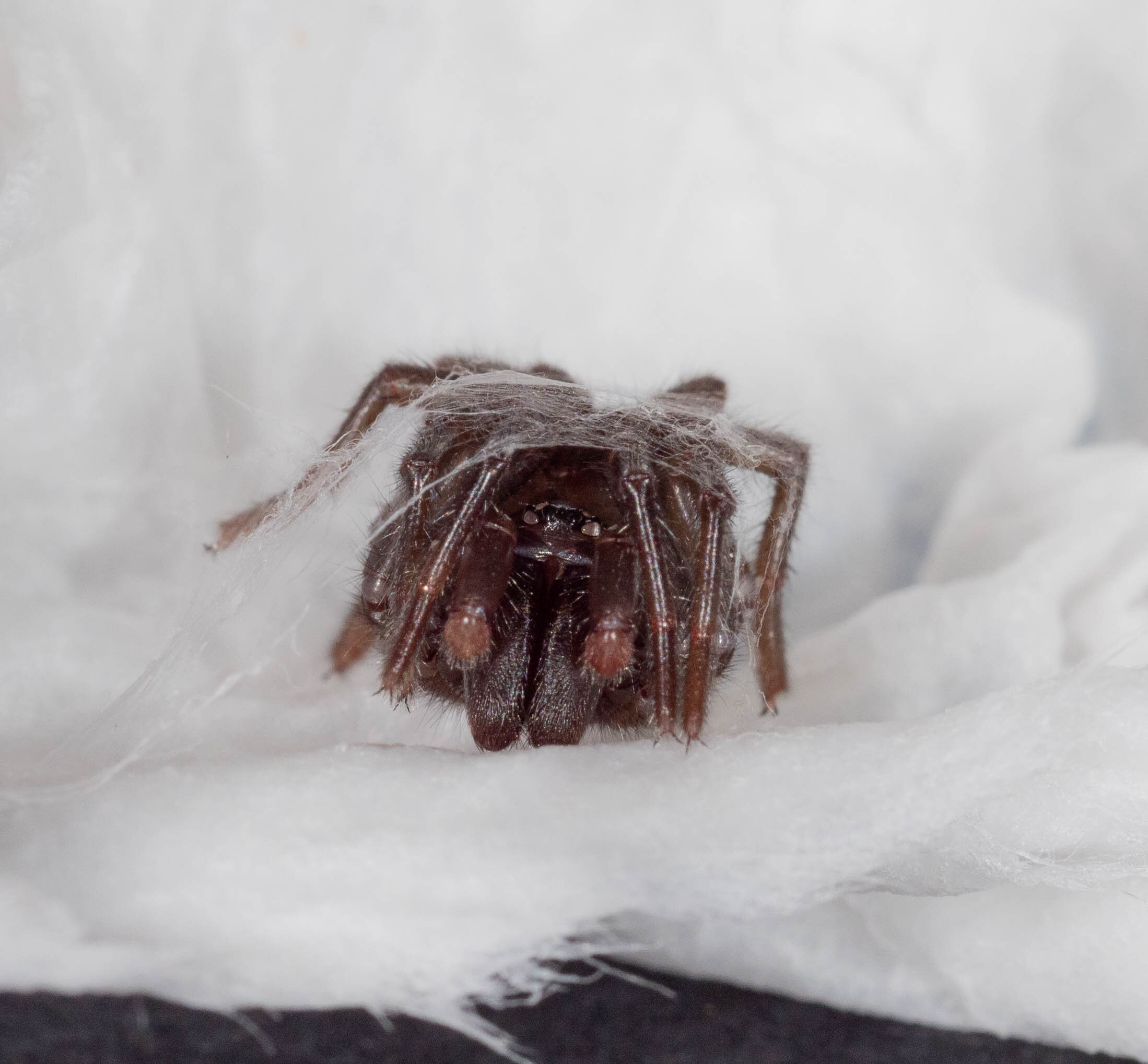
Interestingly, one of them (#1) had woven a silk retreat in a fold of the moistened paper. He needed considerable encouragement to leave his shelter.


Following a final live photo shoot, the remaining 3 spiders were killed and preserved. After 2 hours in the freezer we photographed the spiders again (4pm 24/5). In particular, we closely examined and imaged the pattern of cheliceral teeth. Within an hour of removing the spiders from the freezer, they were preserved in 100% ethanol. At this time Spider #3 (which had been held at room temperature for 2 days) was also preserved in 100% ethanol. All 4 spiders were then stored at 4ºC.
B. Findings
The spiders are unlike any we’ve collected before. They are not Hadronyche nimoola nor Arbanitis melancholicus. Indeed, we are unable to find a match. They do not fit the general family descriptions for any of the Mygalomorphae.
The spiders appear to have recently moulted. That would explain the change in colouration, spinneret size and behaviour during their two days in captivity.
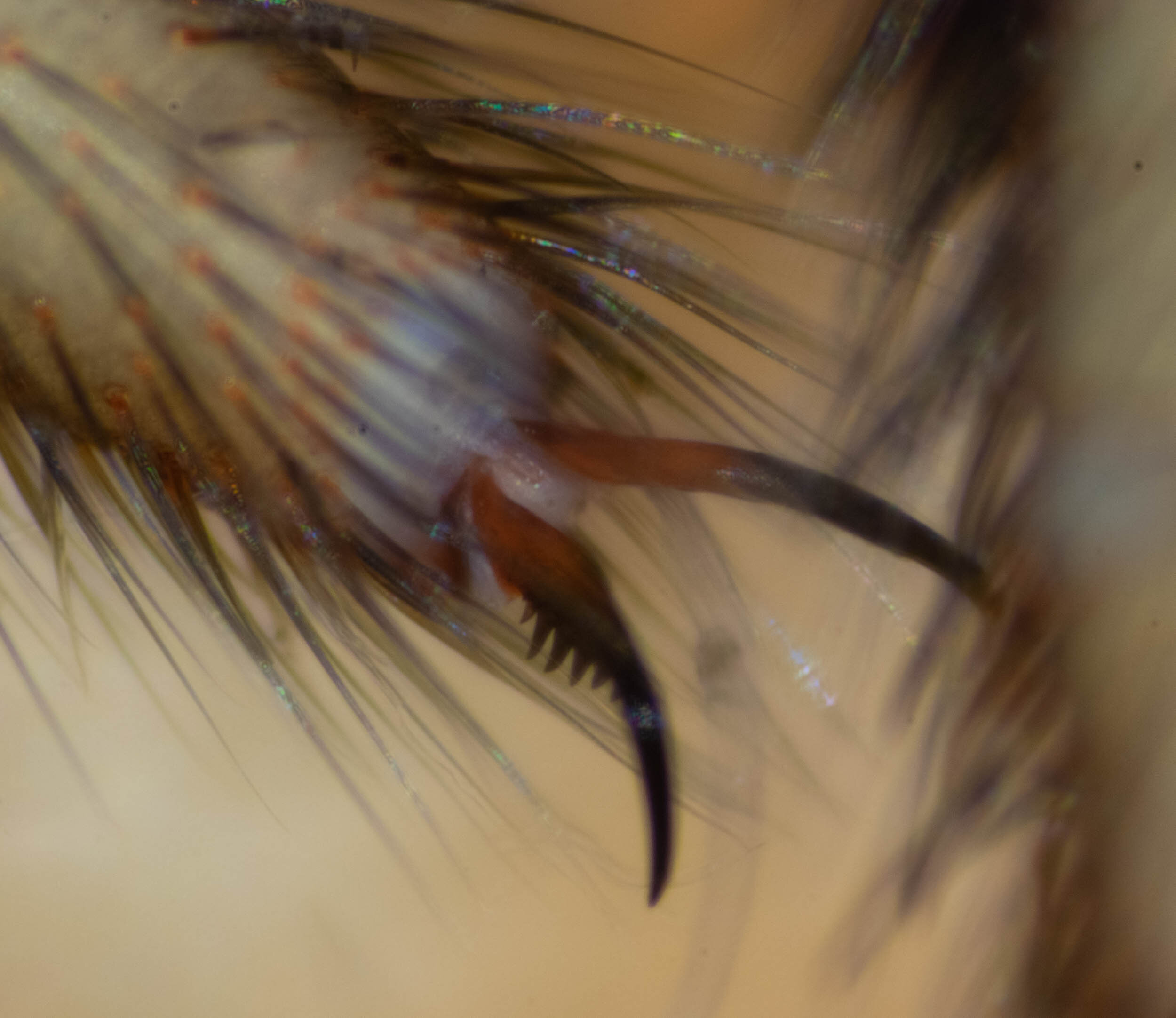
The pattern of cheliceral teeth is a key distinguishing feature. It is a single row, and so precludes Hexathelidae and Atracidae. For this reason we carefully examined each of the 4 spiders. In all there is just one row of teeth, medial to an indistinct groove. There are 8 or 9 teeth, varying between individuals and between chelicerae in two individuals. In some cases there is also a small cluster of tiny teeth basally, but there is no evidence on any of the eight chelicerae of a second row of teeth laterally.
Cheliceral tooth count: Spider #1 (Left 8, Right 8); Spider #2 (Left 9, Right 9); Spider #3 (Left 9, Right 8); Spider #4 (Left 9, Right 8);


Conclusions
There seem to be several possible explanations for our inability to identify the spiders:
We have overlooked or misinterpreted one or more morphological features in the family descriptions we’ve read.
There are known exceptions to the general family or genus descriptions that are not noted in the resources we’ve used.
The spiders are not fully mature (but this seems unlikely as their palps are well developed).
The species is currently undescribed.
Now that we are more familiar with the general appearance of this species, we note another recognisable feature … the pleura is wide and flat above leg #3, in line with the fovea. This is a feature we can easily see in live spiders, so it will be useful for future spider identification in the field.
widened region of pleura level with fovea
C. Logic behind identification
Step 1: Definitely a Mygal
This species is quite unlike any mygalomorph we’ve recorded here before. But it is definitely a mygal. The two pairs of book lungs are unequivocal.


Step 2: what family?
We initially assumed that the spiders would be either Hexathelidae or Idiopidae. However, we decided to check all possibilities.
AFD lists 11 families in the infraorder Mygalomorphae: Actinopodidae; Atracidae; Barychelidae; Ctenizidae; Cyrtaucheniidae; Dipluridae; Hexathelidae; Idiopidae; Migidae; Nemesiidae; Theraphosidae.
For many family descriptions we relied upon field guides. However, for Idiopidae, Hexathelidae and Atracide we checked recent family reviews (see reference list).
Images below are all spider #3, unless otherwise noted.
Not Actinopodidae (Mouse Spiders). The eyes are grouped into a tight rectangle on a slight prominence … not widely spaced across the caput, as they are in Actinopodidae.
Also, the carapace is not as highly elevated as in Actinopodidae.
8 eyes in 2 rows, arranged in a close rectangle on a low tubercle
Not Atracidae. Our spiders have only a single row of cheliceral teeth (8 or 9) plus a cluster of small teeth/bumps lateral to the basal teeth.
The tooth row lies in a position equivalent to the promargin of Atracidae. That is, it runs medial to the fang when the fang is folded against the paturon.
All Atracidae have a cheliceral groove flanked by two rows of large teeth and a middle region of smaller teeth (Gray, 2010).
The only exception, noted by Gray (2010), is Hadronyche anzses, which has the “prolateral tooth row reduced to a few basal teeth” (p. 292).
ventral side chelicerae
Hadronyche anzses was described in 2000 from animals collected in Far North Queensland (Raven, 2000).
“Differs from all other named species of Hadronyche in having teeth for full length of retromargin but only three teeth on the usually heavily dentate promargin …” (Raven, 2000. p226)

Extract from Raven 2000, p226, Figure 1: Hadronyche anzses sp. nov. … G … chelicerae showing fang groove.
We can preclude this as a match for our species. There is no evidence of any second row in our spiders. In addition, the tooth row in our spiders is medial to the fang (‘prolateral’), whereas the complete row in H. anzses is lateral (‘retrolateral’). Plus, the distant highlands of Far North Queensland is an environment very different to ours!
Have we considered all known species of Atracidae? That is, have any new species been described since the 2010 Gray review? No. We cross-checked the AFD listings against those in Gray 2010. The only additional species in AFD are two unplaced, unnamed species that have doubtful origins.
The maxillae in our spiders are consistent with Atracidae in that they have “a strong coniform lobe anteromedially” (Gray, 2010, p.290).
Lobe of maxillae (arrows) (spider #1 in ethanol, ventral view)
Not Barychelidae (Brush-footed Trapdoor Spiders). The tarsi lack obvious tufts of hair … although I find this a difficult character to be sure of.
More convincingly, however, there is only a single row of teeth on the tarsal claws. Most Barychelidae are in the subfamily Barychelinae and they have 2 rows of teeth … there is a single species in the subfamily Sasoninae (Sason colemani) which has a single row, but the eyes of that species do not sit on a tubercle (the eyes in our spiders do).


Not Ctenizidae (Cork-lid Trapdoor Spiders). The spiders lack the “saddle-shaped dent in the middle of the third leg” (Whyte & Anderson, 2017) ascribed to this family (Main, 1985). (Note that there is currently only a single genus in this family, Conotheles, as many other trapdoor genera have been moved to other families in recent revisions.)

Third leg lacks any depression in tibia
Some other features do fit. The ovoid carapace with raised head; the rectangular eye cluster; the U-shaped fovea; the short spinnerets.
However, the chelicera of Conotheles have “teeth of both margins of the furrow” (Main, 1985) … ours has only a single row of teeth and no apparent furrow.
Also, Conotheles lack a suture separating the labium from the sternum (Main, 1985) … ours have a clear separation of these structures.

labium and sternum separated by suture
Not Cyrtaucheniidae … well, not Kiama lachrymoides, anyway.
The only Australian species that AFD lists for this family is Kiama lachrymoides. However, in 2012 the species was moved to Nemesiidae (Bond et al., 2012) and has more recently been assigned to the newly-erected family Microstigmatidae (Opatova et al., 2019).
We compared our spiders to the species description for Kiama lachymoides (Main & Mascord, 1971) … for detailed analysis see Part E, below. In short, our spiders differ from Kiama in many respects including: eye arrangement; spinneret shape; carapace size; tarsal claw teeth; cheliceral teeth.
Not Dipluridae (Curtain-web spiders). Carapace is not low and hairy and spinnerets are not very long.

Two pairs of short spinnerets
Not Hexathelidae (Funnelweb spiders). Our spiders have only two pairs of spinnerets, not 3 as in Hexathelidae.
Note that this family previously included the genera Atrax, Hadronyche, Illawarra. That subfamily (Atracinae) has now been elevated to family level - Atracidae. The remaining members of Hexathelidae are in the old subfamilies of Hexathelinae and Plesiothelinae.
Not Idiopidae. The palps of our spiders lack tibial apophyses.
All* Idiopidae genera have protrusions on the tibia (Rix et al., 2017), which protect and surround the bulb embolus when it is folded back.
*These tibial apophyses are absent in only one genus of Idiopidae, Bungulla. However, our spiders do not fit the description for Bungulla. Ours lack strong dorsal spines on the cymbium. Bungulla have eyes in a trapezoidal arrangement, with the front row strongly procurved. Bungulla is also only known from WA (Rix et al., 2017).
ventral view pedipalp and embolus


Not Migidae (Tree trapdoor spiders). Our spiders are too large and their eyes are not widely spaced, as they are in Migidae.
More convincing, however, is that our spiders lack the “two ridges on the outer edge of the fangs (which) unmistakably identifies the family (Migidae)” (Framenau et al, 2014).

fangs smooth, with no ridges
Not Nemesiidae (Wishbone spiders). Spinnerets of our spiders lack long apical segments.
Also, tarsal claws with single row of teeth (see images for Barychelidae, above) … Nemesiidae have “superior tarsal claws with two rows of teeth” (Framenau et al. 2014)
Not Theraphosidae (Australian Tarantulas). Our spiders are too small and too hairless. Theraphosidae are large and very hairy spiders.
In addition, Theraphodids have hairy tufts concealing their tarsal claws. Our spiders do not.
References
Bond JE, Hendrixson BE, Hamilton CA, Hedin M. 2012. A reconsideration of the classification of the spider infra- order Mygalomorphae (Arachnida: Araneae) based on three nuclear genes and morphology. PLoS One 7: e38753
Felix, R.F. 2011. Biology of Spiders (3rd Edition). Oxford University Press, New York.
Gray. M. 2010. A revision of Australian funnel-web spiders. Records of the Australian Museum, 62: 285=392.
Main, B.Y. & Mascord, R. 1971. A new genus of diplurid spider (Araneae: Mygalomorphae) from New South Wales [online]. Journal of the Entomological Society of Australia (N.S.W.), Vol. 6 (1969): 24-30
Main, B. Y. 1985. Further studies on the systematics of the ctenizid trapdoor spiders: a review of the Australian genera (Araneae : Mygalomorphae : Ctenizidae). Australian Journal of Zoology, Supplementary Series. 108, 1-84.
Opatova, V., Hamilton, C.A., Hedin, M., Montes de Oca, L., Kral, J. & Bond, J.E. 2019. Phylogenetic systematics and evolution of the spider infraorder Mygalomorphae using genomic scale data. Systematic Biology, 1: 1-37
Raven, R.J. 2000. A new species of funnel web spider (Hadronyche : Hexathelidae : Mygalomorphae) from north Queensland. Memoirs of the Queensland Museum, 46, 225-230.
Rix, M.G., Raven, R.J., Main, B.Y., Harrison, S.E., Austin, A.D., Cooper, S.J.B. & Harvey, M.S. 2017. The Australasian spiny trapdoor spiders of the family Idiopidae (Mygalomorphae ; Arbanitinae): a relimitation and revision at the generic level. Invertebrate Systematics, 31, 566-634.
D. UPDATE
We now realise that a spider we collected in June 2018 was also this species. It has a single row of cheliceral teeth.


We (foolishly) pinned the spider and kept it at room temp in a box … so the body is quite fragile now. Luckily the chelicerae are still in good condition (images below).


In addition, another spider we imaged but did not collect at that time also appears to be the same species.


These spiders were part of the ‘Getting to know mygals’ blog of June 2018.
E. UPDATE … comparison to Kiama lachrymoides
On the 1/6/20 we received a copy of Main & Mascord, 1971, the species description of Kiama lachrymoides.
Spinnerets
Our spiders’ spinnerets are different in shape and length. Ours are: shorter overall (1.1mm cf 2.4mm) and the individual segments are much wider relative to their length.
Spinnerets of our spider #1 (in ethanol)
Kiama lachrymoides (male). Extract from Main & York, 1971, Fig 19. Left spinnerets
Carapace size
Our spiders are considerably smaller than K. lachrymoides. The carapace dimensions are a good indicator. Our spider #1: 3.4mm W, 4.3mm L cf. 5.8mm W, 6.0mm L for the male paratype of K. lachrymoides described by Mains & Mascort (1971). In addition, they describe the caput as being 4.8mm wide … ie, significantly narrower than the carapace (83%). In our spiders, the caput spans the anterior edge of the carapace and is 3.0mm wide (88% carapace width). The actual shape of the caput in Kiama is difficult to determine from the diagram.
Carapace of our spider #1 (in ethanol)
Kiama lachrymoides (male). Extract from Main & York, 1971, Fig 13. Dorsal aspect carapace …
Cheliceral teeth
The pattern of cheliceral teeth in Kiama and in our spiders is similar. Both have a single row on the inner ridge of the cheliceral groove.
However, our spiders have fewer teeth (8-9 cf 13 in Kiama). In addition, the teeth row in our spiders extends further apically than is depicted for Kiama. Note: only the female is depicted in the Main & Mascord paper, but there is no sex difference noted in the text.

Kiama lachrymoides (female). Extract from Main & York, 1971, Fig 2.
Tarsal claws
Our spiders have a pair of tarsal claws on each leg, plus a small lower claw.
Each of the upper claws bears a single row of teeth. The row runs from the lateral edge at the claw base to the medial edge at the claw apex. The teeth vary in size and number, typically 8 or 9 per claws.


In contrast, Kiama lachrymoides has two rows of teeth on each claw, with just 5-6 teeth per row.

Kiama lachrymoides (male). Extract from Main & York, 1971, Fig 16. Tarsal claws of first leg (not to scale)
Eye arrangement
The eyes for K. lachrymoides are described as follows: “Eyes in compact group, not on a tubercle, in two rows of four” … “Anterior row slightly recurved in front, straight behind; lateral eyes small; AME the largest” (Main & Mascord, 1971, p25), and, in the male, “length of eye group 0.7mm, width 1.4mm (p.26).
The eye pattern in our spiders is different to K. lachrymoides.
The ALE are as large as the AME.
There is no space between the ALE and AME, but there is a space between the two AME.
The eyes sit on a low tubercle.
Anterior view eye cluster (spider #2, live)
The eye group is 0.5mm long, 1.1–1.3mm wide. This is not very different to the size for the cluster in K. lachrymoides, but it is considerably larger relative to the size of the carapace.


Labium and sternum
The labium in our spiders is different in size and shape to Kiama lachrymoides: ours is smaller (0.6mm L, 0.9mm wide cf 1.5mm long, 1.0mm wide) and ovoid rather than square. The anterior edge is convex rather than concave.
The sternum of our spiders has a less pronounced posterior extension than described for K. lachrymoides. The posterior sigillae are oval in our spiders, rather than the “tear-dropped shaped” sigillae of K. lachrymoides.
Labium of our spider #1 (in ethanol)
Sternum of live spider (#1), showing sigillae
Kiama lachrymoides (male). Extract from Main & York, 1971, Fig 14. Labium and sternum
This is a workbook page … a part of our website where we record the observations and references used in making species identifications. The notes will not necessarily be complete. They are a record for our own use.


Lorem ipsum dolor sit amet, consectetur adipiscing elit. Vestibulum id ligula porta felis euismod semper.