Microgastrinae, Braconidae
Workbook
Family Braconidae
Cell R in hind wing shorter than submarginal vein or absent (image 1);

image 1 - hind wing of parasitoid wasp from 24-26/4/19
2nd recurrent vein (2m-cu) absent from fore wing (in Ichneumonidae, this vein divides 2nd discal cell into two) - image 2

image 2 - fore wing of parasitoid wasp from 24-26/4/19
T3 and T4 rigidly joined
Subfamily Microgastrinae
endodont, i.e. mandibles with 1 or 2 teeth, curved inwards, their tips touching when closed (image 3a)

image 3a - frontal view of head of parasitoid wasp from 24-26/4/19
transverse sulcus between antennal sockets absent (image 3b)

image 3b - frontal view of head of parasitoid wasp from 24-26/4/19
Hypoclypeal depression usually absent; if present then rather shallow, narrow (middle of clypeus not distinctly above upper level of mandibular bases
Spiracles of T2 in weakly sclerotised areas on either side (vs. in or near margins) of sclerotised notum
Cross-vein between pterostigma and 2nd submarginal cell (r) longer (vs. shorter) than anterior length of cell (images 4, 5)

image 4- fore wing of parasitoid wasp from 24-26/4/19

image 5 - fore wing of parasitoid wasp from 24-26/4/19
Antennae 16-segmented (excluding scape and pedicel) - image 6

image 6 - antennae of parasitoid wasp from 24-26/4/19
Genus Cotesia
I have posted several wasps to Erinn Fagan-Jeffries from the University of Adelaide for identification.
10/5/19 She has provisionally identified the wasp as Cotesia sp. This fits with the description of the genus in Austin and Dangerfield (1992) - Ref.1. Now waiting on results of DNA barcoding.
absence of areolet

short inflexible hypogygium, venly sclerotised throughout, never shows lateral creases
short ovipositor, hardly extending past posterior gaster
parallel-sided or posteriorly broadened T1
large rectangular T2
propodeum, T1, T2 rugose
propodeum has a median longitudinal carina
this group contains both solitary and gregarious parasites


Fig.124 Ref. 2 ovipositor and hypopygium of Cotesia (previously Apanteles) acuminatus
However, hindwing vein 2r-m, which is one of the features of Cotesia, is not clearly evident in this wasp.
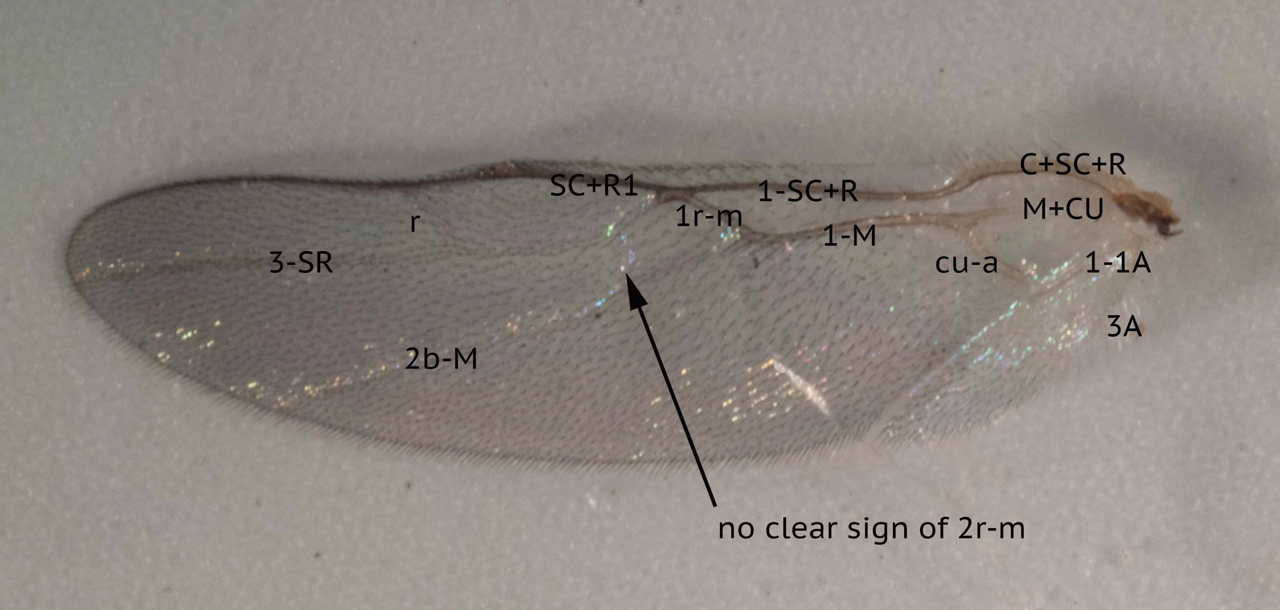
image 17 - hind wing veins

image 18- hind wing cells
Cotesia anthelae (Wilkinson, 1928a) has been shown to have Anthela ocellata as host. The parasitised caterpillar is almost certainly an Anthela sp. and we have sighted Anthela ferruginosa on the block.
Female
Body

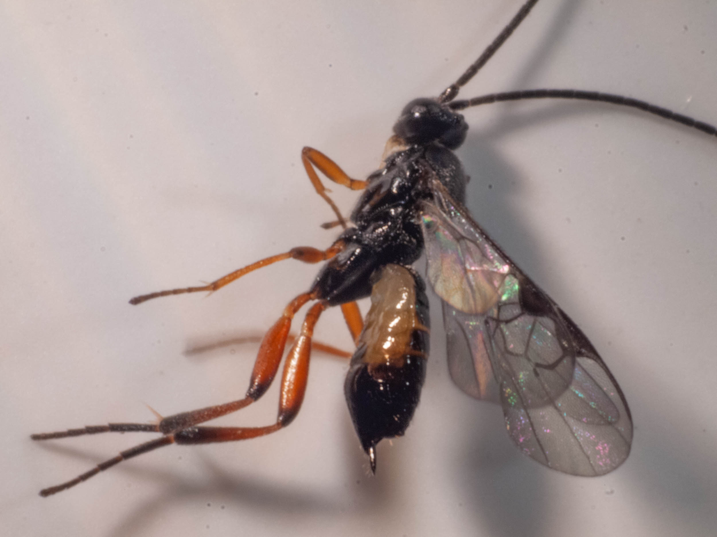
image 7 -parasitoid wasp from 24-26/4/19
Head

image 8a - frontal view of head of parasitoid wasp from 24-26/4/19

image 8b - frontal view of head of parasitoid wasp from 24-26/4/19

image 9 - dorsal view of head showing ocelli

image 10 - antennae of parasitoid wasp from 24-26/4/19
Alitrunk

image 11

image 12
Legs

image 13

image 14
Wings

image 16 - fore wing

image 19

image 20a - posterior end of female

image 20b - posterior end of female
Cotesia sp.
All images below of females that emerged from cocoons on a lepidopteran larva on 24-26/4/19. Body length 3mm.
Female
length 3.1mm (range 2.6-3.7)
Colouration


Head


image 27

image 28a

image 28b

image 29a

image 29b

image 32 - hindwing cells
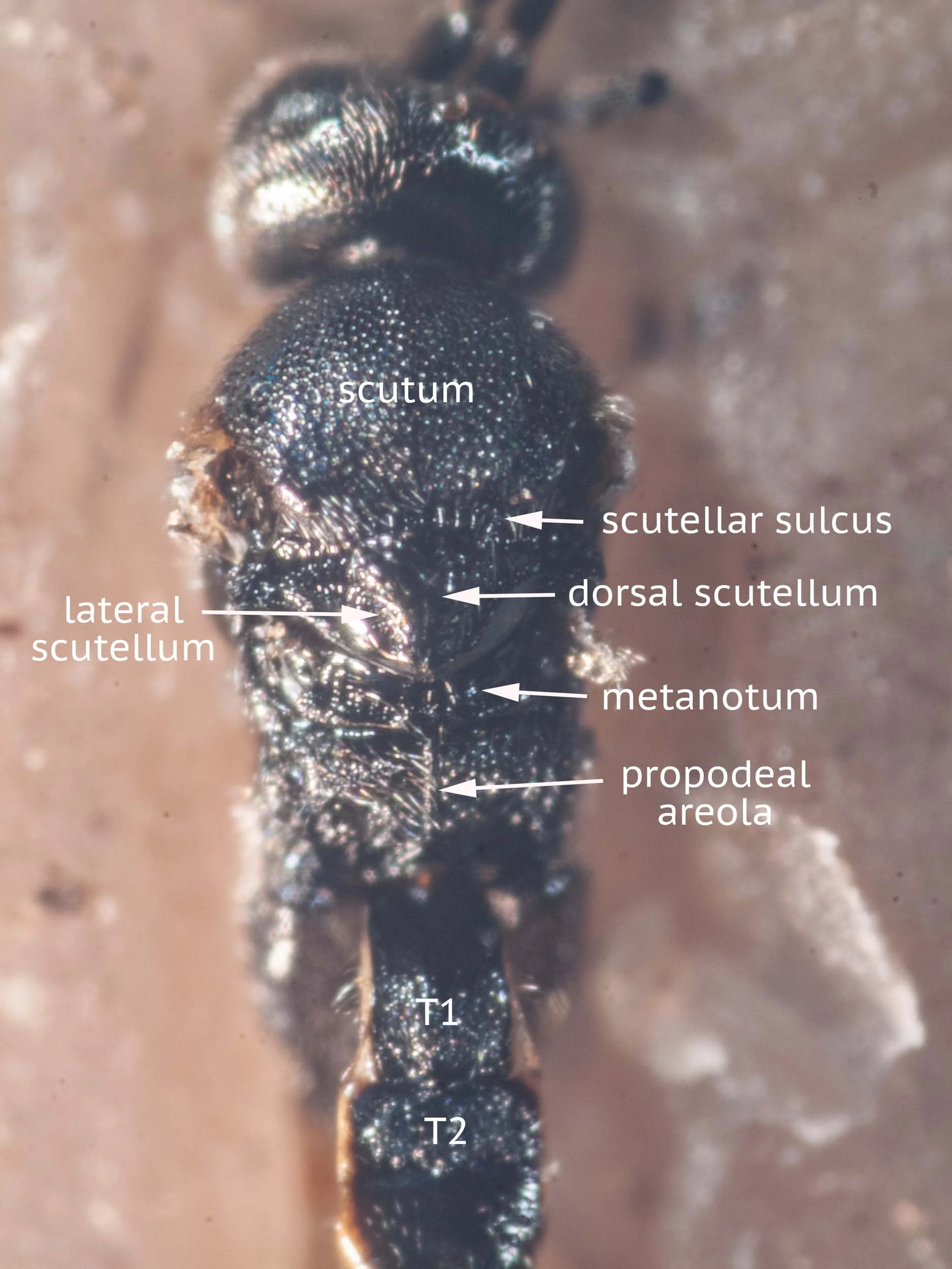


image 37 - lateral view of alitrunk
Mesopleuron with surface varying from finely and densely punctate all over to punctate only in anterior and ventral halves, posterior margin and epicnemial furrow faintly foveate; area of precoxal suture represented by broad shallow furrow (image 37)
Metapleuron smooth except in anterior half, divided by narrow medial furrow, smooth to very finely punctate and pilose in posterior half (image 37)
References
Austin, A.D. and P.C. Dangerfield (1992) Synopsis of Australasian Microgastrinae (Hymenoptera: Braconidae), with a key to genera and description of new taxa. Invertebrate Taxonomy 6: 1-76
Nixon, G.E.J. (1965) A reclassification of the Tribe Microgasterini (Hymenoptera: Braconidae). Bulletin of the British Museum (Natural History) Entomology, Supplement 2, 1-284
This is a workbook page … a part of our website where we record the observations and references used in making species identifications. The notes will not necessarily be complete. They are a record for our own use, but we are happy to share this information with others.