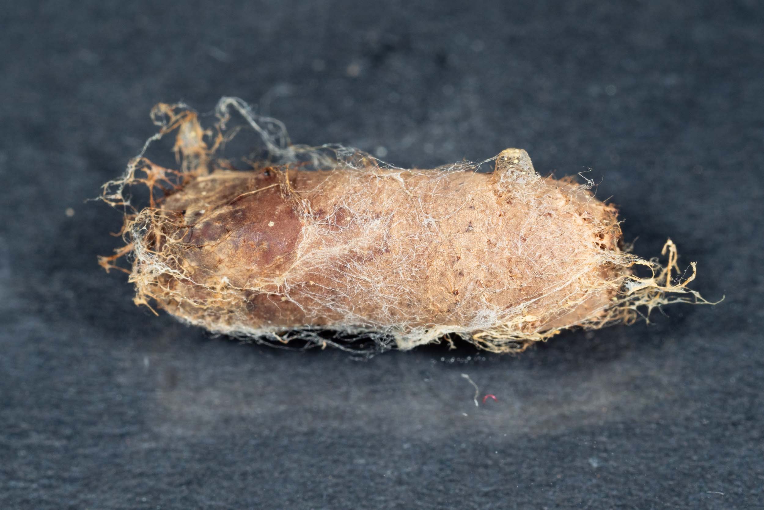
Cocoon B
As it appeared before I tore away the papery cocoon to reveal the contents.
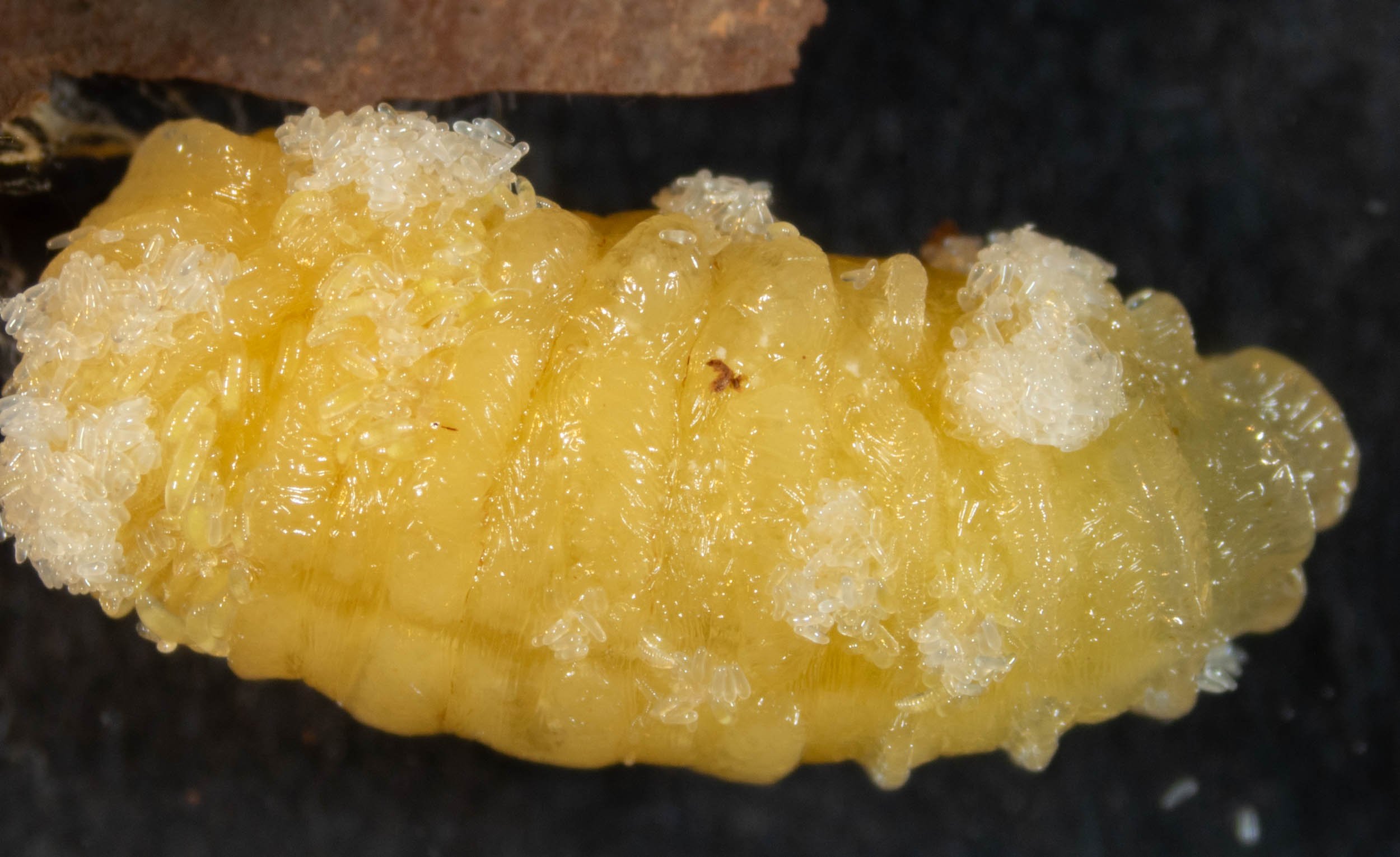
clumps of eggs and tiny larvae
The larva from cocoon B was still relatively undamaged … and no doubt alive. It was motionless. At this prepupal stage, they are largely immobile anyway, but the addition of toxins from the founding parasitoid would have effectively paralysed the host. This ensures that her eggs and larvae are not dislodged or crushed.
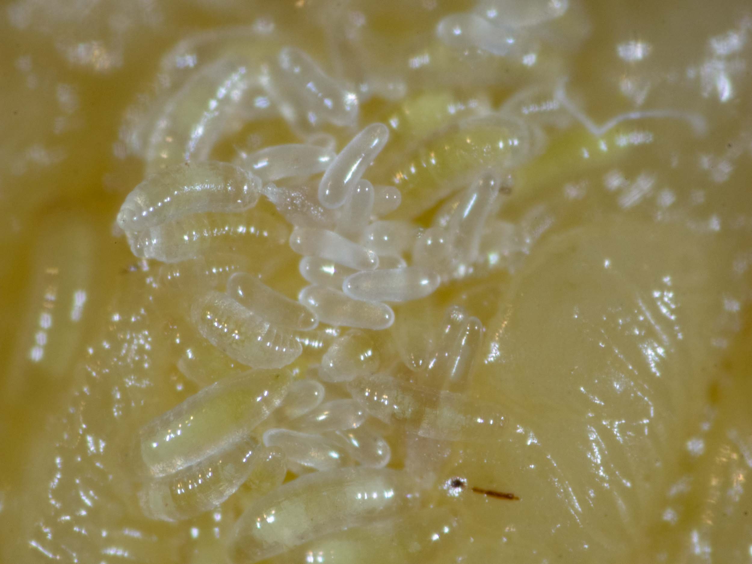
feeding larvae
The hatched larvae, with their segmented bodies, are already feeding. Some are larger than others, their guts appearing yellow through otherwise transparent bodies.
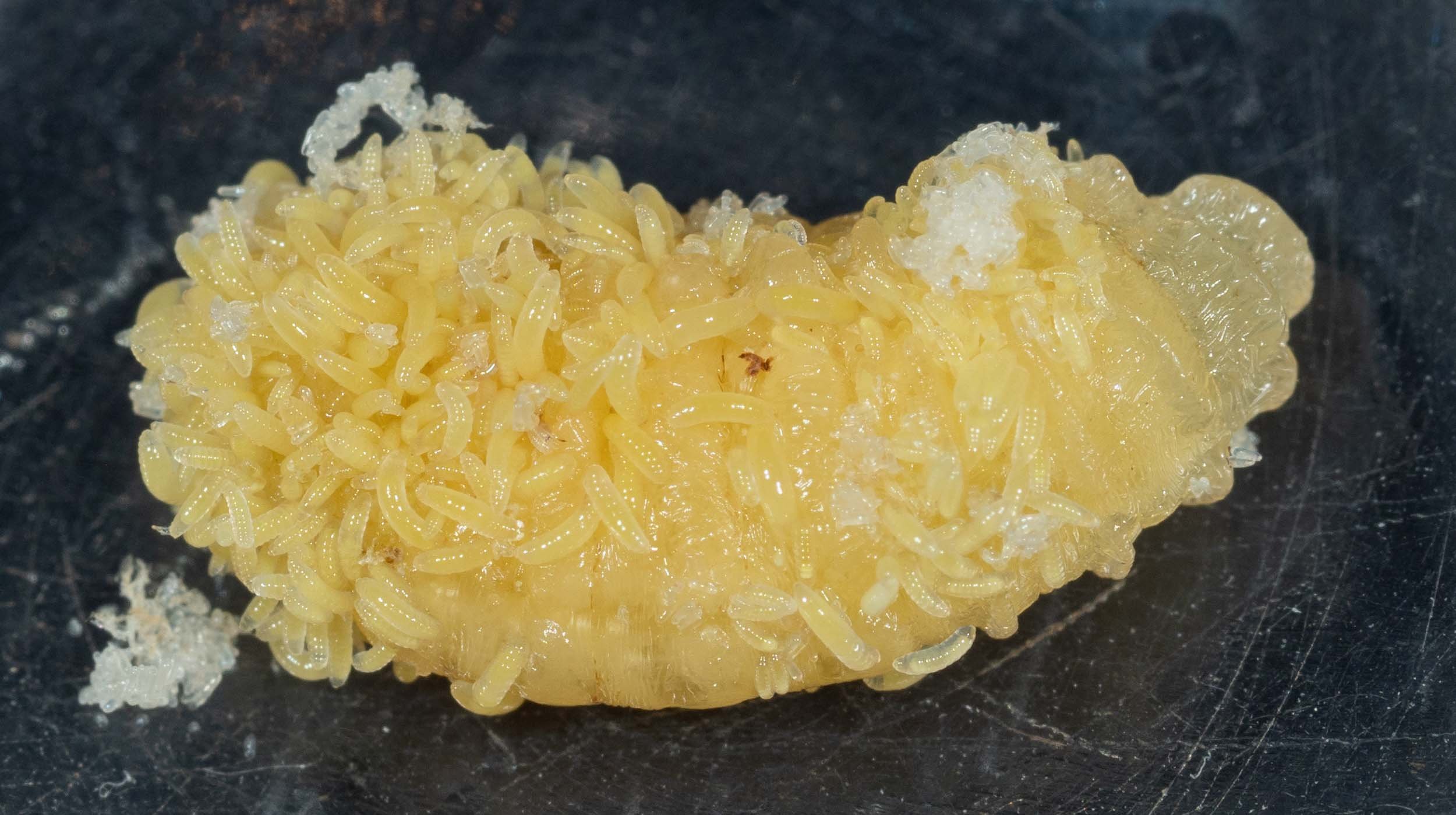
rapidly growing larvae
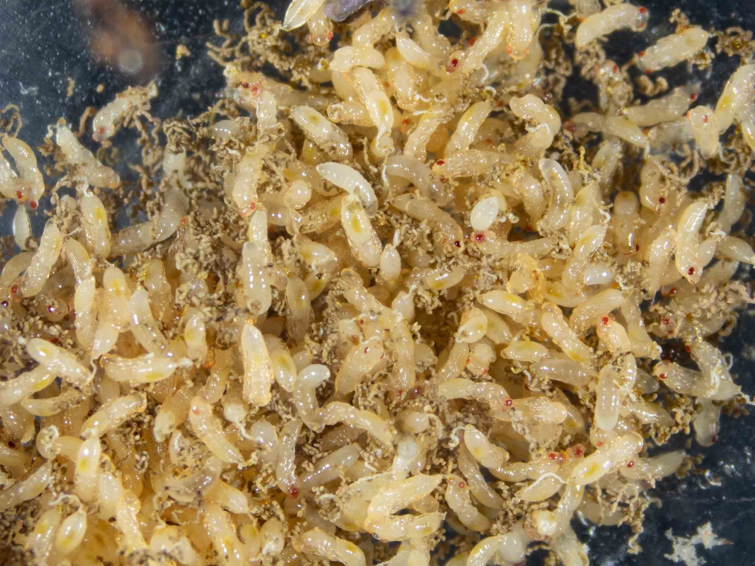
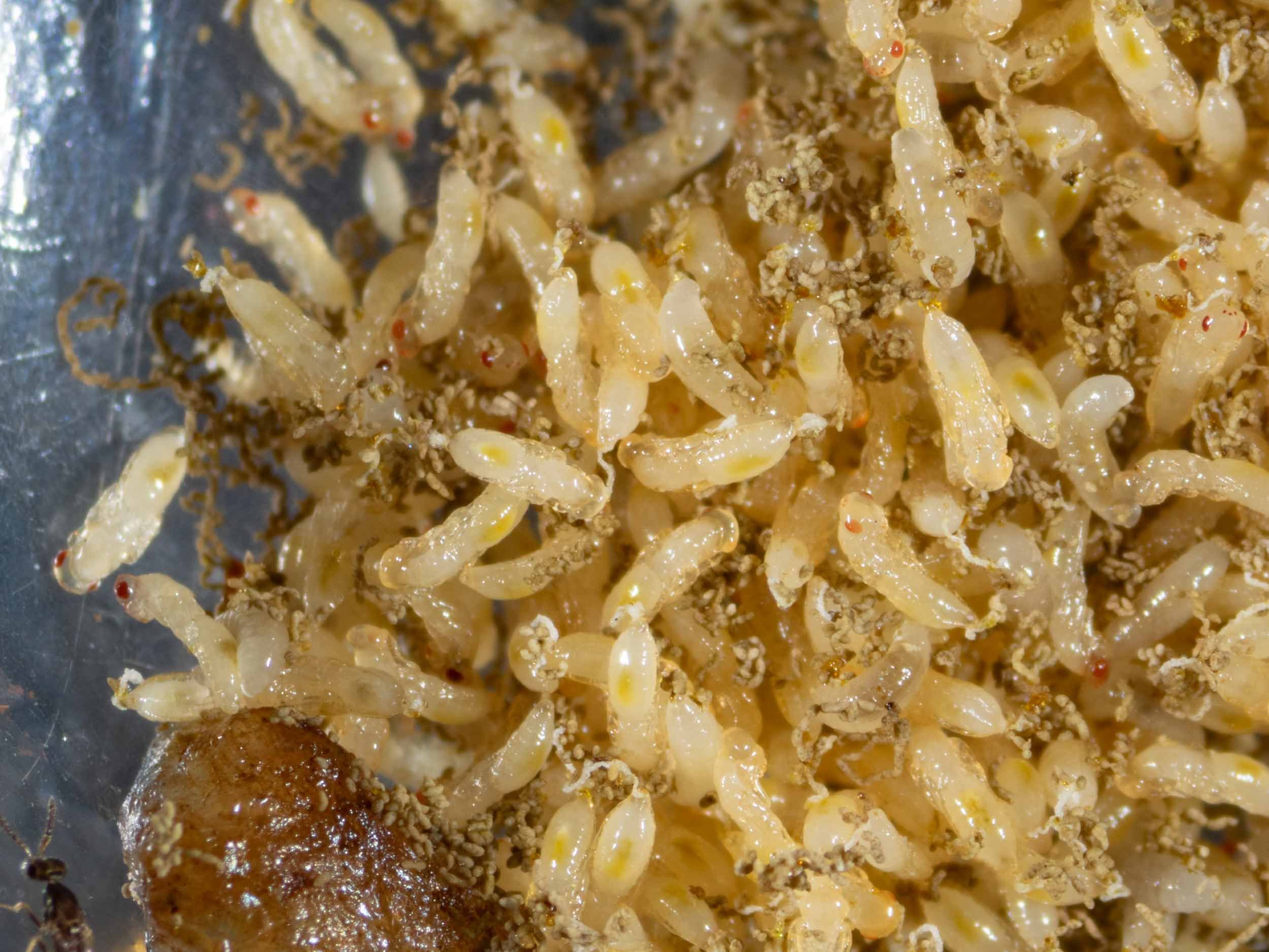
When I first opened Cell B (on 7th May) the larvae were small and not obvious. Two days later it’s a different story!
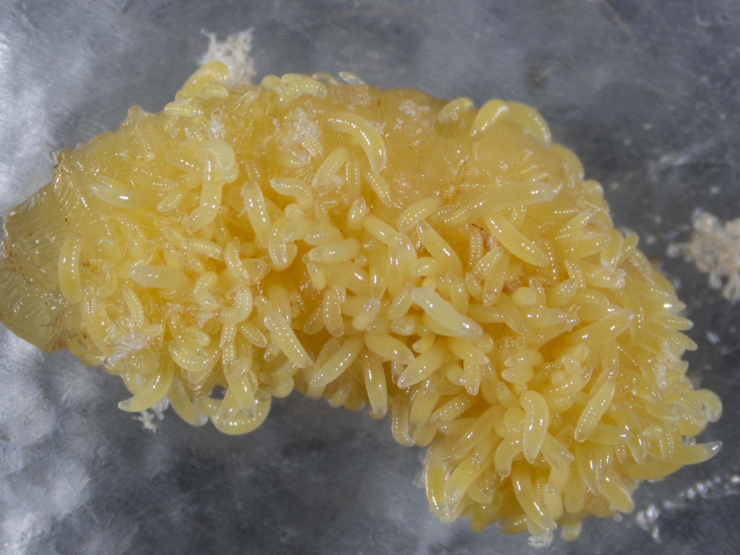
a seething mass
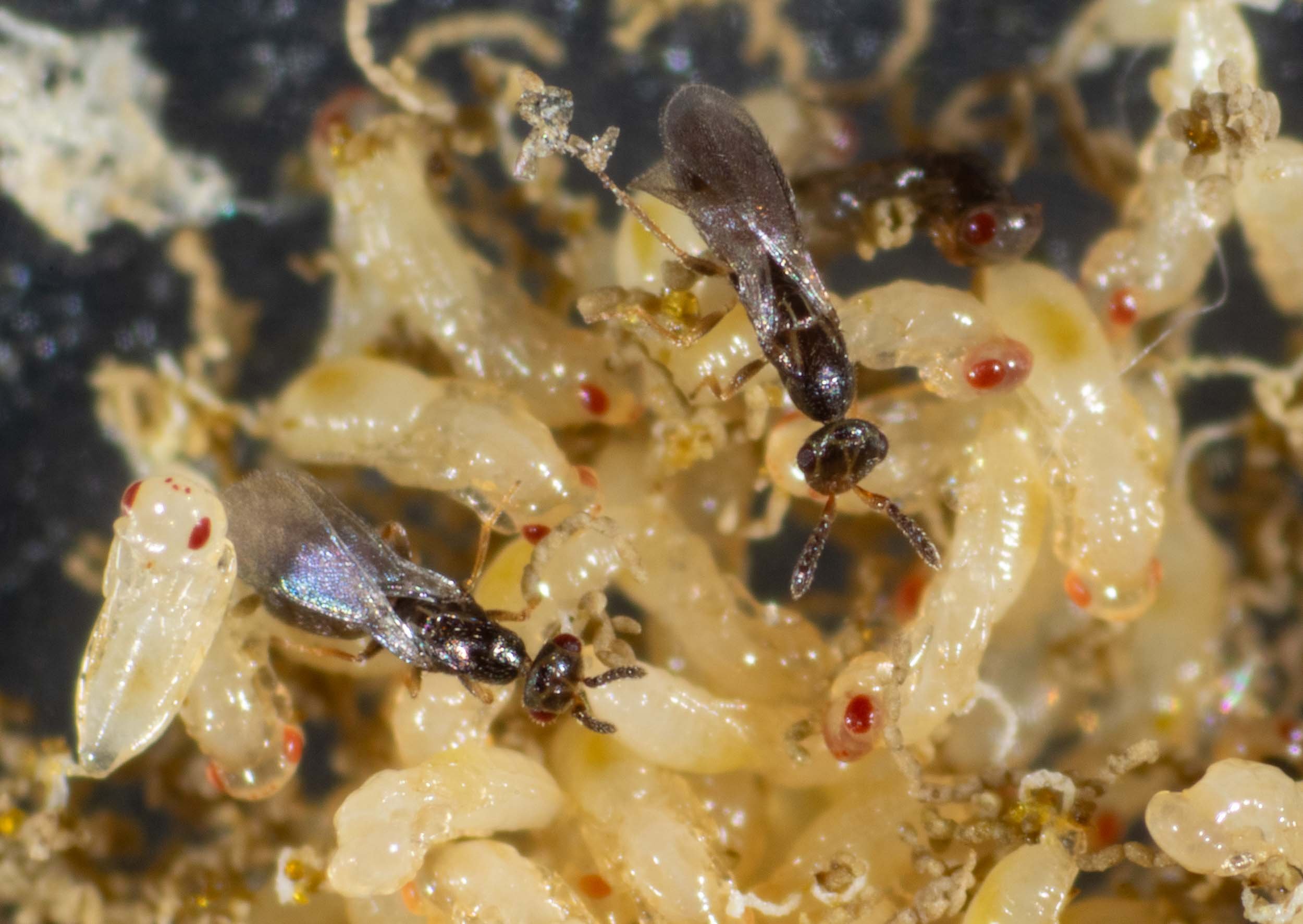
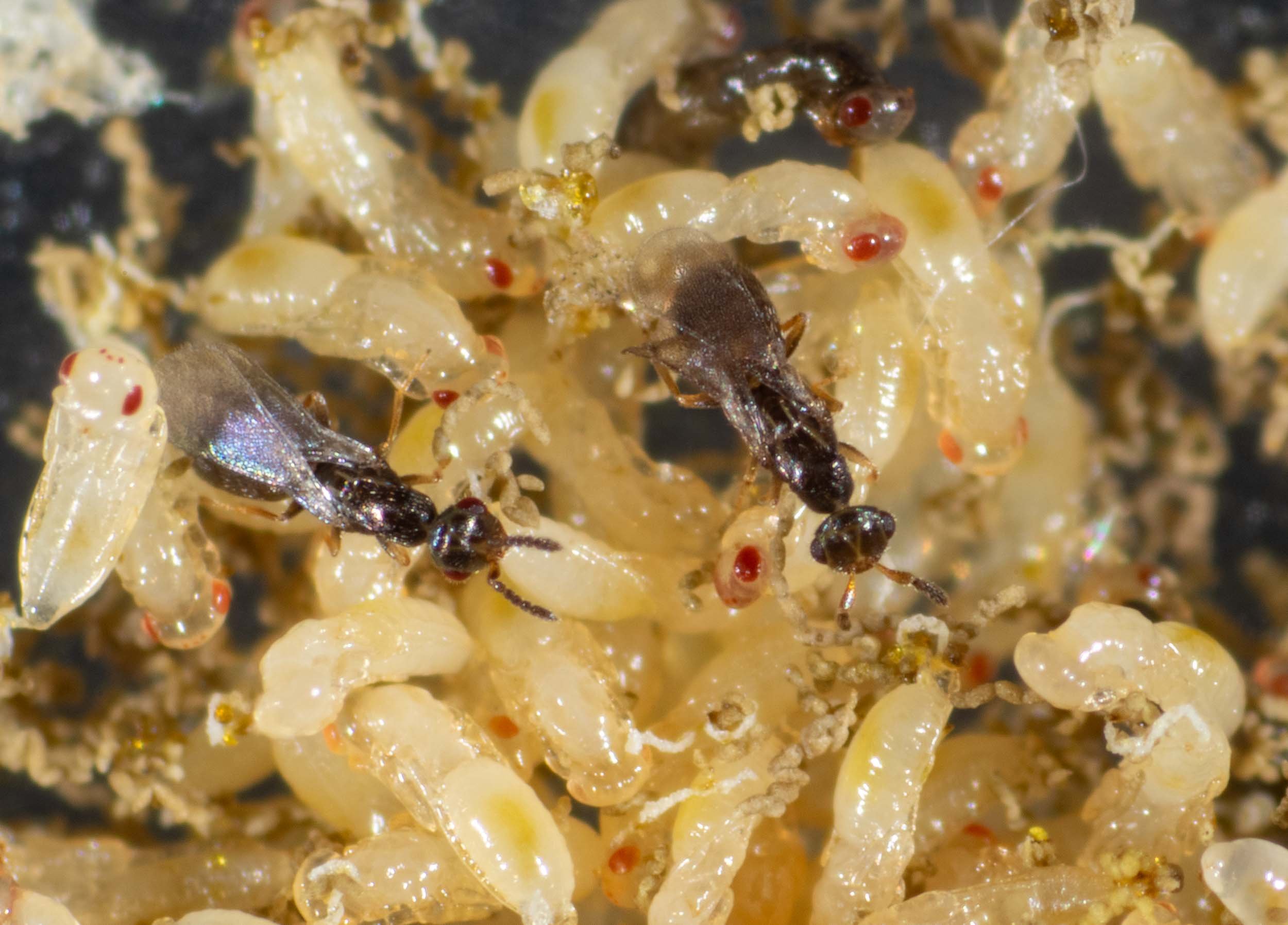
Note that on 7th May I removed all adults from this enclosure. The plan is to monitor these through to maturity. This will give me an estimate of life cycle timing and sex ratios. I expect only around 5 males for every 100 females. The genus has a well documented female bias.

ectoparasites
The larvae remain outside the body of the host as they feed. They have relatively small mouthparts and are quite maggot-like in appearance. All they need do for the next week or two is feed and grow. By the time they are ready to pupate they will be 1.6mm in length … roughly the same size as the adult wasp they will become.
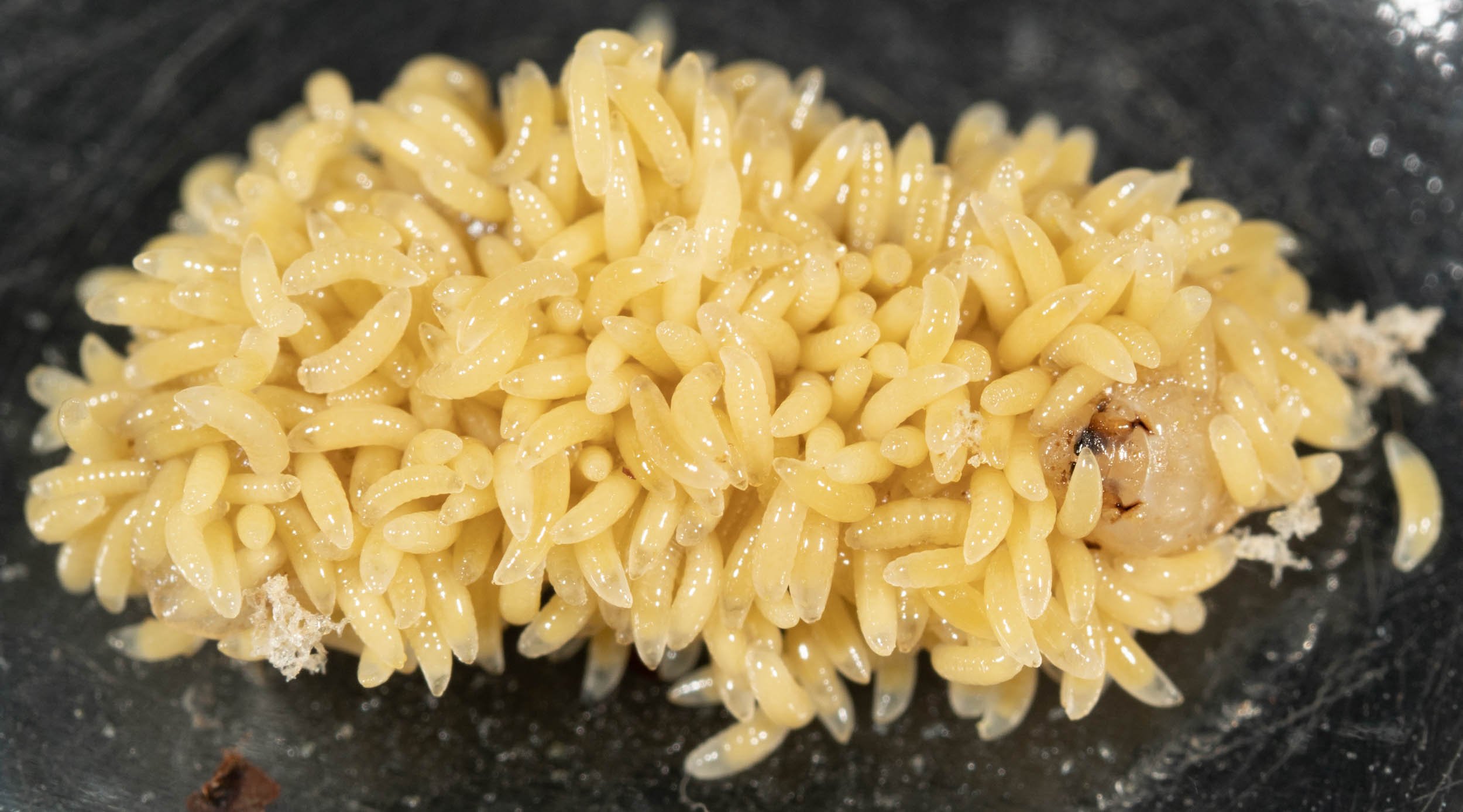
wow!
The host is now completely cloaked in Melittobia larvae … just its head is visible (to the right). Perhaps the feeding parasites can’t effectively penetrate the tougher cuticle of the head capsule. There is so little room that some of the grubs have been pushed away from the host by their siblings.
Compare this scene to that just 6 days before … noting that no new eggs were laid in the interim, as no adults had access to the host. Wow bugs indeed!
