


1
2
3
4
5
6
7
8
9
10
11
12
13
14
15

- gaster constricted between segments
- body coarsely sculptured
- ocelli normal
- 3 submarginal cells in forewing; SCII nearly always petiolate (as here)
- pronotal collar short
- first gastral segment usually pedunculate (sometimes a narrow petiole)
- body coarsely sculptured
- head wide
- eyes widely separated
- ocelli normal
- 3 submarginal cells in forewing; SCII nearly always petiolate (as here)
- pronotal lobe & tegula separated
- head wide
- eyes widely separated
- eyes slightly (as here) or strongly converging above
- antennal sockets divided by a strongly raised ridge
- clypeus trilobed (although not always as obviously as in this species), with the median lobe extending upwards and truncate a short distance below antennal sockets
- gaster constricted between segments
- body coarsely sculptured
- head wide
- hind femur apically expanded, truncate
- gaster pedunculate or petiolate; T1 much narrower than T2
- gaster constricted between segments
- body coarsely sculptured
- head wide
- 3 submarginal cells in forewing; SCII nearly always petiolate (as here)
- well-defined pygidial plate
- gaster constricted between segments
- hind femur apically expanded & flattened
clypeal lamina of female
In Cerceris, the clypeus is trilobed (L= lateral lobe; M = median lobe).
The clypeal lamina of the female is a plate-like elevation arising from the median lobe. It varies significantly in different species, and is therefore of considerable diagnostic value.
In this species, Evans (1982) describes the clypeal lamina as “recumbent, nearly as wide apically as median lobe” (p. 357).
clypeal lamina of female
Evans variously describes the clypeal lamina as porrect, subporrect, subrecumbent, recumbent, or absent. These categories are not absolute, and I find the distinction between subrecumbent and subporrect can be quite tricky. But they are useful descriptors (see illustrative figures extracted from Evans, included in the ‘Bits & pieces’ section of this page).
A lateral or near-lateral view is helpful in assessing the shape of the clypeus in detail.
clypeal lamina of female
In addition to the angle of the lamina (porrect, recumbent, etc), some are distinctively shaped and/or bear prominent teeth.
https://inaturalist.ala.org.au/observations/267493072
(image courtesy of Nick Lambert)
clypeal lamina of female
The clypeal lamina can be quite extraordinary, as highlighted here. This museum specimen was identified by H. Evans as Cerceris aurantiaca, and he describes the clypeus as follows:
“clypeal lamina much longer than wide, porrect but strongly curved downward, apex truncate”
Image kindly provided by Dr Ben Parslow, Collection Manager, South Australian Museum (SAMA)
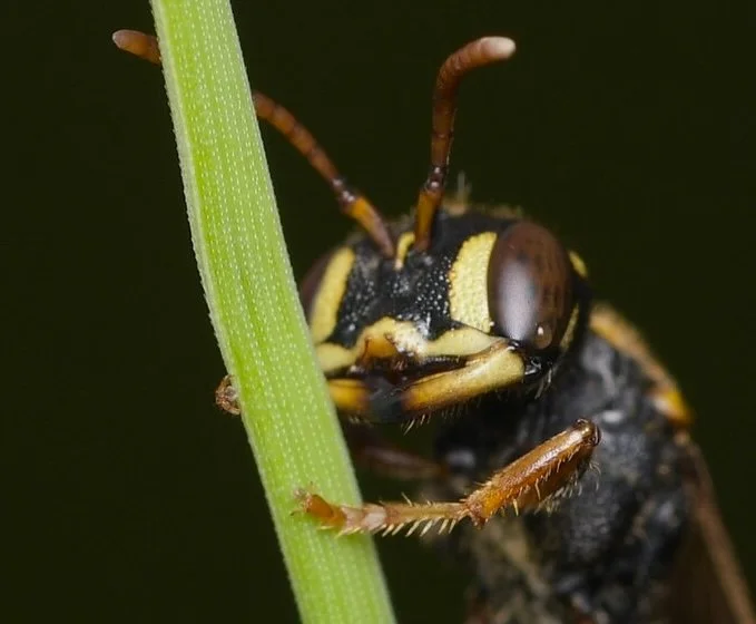
mandibles
The shape of the mandibles varies considerably between Cerceris species, but they are subject to wear so interpret with caution. For example, this female photographed in February has extremely eroded mandibles. Cerceris females can live for months, and their mandibles do a lot of digging in that time.
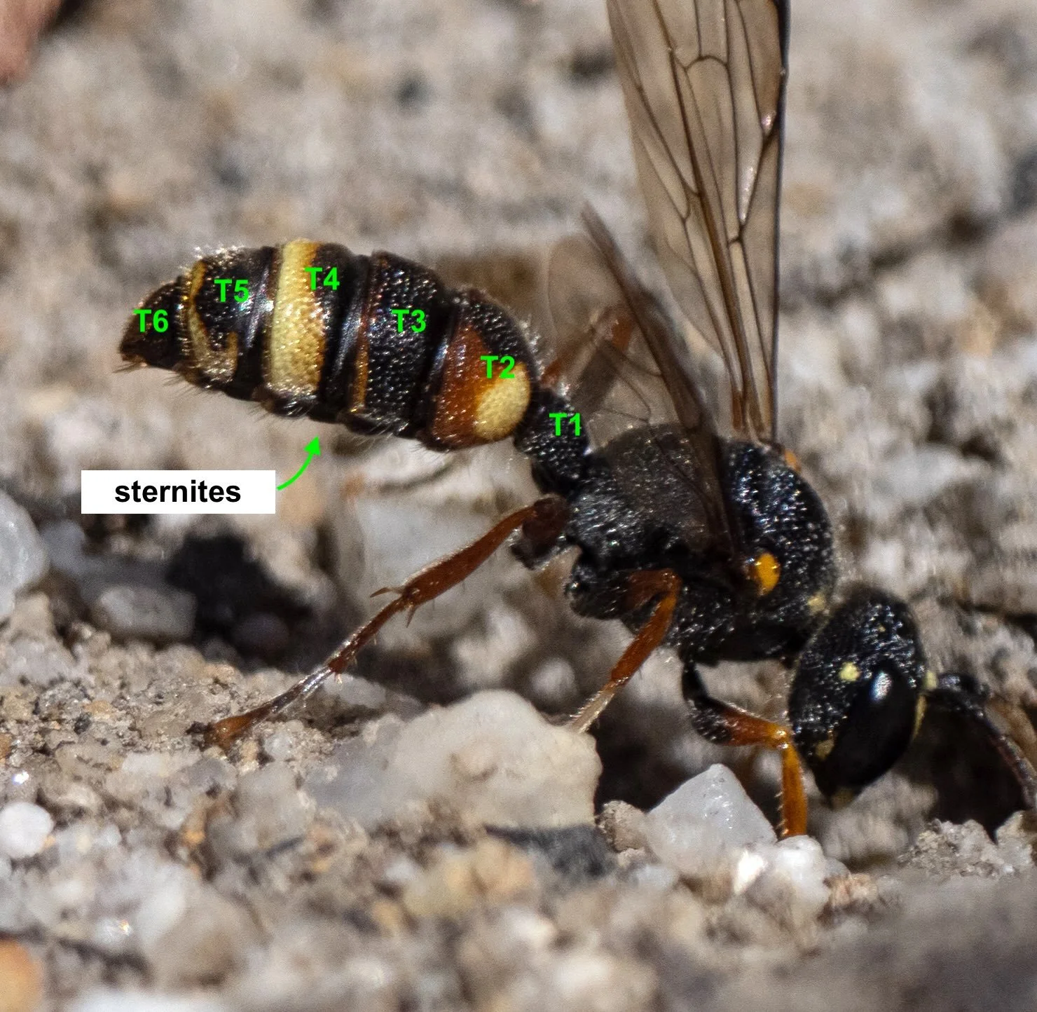
metasoma colour
The colour pattern of the metasoma is remarkably diagnostic. This is particularly true for the colour of tergites (T1/petiole – T6 above). Note that the tergites are visible in dorsal and lateral views, as they wrap well around the segments. The corresponding sternites are best seen in a ventral view, and as such are less often captured in field photos.
colour pattern
C. antipodes is a particularly widespread species, and is one of the most variable in terms of colour pattern. Despite this, the large, yellow, anterolateral spots of T2 are diagnostic.
Note that in this example, the spots are surrounded by a reddish-brown colour.
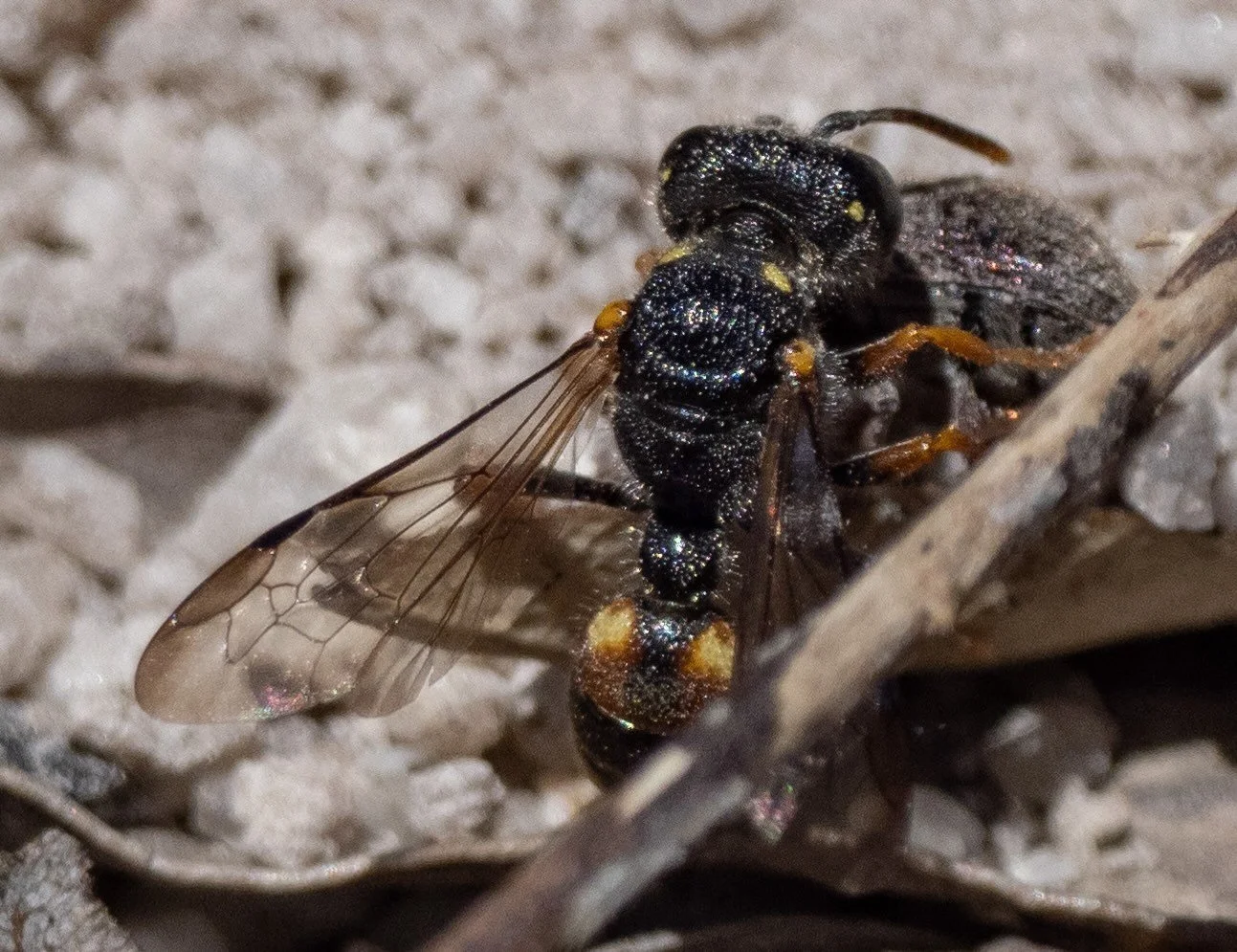
colour pattern
The extent of melanisation (‘blackness’) is a common contributor to intraspecies colour variation.
Compare T2 in this individual with the previous image. The yellow anterolateral spots are present, but otherwise T2 is entirely black. Note also the absence of yellow spots on the pronotum.
Different colour morphs can tend to reflect location. For example, Evans states that southern populations of C. antipodes tend to be darker than those from northern NSW and southeast QLD.
Colour often also varies within populations. These two wasps were part of the same nesting aggregation … observed at the exact same location, just a couple of weeks apart.
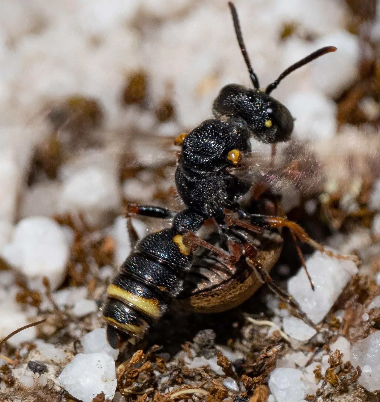
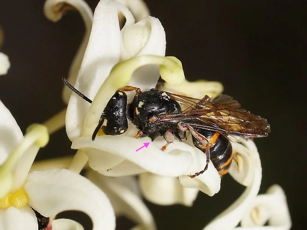
mesopleuron shape
In most Australian Cerceris the mesopleuron is rounded in shape. However, in a few species the lower part bears small tubercles or even large spines … as here (arrow).
https://inaturalist.ala.org.au/observations/25743224
(image courtesy of Reiner Richter)