WING VENATION: Braconidae
2nd recurrent vein (2 m-cu) absent from fore wing; radial cell of hind wing short (or absent in some Braconidae).

FACE & MOUTHPARTS
Hypoclypeal depression wide & deep, with the clypeal margin well above the upper level of the mandible bases. Mandibles curved inwards, tips touching when closed.

SHAPE OF SCUTUM
Scutum not protruding above the pronotum.

HEAD SCULPTURING
No transverse sulcus apparent between antennal sockets (dotted arrow).
No occipital carina (solid arrows).

METASOMA (LATERAL VIEW)
First two metasomal segments less sclerotised laterally (white regions, soft & flexible) than dorsally (pigmented, thickened). Third segment strongly sclerotised dorsally and lateroposteriorly.

WING VENATION: Ichneumonidae
2nd recurrent vein (2 m-cu) present in fore wing; radial cell on hind wing longer than submarginal vein.

1: SCAPE (medial surface, viewed laterally)
Slightly longer ventrally than dorsally. Cylindrical.
However, without apparent emargination.

1: SCAPE (lateral surface, viewed antero-laterally)
Evident emargination on lateral face of scape (dotted arrow).

1: SCAPE (lateral surface, viewed laterally)
Slightly longer ventrally than dorsally (solid arrows). Cylindrical. Emarginate (dotted arrow).

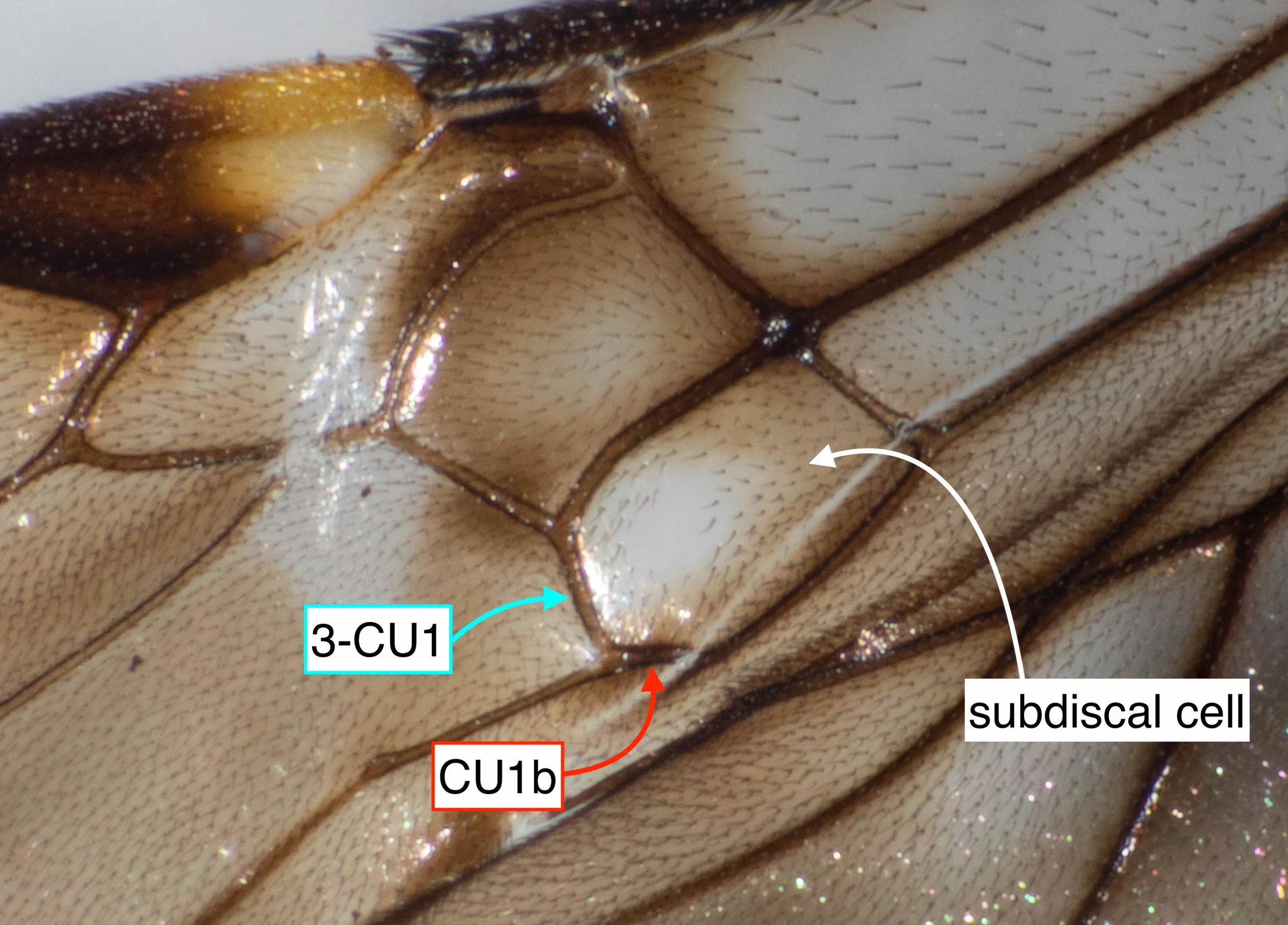
32: FOREWING (right wing, viewed ventrally)
3-CU1 not markedly narrowed posteriorly, and not narrower than vein CU1b.
However, subdiscal cell with medio-distal glabrous area.

32: FOREWING (right wing, viewed ventrally)
Forewing vein 2-1A nearly straight (not markedly curved or angled).

39: HINDWING (right side, base, viewed ventrally)
At least 4 thickened setae (arrow) at the apex of C+SC+R.

HINDWING (right side, base, viewed ventrally)
Four thickened setae (arrow) at the apex of C+SC+R.

PEDICEL (right, viewed laterally)
Sides of pedicel parallel, not petiolate or strongly narrowed basally.

39: PEDICELS (viewed dorsally)
Pedicels not strongly protruding medially, and without specialised sculpturing.

42: TARSAL CLAWS (fore leg, ventral uppermost)
Claws simple, not bifurcate.

42: TARSAL CLAWS (middle leg, ventral uppermost)
Claws simple, not bifurcate.

43: SUBDISCAL CELL DIMENSIONS (left forewing, viewed ventrally)
Length (maximal - pink arrow) 1.5 times width (yellow arrow). That is, not more than twice as long as wide.

43: SUBDISCAL CELL DIMENSIONS (right forewing, viewed ventrally)
Length (maximal - pink arrow) 1.6 times width (yellow arrow).
That is, not more than twice as long as wide.

43: VEIN r-m (left forewing, viewed dorsally)
Tubular, sclerotised for entire length except a single, small bulla posteriorly.

43 & 47: OVIPOSITOR (exposed, viewed laterally)
Ovipositor smooth, without pre-apical notch or nodus.

44: SHAPE OF DISCAL CELL (right forewing, viewed dorsally)
Vein 1=SR+M distinctly curved posteriorly after arising from 1-SR. Also, angle between veins 1-SR and C+SC+R about 90 degrees (i.e. much greater than 55 degrees).

44: SHAPE OF DISCAL CELL (right forewing, viewed ventrally)
(as for 2309L)

45: OVIPOSITOR LENGTH (ventral view)
Short ovipositor … just 25% forewing length.

45: OVIPOSITOR LENGTH (dorsal view)
(as for 2309N)

48: SHAPE OF T2 (dorsal view)
Posteriorly narrowing, mid-basal raised triangular area (dotted arrow).

48: SHAPE OF T2 (lateral view)
Posteriorly narrowing, mid-basal raised triangular area (dotted arrow).

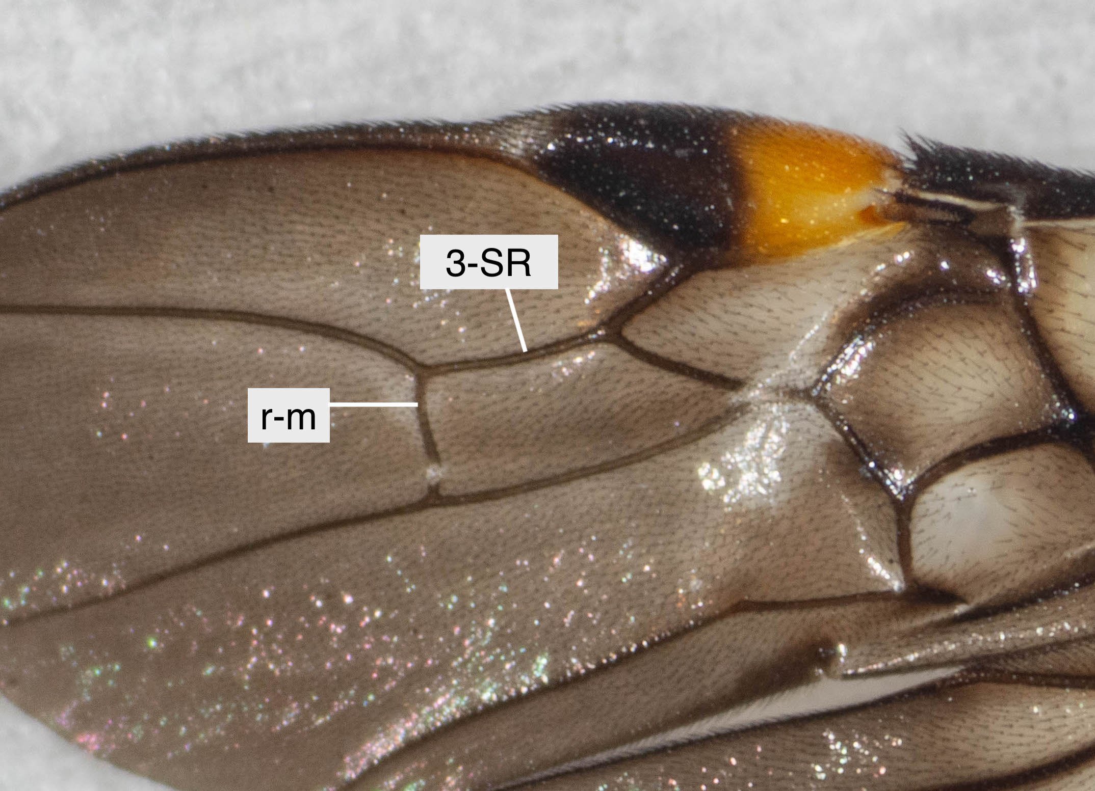
48: 2nd SUBMARGINAL CELL DIMENSIONS (left forewing, viewed dorsally)
Vein 3-SR 1.6 times length of r-m. That is, not more than twice length of r-m.

48: TERGITE MARGINS (lateral view)
Posterior margins of metasomal tergites 3-5 membranous (not sclerotised, not convex in lateral profile), without transverse sub-posterior grooves.

SUMMARY DESCRIPTION OF Vipiellus
extracted from page 135: Quicke, D.L.J. 1987. The Old World genera of braconine wasps (Hymenoptera: Braconidae). Journal of Natural History, 21(1): 43-157

SUMMARY DESCRIPTION OF Vipiellus
extracted from page 332: Quicke, D.L.J. & Ingram, S.N. 1993. Braconine wasps of Australia. Memoirs of the Queensland Museum, 33(1): 299-336

WINGS Vipiellus sp.
extracted from Figure 25. page 311:
Quicke, D.L.J. & Ingram, S.N. 1993. Braconine wasps of Australia. Memoirs of the Queensland Museum, 33(1): 299-336

SUBDISCAL CELL OF FOREWING Vipiellus sp.
extracted from page 329: Quicke, D.L.J. & Ingram, S.N. 1993. Braconine wasps of Australia. Memoirs of the Queensland Museum, 33(1): 299-336

Vipiellus SPECIMEN, IDENTIFIED BY D.L.J. QUICKE
Specimen Depository: Centre for Biodiversity Genomics
Photography: CBG Photography Group, Centre for Biodiversity Genomics
Collectors: Neil Brougham
Specimen Identification: Donald l.J. Quicke
Project Manager: CBG Collections Unit
Collected in Western Australia (5/9/2014)

original description V. australiensis
extract from:
Szépligeti, G.V. 1905. Exotische Braconiden aus den Aethopischen, Orientalischen und Australischen Regionen. Annales Historico-Naturales Musei Nationalis Hungarici (Zoologica) 3: 25-55 [33].

Quick (& rather rough) translation of the original description by Szépligeti.


