1. SCAPE (viewed laterally)
Scape considerably longer ventrally than dorsally. Only slightly emarginate laterally (dotted arrow).

32: FOREWING VENATION (removed & flattened, viewed ventrally)
Forewing vein 3-CU1 not markedly narrowed posteriorly and not narrower than vein CU1b.
1st subdiscal cell evenly setose.
Vein 2-1A not strongly curved or angled.

39. SCAPES & PEDICELS (viewed dorsally)
Scape not angularly narrowed basally (i.e. not petiolate).

39: BASE OF HINDWING (removed & flattened; viewed dorsally)
Four thickened setae at apex of C+SC+R. Note also, a single slightly thickened bristle sub-apically (dotted arrow).

39: BASE OF HINDWING (removed & flattened; viewed ventrally)
Four thickened setae at apex of hind wing vein C+SR+R.

42. TARSAL CLAW
Claw simple, not bifurcate.

43 & 44. SUBDISCAL CELL OF FOREWING (removed & flattened; viewed ventrally)
1st subdiscal cell 1.8 times longer than wide.
Vein r 0.4 times length of vein m-cu

43: FOREWING VEIN r-m (wing removed & flattened; viewed dorsally)
r-m vein largely sclerotized, with 2 distinct bullae.

44. FOREWING (wing removed & flattened; viewed ventrally)
1-SR+M curved/bent posteriorly after arising from 1-SR.
Angle between veins 1-SR and C+SC+R approximately 60 degrees (yellow).
See inset for comparison (Fig. 114, p. 59 in Quicke, 1987), showing Stenobracon, and illustrating method used for estimating the angle. Note that in Stenobracon, the angle is less than 50 degrees.

45. OVIPOSITOR LENGTH (scale in mm)
Ovipositor shorter than forewing.
Ovipositor length (the part extending beyond apex of metasoma) = 9mm.
Forewing length = nearly 10mm.

47. TIP OF OVIPOSITOR (sheath removed; viewed laterally)
Small, pre-apical, dorsal bump (‘nodus’) (green arrow).
Series of ventral serrations (white arrows).
Note: the pre-apical nodus is so small that I find it a bit unconvincing. However, the alternative in the key leads to Vipiellus (& it’s not that) and Iphiaulax. Quicke (1991) describes the ovipositor apex of Iphiaulax as “without distinct preapical ventral serrations” (p. 65) … so I conclude that the tiny bump at the green arrow (above), is indeed a ‘pre-apical nodus’.

47. TIP OF OVIPOSITOR (sheath removed; viewed laterally)
Ventral serrations are more apparent in this image.

49. METASOMA (viewed dorsally)
Moderately elongate, smooth and shiny.

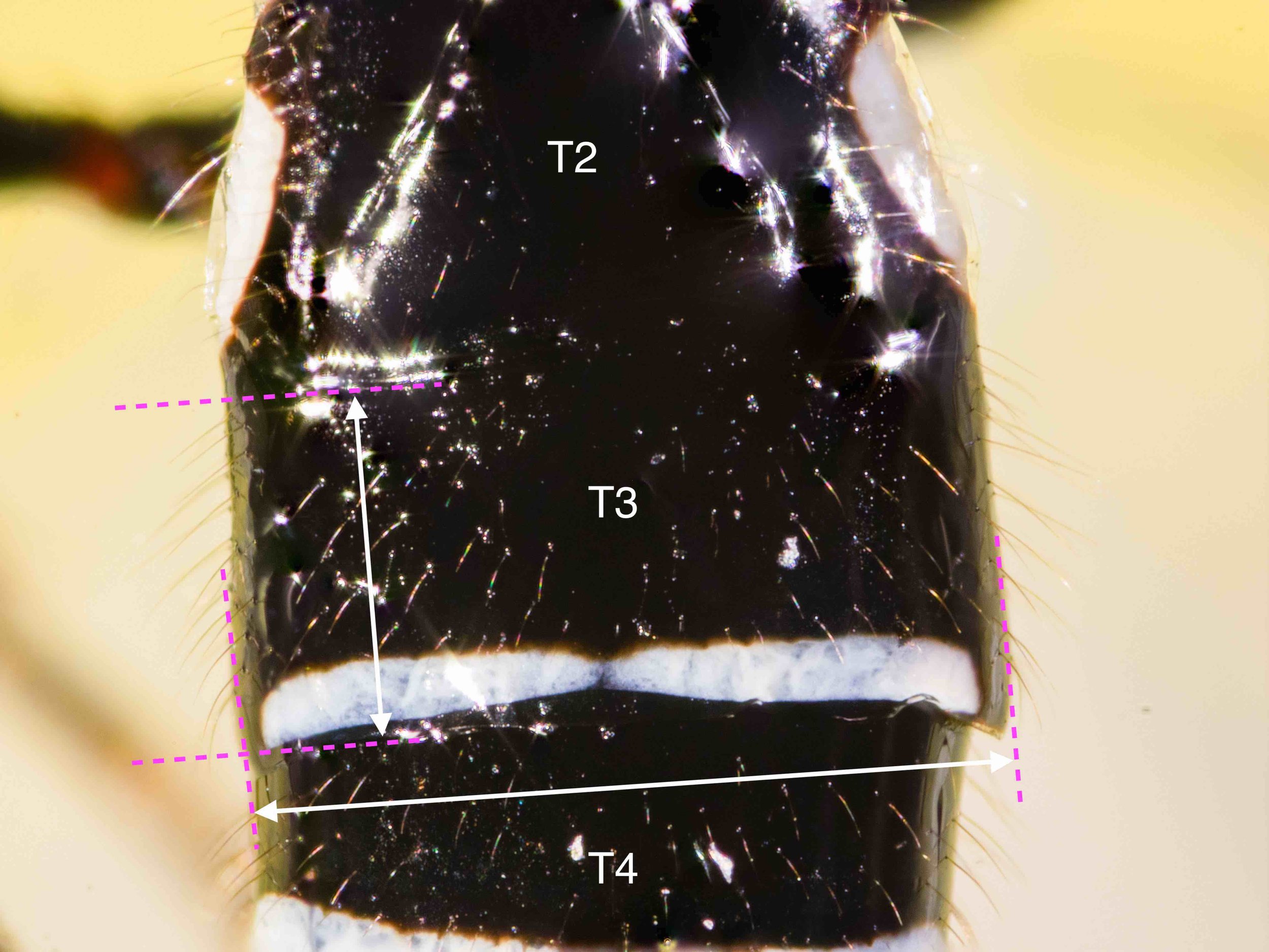
49. 3rd SEGMENT OF METASOMA (viewed dorsally)
3rd metasomal tergite 2.16 times wider than minimally long.

131. METASOMAL, T1 & 2 (viewed dorsally)
Pair of posteriorly diverging, sub-lateral grooves (dotted green arrows).
Well-developed pair of posteriorly converging grooves (solid white arrows) defining a mid-basal triangular are which almost reaches the posterior margin of the tergite (see Fig. 220).

131. METASOMA TERGITES 1-3 (viewed dorso-laterally)
Pair of posteriorly diverging, sub-lateral grooves (dotted green arrows).
Well-developed pair of posteriorly converging grooves (solid white arrow) defining a mid-basal triangular are which almost reaches the posterior margin of the tergite (see Fig. 220).
Suture between T2 and T3 well-defined, but not obviously crenulate.
T3 without antero-lateral areas (see Fig. 239).

130. METASOMA T2 (viewed dorsally)
Lateral margins of the sclerotized part of the 2nd metasomal tergite (arrows) concave behind the spiracles (see Fig. 239).

130. METASOMA T2 (viewed dorso-laterally)
Lateral margins of the sclerotized part of the 2nd metasomal tergite (arrow) concave behind the spiracles (see Fig. 239).

128. FOREWING VEIN cu-a & 1st SUB-DISCAL CELL (viewed ventrally)
Forewing vein cu-a interstitial or nearly so.
Based on the couplet, I interpret ‘interstitial’ to mean in line with 1-M, as the other option is ‘post-furcal’.

HYPOPYGIUM (viewed laterally)
Hypopygium sharply pointed.

HYPOPYGIUM (viewed ventrally)
Hypopygium sharply pointed.

T2-T3 (viewed dorsally)
Join between T2 and T3 (arrows) is narrow, shallow and smooth. It is also sinuous, distinctly curved anteriorly in the middle.
Posterior margin of T3 is banded white.

FOREWING SUBDISCAL CELL (wing removed, flattened, viewed ventrally)
Vein 1-SR+M (arrows) sharply angled posteriorly, then more or less straight to junction with m-cu.

HEAD COLOUR (viewed dorsally)
Orange throughout, including in ocellar area (the ‘stemmaticum’).

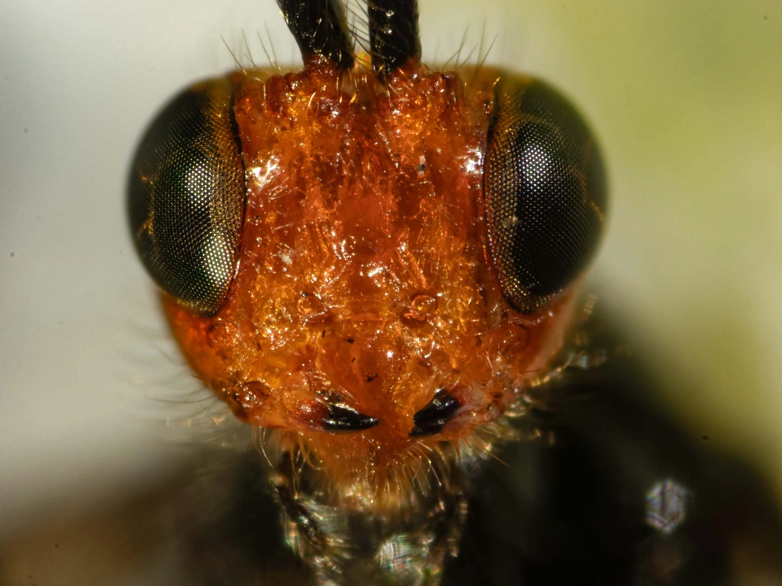
HEAD COLOUR (viewed anteriorly)
Frons orange throughout. Tips of mandibles black.

HEAD & ANTERIOR METATHORAX COLOUR (viewed laterally)
Head orange. Palps black. Propleuron (arrow) black.

SCAPES (viewed dorsally)
Long and narrow. Around 3 times longer than wide.

SCAPE (viewed laterally)
Long and narrow. Around 3 times longer than wide.

Callibracon limbatus
Figures from Austin et. al. 1994


Flattened, viewed dorsally

Flattened, viewed dorsally

Flattened, viewed ventrally

Flattened, viewed ventrally


